. 2017 Jun;31(6):495-509.
doi: 10.1007/s40263-017-0434-z.

Elucidating the Aβ42 Anti-Aggregation Mechanism of Action of Tramiprosate in Alzheimer's Disease: Integrating Molecular Analytical Methods, Pharmacokinetic and Clinical Data

Affiliations

Elucidating the Aβ42 Anti-Aggregation Mechanism of Action of Tramiprosate in Alzheimer's Disease: Integrating Molecular Analytical Methods, Pharmacokinetic and Clinical Data

Petr Kocis et al. CNS Drugs. 2017 Jun.

Abstract

Background: Amyloid beta (Aβ) oligomers play a critical role in the pathogenesis of Alzheimer's disease (AD) and represent a promising target for drug development. Tramiprosate is a small-molecule Aβ anti-aggregation agent that was evaluated in phase III clinical trials for AD but did not meet the primary efficacy endpoints; however, a pre-specified subgroup analysis revealed robust, sustained, and clinically meaningful cognitive and functional effects in patients with AD homozygous for the ε4 allele of apolipoprotein E4 (APOE4/4 homozygotes), who carry an increased risk for the disease. Therefore, to build on this important efficacy attribute and to further improve its pharmaceutical properties, we have developed a prodrug of tramiprosate ALZ-801 that is in advanced stages of clinical development. To elucidate how tramiprosate works, we investigated its molecular mechanism of action (MOA) and the translation to observed clinical outcomes.

Objective: The two main objectives of this research were to (1) elucidate and characterize the MOA of tramiprosate via an integrated application of three independent molecular methodologies and (2) present an integrated translational analysis that links the MOA, conformation of the target, stoichiometry, and pharmacokinetic dose exposure to the observed clinical outcome in APOE4/4 homozygote subjects.

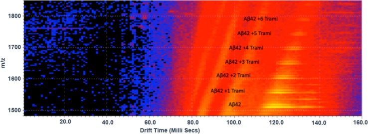
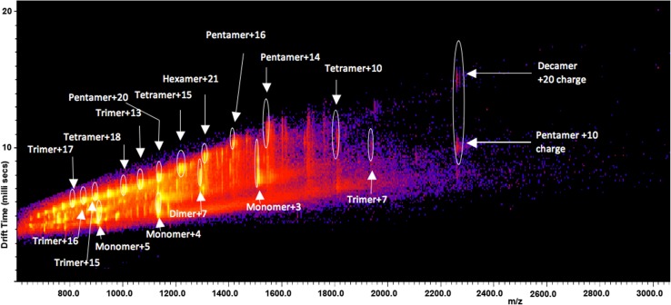
Method: We used three molecular analytical methods-ion mobility spectrometry-mass spectrometry (IMS-MS), nuclear magnetic resonance (NMR), and molecular dynamics-to characterize the concentration-related interactions of tramiprosate versus Aβ42 monomers and the resultant conformational alterations affecting aggregation into oligomers. The molecular stoichiometry of the tramiprosate versus Aβ42 interaction was further analyzed in the context of clinical pharmacokinetic dose exposure and central nervous system Aβ42 levels (i.e., pharmacokinetic-pharmacodynamic translation in humans).

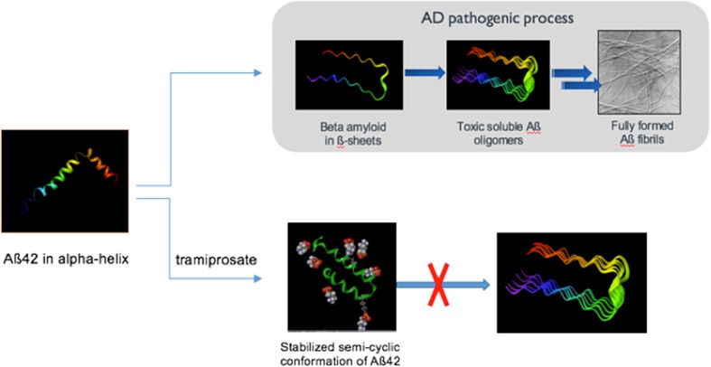
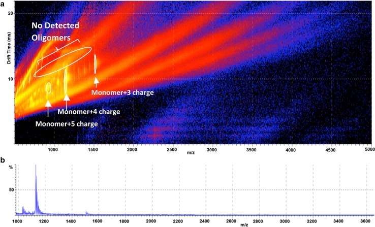
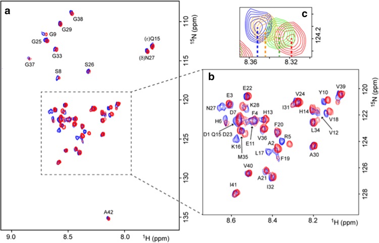
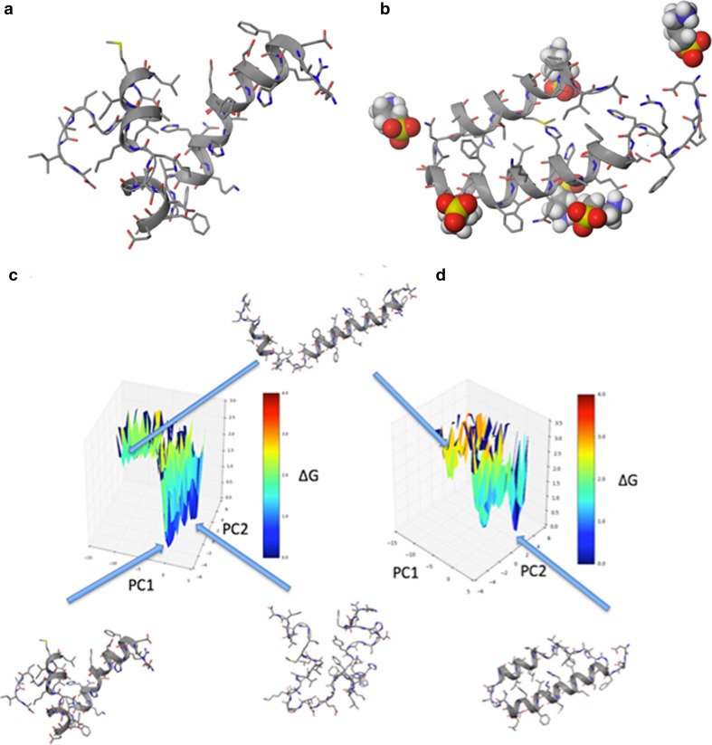
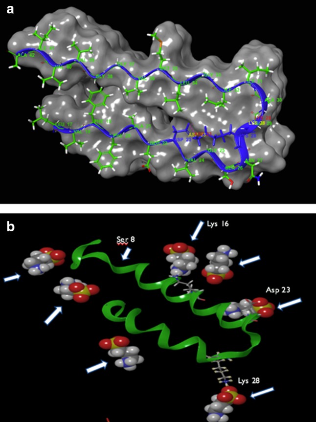
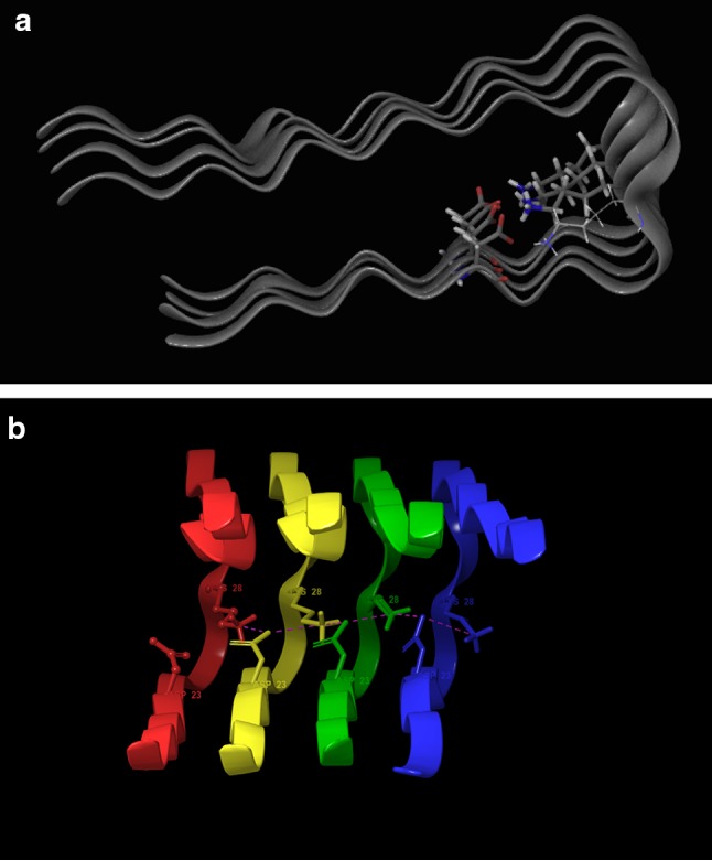
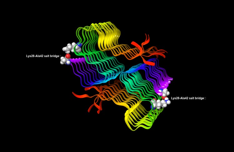
Results: We observed a multi-ligand interaction of tramiprosate with monomeric Aβ42, which differs from the traditional 1:1 binding. This resulted in the stabilization of Aβ42 monomers and inhibition of oligomer formation and elongation, as demonstrated by IMS-MS and molecular dynamics. Using NMR spectroscopy and molecular dynamics, we also showed that tramiprosate bound to Lys16, Lys28, and Asp23, the key amino acid side chains of Aβ42 that are responsible for both conformational seed formation and neuronal toxicity. The projected molar excess of tramiprosate versus Aβ42 in humans using the dose effective in patients with AD aligned with the molecular stoichiometry of the interaction, providing a clear clinical translation of the MOA. A consistent alignment of these preclinical-to-clinical elements describes a unique example of translational medicine and supports the efficacy seen in symptomatic patients with AD. This unique "enveloping mechanism" of tramiprosate also provides a potential basis for tramiprosate dose selection for patients with homozygous AD at earlier stages of disease.

Conclusion: We have identified the molecular mechanism that may account for the observed clinical efficacy of tramiprosate in patients with APOE4/4 homozygous AD. In addition, the integrated application of the molecular methodologies (i.e., IMS-MS, NMR, and thermodynamics analysis) indicates that it is feasible to modulate and control the Aβ42 conformational dynamics landscape by a small molecule, resulting in a favorable Aβ42 conformational change that leads to a clinically relevant amyloid anti-aggregation effect and inhibition of oligomer formation. This novel enveloping MOA of tramiprosate has potential utility in the development of disease-modifying therapies for AD and other neurodegenerative diseases caused by misfolded proteins.


Conflict of interest statement

Funding

Alzheon Inc. sponsored the research described in this article and paid the open access fee.

Conflict of interest

PK, MT, JH, and JY are employees of Alzheon Inc. WS and SR are employees of Schrödinger, which had a scientific services agreement in place to perform some of the work in this manuscript. KB has served as a consultant or on advisory boards for Alzheon, Eli Lilly, Fujirebio Europe, IBL International, Novartis, and Roche Diagnostics, and is a co-founder of Brain Biomarker Solutions in Gothenburg AB, a GU Venture-based platform company at the University of Gothenburg. HF has no competing interests.

Figures

Fig. 1
Fig. 1
Chemical structure of tramiprosate (left) and amino acid sequence of amyloid beta Aβ42 (right)
Fig. 2
Fig. 2
An illustration of the impact of tramiprosate on amyloid beta Aβ42 conformation and the resultant anti-aggregation effects. Comparison of Aβ42 conformation of β-sheets in the pathological process leading to Alzheimer’s disease with the semi-cyclic conformation of Aβ42 under multi-ligand tramiprosate effect, which prevents the formation of Aβ42 oligomers. AD Alzheimer’s disease
Fig. 3
Fig. 3
Ion mobility spectrometry–mass spectrometry (IMS–MS) driftscope plot of the IMS drift time versus mass/charge (m/z) of amyloid beta Aβ42-tramiprosate stoichiometry. Aβ42 alone shows long time drifts (yellow zone), indicating many different populations of conformers. With an increasing number of bound tramiprosate molecules, the drift time of the Aβ42 conformers changes, indicating the presence of fewer and more stabilized conformations. Some of the extended conformers on the right completely disappear
Fig. 4
Fig. 4
Ion mobility spectrometry–mass spectrometry (IMS–MS) 2D arrival time data showing annotated detection of oligomers of amyloid beta Aβ42 after 24-h incubation in the absence of tramiprosate. m/z mass/charge
Fig. 5
Fig. 5
Ion mobility spectrometry–mass spectrometry (IMS–MS) 2D arrival time data. a Detection of no oligomers in the amyloid beta Aβ42 + 1000 × tramiprosate sample after 24-h incubation. b The corresponding mass spectrum detecting only Aβ42 monomers in different charge states. m/z mass/charge
Fig. 6
Fig. 6
2D 1H-15N heteronuclear multiple quantum correlation (HMQC) nuclear magnetic resonance (NMR) spectrum showing interactions of tramiprosate with amyloid beta Aβ42. a 2D 1H-15N HMQC NMR spectrum with assignments. Aβ42 alone is shown in blue, and Aβ42 with tramiprosate at a ratio of 1:1000 is overlaid in red. b An expanded view of part of a. Assignments in red indicate a significant observed chemical shift perturbation. c Example of a chemical shift perturbation of R5 residue observed when Aβ42 was incubated with tramiprosate at a ratio of 1:1000. Residue R5 is an isolated peak that clearly shows a chemical shift as the tramiprosate concentration is increased. The dotted lines illustrate the center of each peak to gauge the change in chemical shift at each concentration level. The color coding represents no tramiprosate (blue), 100:1 (gold), 500:1 (green) and 1000:1 (red) tramiprosate to Aβ42
Fig. 7
Fig. 7
Analysis of molecular dynamics simulations with and without 1% tramiprosate. a Representative disordered structure of amyloid beta Aβ42. b Representative Aβ42 semicyclic ordered structure with six tramiprosate molecules bound. c Principle component analysis of simulation of Aβ42 folding alone in water. d Principle component analysis of simulation of Aβ42 folding in the presence of 1% tramiprosate
Fig. 8
Fig. 8
a Amyloid beta Aβ42 trimer with the first molecule depicted as a β-sheet in blue with Lys28–Asp23 salt bridge also in blue (PDB source 2BEG). The formation of this stabilizing Lys28–Asp23 salt bridge is disrupted by tramiprosate and, consequently, tramiprosate inhibits the formation of not only the critical seeding conformation but also the stabilizing structural element of the otherwise forming oligomers. b Aβ42 conformation adopted under excess tramiprosate conditions. Tramiprosate binds to a number of residues, most prevalently to Lys16 and Lys28 but also to Asp23, and thus prevents the formation of the Lys28–Asp23 salt bridge. Note that Lys28 as well as Asp23 point outward the conformer
Fig. 9
Fig. 9
Solid-state nuclear magnetic resonance (NMR) of amyloid fibril where tetramer is depicted with each individual amyloid beta Aβ42 molecule colored differently to highlight the crosslinking intermolecular salt bridge Lys28–Asp23 (source PDB 2BEG). Thus, red Lys28 forms a salt bridge with yellow Asp23. This intermolecular salt bridge stabilizes the growing superstructure
Fig. 10
Fig. 10
Recently published high atomic resolution of full molecular structures of amyloid beta Aβ42 aggregates [42, 43] illustrating the salt bridge between Lys28 and C-terminal Ala42 (highlighted and annotated). The figure highlights Lys28–Ala42 as examples of salt bridges in the structures that are to be disrupted by tramiprosate binding

Similar articles

Cited by

References

    1. Scheltens P, Blennow K, Breteler MMB, De Strooper B, Frisoni GB, Salloway S, et al. Alzheimer’s disease. Lancet. 2016;388:505–517. doi: 10.1016/S0140-6736(15)01124-1. - DOI - PubMed
    1. Selkoe DJ, Hardy J. The amyloid hypothesis of Alzheimer’s disease at 25 years. EMBO Mol Med. 2016;8:595–608. doi: 10.15252/emmm.201606210. - DOI - PMC - PubMed
    1. Lambert MP, Barlow AK, Chromy BA, Edwards C, Freed R, Liosatos M, et al. Diffusible, nonfibrillar ligands derived from Aβ 1-42 are potent central nervous system neurotoxins. Proc Natl Acad Sci. 1998;95:6448–6453. doi: 10.1073/pnas.95.11.6448. - DOI - PMC - PubMed
    1. Lue L-F, Kuo YM, Roher AE, Brachova L, Shen Y, Sue L, et al. Soluble amyloid beta peptide concentration as a predictor of synaptic change in Alzheimer’s disease. Am J Pathol. 1999;155:853–862. doi: 10.1016/S0002-9440(10)65184-X. - DOI - PMC - PubMed
    1. Viola KL, Klein WL. Amyloid β oligomers in Alzheimer’s disease pathogenesis, treatment, and diagnosis. Acta Neuropathol. 2015;129:183–206. doi: 10.1007/s00401-015-1386-3. - DOI - PMC - PubMed

MeSH terms