. 2017 Jun 1;595(11):3535-3547.
doi: 10.1113/JP273005. Epub 2017 Apr 23.

Direct current stimulation boosts synaptic gain and cooperativity in vitro

Affiliations

Direct current stimulation boosts synaptic gain and cooperativity in vitro

Asif Rahman et al. J Physiol. .

Abstract

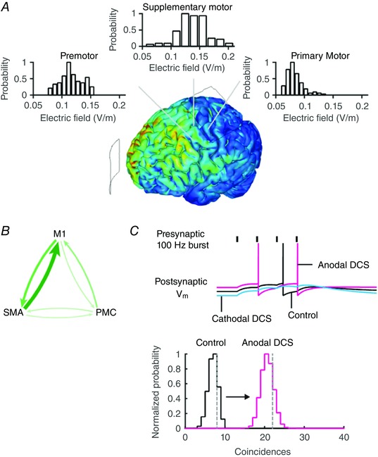
Key points: Direct current stimulation (DCS) polarity specifically modulates synaptic efficacy during a continuous train of presynaptic inputs, despite synaptic depression. DCS polarizes afferent axons and postsynaptic neurons, boosting cooperativity between synaptic inputs. Polarization of afferent neurons in upstream brain regions may modulate activity in the target brain region during transcranial DCS (tDCS). A statistical theory of coincident activity predicts that the diffuse and weak profile of current flow can be advantageous in enhancing connectivity between co-active brain regions.

Abstract: Transcranial direct current stimulation (tDCS) produces sustained and diffuse current flow in the brain with effects that are state dependent and outlast stimulation. A mechanistic explanation for tDCS should capture these spatiotemporal features. It remains unclear how sustained DCS affects ongoing synaptic dynamics and how modulation of afferent inputs by diffuse stimulation changes synaptic activity at the target brain region. We tested the effect of acute DCS (10-20 V m-1 for 3-5 s) on synaptic dynamics with constant rate (5-40 Hz) and Poisson-distributed (4 Hz mean) trains of presynaptic inputs. Across tested frequencies, sustained synaptic activity was modulated by DCS with polarity-specific effects. Synaptic depression attenuates the sensitivity to DCS from 1.1% per V m-1 to 0.55%. DCS applied during synaptic activity facilitates cumulative neuromodulation, potentially reversing endogenous synaptic depression. We establish these effects are mediated by both postsynaptic membrane polarization and afferent axon fibre polarization, which boosts cooperativity between synaptic inputs. This potentially extends the locus of neuromodulation from the nominal target to afferent brain regions. Based on these results we hypothesized the polarization of afferent neurons in upstream brain regions may modulate activity in the target brain region during tDCS. A multiscale model of transcranial electrical stimulation including a finite element model of brain current flow, numerical simulations of neuronal activity, and a statistical theory of coincident activity predicts that the diffuse and weak profile of current flow can be advantageous. Thus, we propose that specifically because tDCS is diffuse, weak and sustained it can boost connectivity between co-active brain regions.

Keywords: electrical brain stimulation; synaptic depression; transcranial direct current stimulation.


Figures

Figure 1
Figure 1. The postsynaptic voltage response during DCS and ongoing presynaptic activity results in sustained and cumulative changes that are regulated by synaptic efficacy, number of active inputs, and rate of presynaptic activity
A, schematic diagram representing the voltage output of a postsynaptic cell during a train of presynaptic spikes arriving at times {t nk } down N f input fibres. Synaptic transmission at cortical synapses are not static, but are dynamically regulated by the available pool of releasable vesicles. B, synaptic efficacy during activity is controlled by short‐term plasticity based on the initial vesicle release probabilities (P release) and vesicle depletion and recovery times. C, simulation of the postsynaptic response during DCS in a conductance‐based model with dynamic synapses. Anodal DCS (magenta), modelled as a postsynaptic depolarization, facilitates EPSP amplitudes and cathodal DCS (blue), modelled as a postsynaptic hyperpolarization, depresses EPSP amplitudes. The postsynaptic response during DCS is sustained for the duration of DCS. [Color figure can be viewed at wileyonlinelibrary.com]
Figure 2
Figure 2. DCS in rat primary motor cortex results in a sustained modulation of synaptic efficacy
A, schematic diagram of anodal (left) and cathodal (right) DCS with current flow along the somato‐dendritic axis and the effective membrane polarization of cells and axons in false colour. B, orthodromic stimulation of the L5 → 2/3 pathway at a constant frequency (20 Hz shown) in M1 facilitates synaptic efficacy during anodal DCS and depresses efficacy during cathodal DCS, compared to control cases without DCS (red: anodal, blue: cathodal, black: control, no DCS). The DCS effects are sustained for the duration of the electric field (EF). C, DCS modulates synaptic efficacy during continuous afferent inputs despite synaptic depression arising from vesicle depletion in the presynaptic terminals. D, the immediate change in field potential (FP) amplitude during DCS is correlated with the last FP amplitude during the train. Panel D shows FP amplitudes normalized by the corresponding n‐th FP amplitude during a train without DCS. E, the sensitivity (% change in FP amplitude per V m−1) to DCS of the last FP is less than the sensitivity of the first FP (n = 124) during the train. Grey best fit lines are for each of the tested frequencies (5, 10, 20, and 40 Hz) and red is the combined average across frequencies. There was no significant difference in sensitivities in either first or last FP across frequencies. F, the rate of cumulative change in FP amplitude (the cumulative % change from baseline) is greater during anodal DCS and less during cathodal DCS compared to control. Dark points indicate 20 V m−1 and light points are 10 V m−1. In all cases, magenta indicates anodal DCS and blue is cathodal DCS (error bars are standard error). [Color figure can be viewed at wileyonlinelibrary.com]
Figure 3
Figure 3. Synaptic efficacy is frequency‐independently modulated during DCS, sustained for the duration of the electric field (EF), and is cumulative
A, the effect of DCS on synaptic efficacy is sustained during the trains at a constant rate of input. Here, FP amplitudes during DCS are normalized by the corresponding n‐th FP amplitude in a control train, within slice (filled circles: anodal DCS, +20 V m−1; open circles: cathodal DCS, −20 V m−1). B, the immediate change in FP amplitude (x‐axis, FP1,DCS/ FP1,Control) is correlated with the steady‐state change in FP amplitude (y‐axis, FPSS,DCS/ FPSS,Control, where SS refers to the FP amplitude at steady state). C, anodal DCS attenuates synaptic depression. The cumulative percentage change from baseline is plotted for anodal (magenta), cathodal (blue), and control (black) conditions. DCS can, in some cases, reverse synaptic depression. [Color figure can be viewed at wileyonlinelibrary.com]
Figure 4
Figure 4. DCS produces a sustained change in synaptic efficacy during a naturalistic pattern of presynaptic activity
Afferent inputs reflecting the Poisson distributed spike train pattern in vivo (mean rate 4 Hz) is acutely facilitated by anodal DCS (magenta) and depressed by cathodal DCS (blue). [Color figure can be viewed at wileyonlinelibrary.com]
Figure 5
Figure 5. DCS post adaptation modulates synaptic efficacy and is driven by recruitment of afferent axons
A, cathodal (blue) and anodal DCS (magenta) during a train of synaptic inputs at 10 Hz modulates synaptic efficacy for the duration of the electric field (EF) despite adaptation. B, DCS polarity specifically modulates the first field potential (FP) amplitude relative to pre‐DCS. C, there is a correlation between the change in the first FP amplitude and the amount of recovery from facilitation or depression during DCS. D, changing the stimulus intensity, instead of applying DCS, results in similar dynamics on FP amplitude. Decreasing stimulus intensity by a half (left) decreases FP amplitudes. There is a pronounced rebound in FP amplitude when the stimulus intensity is returned to the probe intensity. E, a simple dynamic synapse model incorporating recruitment of presynaptic inputs is able to reproduce the effects of DCS on synaptic efficacy and the post‐DCS rebound effect. [Color figure can be viewed at wileyonlinelibrary.com]
Figure 6
Figure 6. DCS modulates synaptic efficacy post adaptation through somatic polarization alone
A, schematic diagram of orthodromic stimulation of L2/3 during DCS. Current flow along the somato‐dendritic axis and perpendicular to the horizontally oriented axons selectively polarizes the postsynaptic cells and not the axons. B, current flow along the somato‐dendritic axis during orthodromic stimulation of the horizontal L2/3 pathway modulates synaptic efficacy independently of axonal recruitment. C, anodal DCS facilitates synaptic efficacy and cathodal DCS depresses efficacy. [Color figure can be viewed at wileyonlinelibrary.com]
Figure 7
Figure 7. Current flow along axons during DCS modulates fibre volley in rat primary motor cortex and hippocampus
A, current flow along the somato‐dendritic axis during orthodromic stimulation of the ascending L5 → 2/3 pathway polarizes afferent axons and modulates axonal recruitment during DCS. The amplitude of the fibre volley (FV) reflects the number of axons firing action potentials during orthodromic stimulation. B, similarly, current flow along the afferent axons of the Schaffer collateral pathway modulates the fibre volley amplitude. C, the change in fibre volley amplitudes is directly correlated with the change in postsynaptic field potential (FP) amplitude in the rat primary motor cortex. These results show polarizing afferent axons changes the number of active presynaptic inputs and directly modulates synaptic current. [Color figure can be viewed at wileyonlinelibrary.com]
Figure 8
Figure 8. tDCS produces diffuse current flow in the brain which increases the probability of coincident pre‐ and postsynaptic inputs
A, the electric field magnitude is plotted in a finite element model of tDCS with anode over the primary motor cortex (M1) and cathode over supraorbital region (SO). The electric field magnitude distribution in brain regions adjacent and upstream to M1 is comparable to or higher than in M1. B, the primary motor cortex is synaptically connected to the supplementary motor area (SMA) and premotor cortex (PMC), which drives motor activity. The thickness of the arrows indicates the relative connection strength from fMRI studies of this functional network. Modulating presynaptic activity in these upstream brain regions during tDCS may drive postsynaptic activity in M1. C, DCS‐induced membrane polarization changes the likelihood of firing. An increase in the presynaptic activity (or postsynaptic activity) increases the probability of coincident pre‐ and postsynaptic action potentials. The vertical grey dashed lines in the graph indicate the simulated number of coincidences in a model Integrate & Fire (I&F) neuron receiving Poisson distributed synaptic inputs. The hypergeometric probability distribution can be used to approximate the estimated number of coincidences. Combined, these results suggest the probability of coincident inputs can be directly modulated when presynaptic or postsynaptic activity is modulated by diffuse current flow. [Color figure can be viewed at wileyonlinelibrary.com]

References

    1. Abbott LF & Regehr WG (2004). Synaptic computation. Nature 431, 796–803. - PubMed
    1. Abbott LF, Varela JA, Sen K & Nelson SB (1997). Synaptic depression and cortical gain control. Science 275, 220–224. - PubMed
    1. Antal A, Bikson M, Datta A, Lafon B, Dechent P, Parra LC & Paulus W (2012). Imaging artifacts induced by electrical stimulation during conventional fMRI of the brain. Neuroimage 85, 1040–1047. - PMC - PubMed
    1. Antal A, Varga ET, Kincses TZ, Nitsche MA & Paulus W (2004). Oscillatory brain activity and transcranial direct current stimulation in humans. Neuroreport 15, 1307–1310. - PubMed
    1. Baczyk M & Jankowska E (2014). Presynaptic actions of transcranial and local direct current stimulation in the red nucleus. J Physiol 592, 4313–4328. - PMC - PubMed

Publication types

LinkOut - more resources