

Brain temperature but not core temperature increases during spreading depolarizations in patients with spontaneous intracerebral hemorrhage
- PMID: 28436257
- PMCID: PMC5851146
- DOI: 10.1177/0271678X17703940
Brain temperature but not core temperature increases during spreading depolarizations in patients with spontaneous intracerebral hemorrhage
Abstract
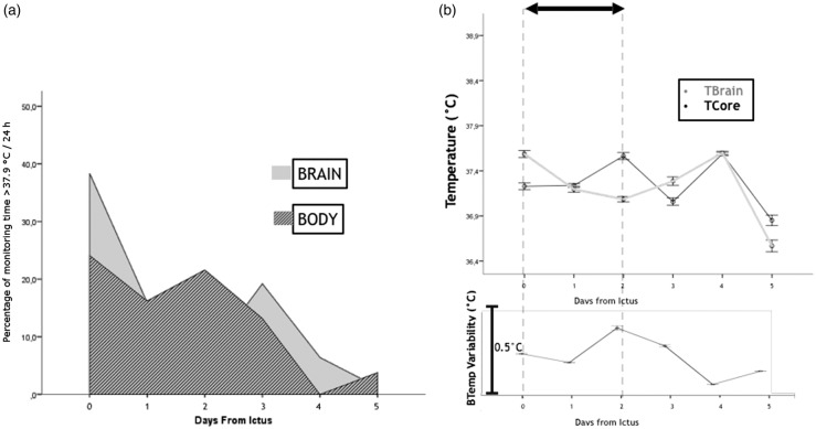
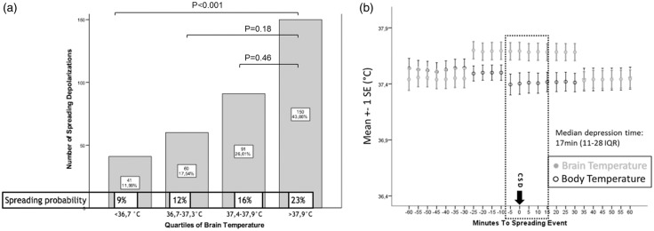
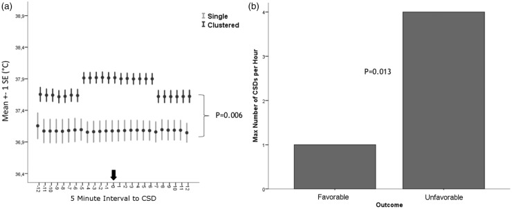
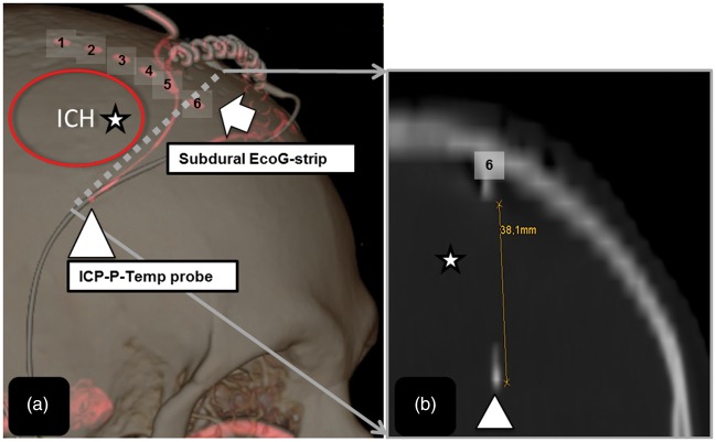
Spreading depolarizations (SDs) are highly active metabolic events, commonly occur in patients with intracerebral hemorrhage (ICH) and may be triggered by fever. We investigated the dynamics of brain-temperature (Tbrain) and core-temperature (Tcore) relative to the occurrence of SDs. Twenty consecutive comatose ICH patients with multimodal electrocorticograpy (ECoG) and Tbrain monitoring of the perihematomal area were prospectively enrolled. Clusters of SDs were defined as ≥2 SDs/h. Generalized estimating equations were used for statistical calculations. Data are presented as median and interquartile range. During 3097 h (173 h [81-223]/patient) of ECoG monitoring, 342 SDs were analyzed of which 51 (15%) occurred in clusters. Baseline Tcore and Tbrain was 37.3℃ (36.9-37.8) and 37.4℃ (36.7-37.9), respectively. Tbrain but not Tcore significantly increased 25 min preceding the onset of SDs by 0.2℃ (0.1-0.2; p < 0.001) and returned to baseline 35 min following SDs. During clusters, Tbrain increased to a higher level (+0.4℃ [0.1-0.4]; p = 0.006) when compared to single SDs. A higher probability (OR = 36.9; CI = 36.8-37.1; p < 0.001) of developing SDs was observed during episodes of Tbrain ≥ 38.0℃ (23% probability), than during Tbrain ≤ 36.6℃ (9% probability). Spreading depolarizations - and in particular clusters of SDs - may increase brain temperature following ICH.
Keywords: Cortical spreading depolarizations; body temperature; brain temperature; brain temperature regulation; spontaneous intracerebral hemorrhage.
Figures
Similar articles
-
Spreading depolarizations in patients with spontaneous intracerebral hemorrhage: Association with perihematomal edema progression.J Cereb Blood Flow Metab. 2017 May;37(5):1871-1882. doi: 10.1177/0271678X16651269. Epub 2016 Jan 1. J Cereb Blood Flow Metab. 2017. PMID: 27207168 Free PMC article.
-
Prognostic Value of Spreading Depolarizations in Patients With Severe Traumatic Brain Injury.JAMA Neurol. 2020 Apr 1;77(4):489-499. doi: 10.1001/jamaneurol.2019.4476. JAMA Neurol. 2020. PMID: 31886870 Free PMC article.
-
Neurostereologic Lesion Volumes and Spreading Depolarizations in Severe Traumatic Brain Injury Patients: A Pilot Study.Neurocrit Care. 2019 Jun;30(3):557-568. doi: 10.1007/s12028-019-00692-w. Neurocrit Care. 2019. PMID: 30972614
-
Correlates of spreading depolarization in human scalp electroencephalography.Brain. 2012 Mar;135(Pt 3):853-68. doi: 10.1093/brain/aws010. Brain. 2012. PMID: 22366798 Free PMC article.
-
Transhemispheric depolarizations persist in the intracerebral hemorrhage swine brain following corpus callosal transection.Brain Res. 2006 Feb 16;1073-1074:481-90. doi: 10.1016/j.brainres.2005.12.071. Epub 2006 Jan 27. Brain Res. 2006. PMID: 16443194
Cited by
-
The focus of temperature monitoring with zero-heat-flux technology (3M Bair-Hugger): a clinical study with patients undergoing craniotomy.J Clin Monit Comput. 2019 Oct;33(5):917-923. doi: 10.1007/s10877-018-0227-z. Epub 2018 Nov 22. J Clin Monit Comput. 2019. PMID: 30467673 Free PMC article.
-
The neurophysiological effect of mild hypothermia in gyrencephalic brains submitted to ischemic stroke and spreading depolarizations.Front Neurosci. 2024 Mar 14;18:1302767. doi: 10.3389/fnins.2024.1302767. eCollection 2024. Front Neurosci. 2024. PMID: 38567280 Free PMC article.
-
The negative ultraslow potential, electrophysiological correlate of infarction in the human cortex.Brain. 2018 Jun 1;141(6):1734-1752. doi: 10.1093/brain/awy102. Brain. 2018. PMID: 29668855 Free PMC article.
-
Brain temperature regulation in poor-grade subarachnoid hemorrhage patients - A multimodal neuromonitoring study.J Cereb Blood Flow Metab. 2021 Feb;41(2):359-368. doi: 10.1177/0271678X20910405. Epub 2020 Mar 9. J Cereb Blood Flow Metab. 2021. PMID: 32151225 Free PMC article.
-
Spreading Depolarizations Suppress Hematoma Growth in Hyperacute Intracerebral Hemorrhage in Mice.Stroke. 2023 Oct;54(10):2640-2651. doi: 10.1161/STROKEAHA.123.042632. Epub 2023 Aug 23. Stroke. 2023. PMID: 37610105 Free PMC article.
References
-
- Hemphill JC, 3rd, Bonovich DC, Besmertis L, et al. The ICH score: a simple, reliable grading scale for intracerebral hemorrhage. Stroke 2001; 32: 891–897. - PubMed
-
- Rincon F, Patel U, Schorr C, et al. Brain injury as a risk factor for fever upon admission to the intensive care unit and association with in-hospital case fatality: a matched cohort study. J Intensive Care Med 2015; 30: 107–114. - PubMed
-
- Badjatia N. Hyperthermia and fever control in brain injury. Crit Care Med 2009; 37(7 Suppl): S250–S257. - PubMed
-
- Sessler DI. Perioperative thermoregulation and heat balance. Lancet 2016; 387: 2655–2664. - PubMed
Publication types
MeSH terms
LinkOut - more resources
Full Text Sources
Other Literature Sources
