. 2017 Apr 24:7:46683.
doi: 10.1038/srep46683.

Interactions of AMTN, ODAM and SCPPPQ1 proteins of a specialized basal lamina that attaches epithelial cells to tooth mineral

Affiliations

Interactions of AMTN, ODAM and SCPPPQ1 proteins of a specialized basal lamina that attaches epithelial cells to tooth mineral

Aurélien Fouillen et al. Sci Rep. .

Abstract

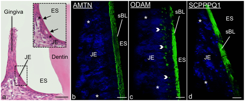
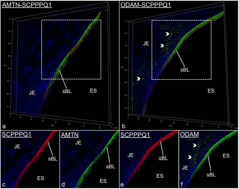
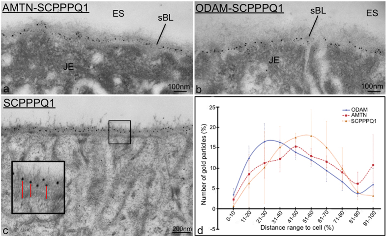
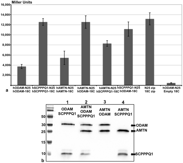
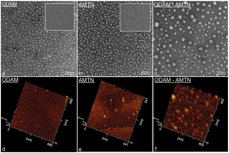
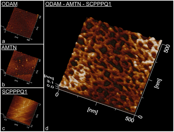
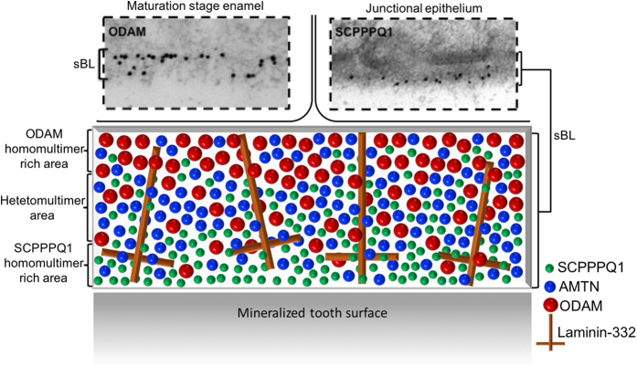
A specialized basal lamina (sBL) mediates adhesion of certain epithelial cells to the tooth. It is distinct because it does not contain collagens type IV and VII, is enriched in laminin-332, and includes three novel constituents called amelotin (AMTN), odontogenic ameloblast-associated (ODAM), and secretory calcium-binding phosphoprotein proline-glutamine rich 1 (SCPPPQ1). The objective of this study was to clarify the structural organization of the sBL. Fluorescence and immunogold labeling showed that the three proteins co-localize. Quantitative analysis of the relative position of gold particles on the sBL demonstrates that the distribution of ODAM is skewed towards the cell while that of AMTN and SCPPPQ1 tends towards the tooth surface. Bacterial two-hybrid analysis and co-immunoprecipitation, gel filtration of purified proteins and transmission electron and atomic force microscopies highlight the propensity of AMTN, ODAM, and SCPPPQ1 to interact with and among themselves and form supramolecular aggregates. These data suggest that AMTN, ODAM and SCPPPQ1 participate in structuring an extracellular matrix with the distinctive capacity of attaching epithelial cells to mineralized surfaces. This unique feature is particularly relevant for the adhesion of gingival epithelial cells to the tooth surface, which forms a protective seal that is the first line of defense against bacterial invasion.


Conflict of interest statement

The authors declare no competing financial interests.

Figures

Figure 1
Figure 1. AMTN, ODAM and SCPPPQ1 localize at the cell-tooth interface.
(a) Micrograph from a histological section stained with hematoxylin and eosin showing apposition of the junctional epithelium (JE) to the enamel, seen here as a space (ES) following decalcification. The inset is a higher magnification of the JE cells (*). The arrows point to the position of the associated specialized basal lamina (sBL). (bd) Immunofluorescence preparations for AMTN, ODAM and SCPPPQ1 imaged by Structured Illumination Microscopy (SIM) indicate that the three proteins reside in the sBL. ODAM also exhibits a cell-associated labeling (arrowheads). Proteins stained in green, nuclei in blue using DAPI. Scale bars = 10 μm.
Figure 2
Figure 2. Immunofluorescence labeling for AMTN, ODAM and SCPPPQ1 visualized in junctional epithelium (JE) preparations by Structured Illumination Microscopy.
(a and b) Illustrate dual-labelled preparations. (cf) Are the corresponding single channel images at higher magnification. These dual preparations validate that the proteins co-localize at the cell-tooth interface where the specialized basal lamina (sBL) is found. AMTN/ODAM in green; SCPPPQ1 in red; nuclei in blue using DAPI.
Figure 3
Figure 3. High resolution immunogold labeling demonstrates that AMTN, ODAM and SCPPPQ1 distribute differentially across the specialized basal lamina (sBL).
(a,b) As exemplified here with the maturation stage enamel organ (EO), dual labeling evidences with high resolution the co-localisation of the three proteins in the specialized basal lamina (sBL). (c,d) Single immunogold preparations; Quantification of the distribution of particles relative to the cell surface reveals that the three proteins distribute differentially across the sBL. AMTN/ODAM 10 nm gold particles; SCPPPQ1 20 nm gold particles.
Figure 4
Figure 4. Interaction analysis between AMTN, ODAM and SCPPPQ1.
(a) Bacterial two-hybrid analysis between human proteins after fusions to the PKNT25 and PUT18C domains of adenylate cyclase. All 3 proteins have the propensity to interact, with AMTN-ODAM and SCPPPQ1-ODAM interactions showing the highest level of interaction. Positive control = PUT18Czip +PKNT25zip; negative control = hSCPPPQ1-PKNT25 + empty PUT18C vector. (b) These interactions are confirmed by co-immunoprecipitation followed by Western blotting.
Figure 5
Figure 5. Visual characterization of the aggregation behaviour of AMTN and ODAM.
(a–c) Transmission electron microscopy (TEM) of negative stain and (d–f) atomic force microscopy preparations of purified AMTN and ODAM demonstrate the propensity of the proteins to self-interact into globular complexes. Mixing the two proteins together generates larger complexes suggesting heterologous interactions. (Insets in a,b) Observation of the complexes under native conditions by cryo-TEM confirms that they do not result from preparation artefacts.
Figure 6
Figure 6. Atomic force microscope characterization of AMTN, ODAM and SCPPPQ1 proteins.
(ac) The discrete profiles formed individually by each protein differ dramatically from (d) the intricate network formed created when they are mixed together.
Figure 7
Figure 7. Schematic model of the distribution of AMTN, ODAM and SCPPPQ1 within the supramolecular organisation of the specialized basal lamina (sBL).
The two boxed above in images are representative immunogold labelings for ODAM (skewed toward the cells) and SCPPPQ1 (skewed toward the tooth surface) over the sBL from the maturation stage enamel organ and the junctional epithelium. The model attempts to integrate the observed differential distribution of the three proteins within a laminin-332 background whose arrangement within the unique extracellular matrix constituted by the sBL remains to be clarified.

References

    1. Ekblom M., Falk M., Salmivirta K., Durbeej M. & Ekblom P. Laminin isoforms and epithelial development. Annals of the New York Academy of Sciences 857, 194–211 (1998). - PubMed
    1. McGuire J. D., Walker M. P., Dusevich V., Wang Y. & Gorski J. P. Enamel organic matrix: potential structural role in enamel and relationship to residual basement membrane constituents at the dentin enamel junction. Connect. Tissue Res. 55 Suppl 1, 33–37, doi: 10.3109/03008207.2014.923883 (2014). - DOI - PMC - PubMed
    1. Scheele S. et al.. Laminin isoforms in development and disease. J. Mol. Med. (Berl.) 85, 825–836, doi: 10.1007/s00109-007-0182-5 (2007). - DOI - PubMed
    1. Kruegel J. & Miosge N. Basement membrane components are key players in specialized extracellular matrices. Cellular and molecular life sciences: CMLS 67, 2879–2895, doi: 10.1007/s00018-010-0367-x (2010). - DOI - PMC - PubMed
    1. Takata T., Nikai H., Ijuhin N. & Okamoto H. Ultrastructure of regenerated junctional epithelium after surgery of the rat molar gingiva. J Periodontol. 57, 776–783 (1986). - PubMed

Publication types