

PathoSpotter-K: A computational tool for the automatic identification of glomerular lesions in histological images of kidneys
- PMID: 28436482
- PMCID: PMC5402276
- DOI: 10.1038/srep46769
PathoSpotter-K: A computational tool for the automatic identification of glomerular lesions in histological images of kidneys
Abstract
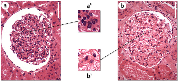
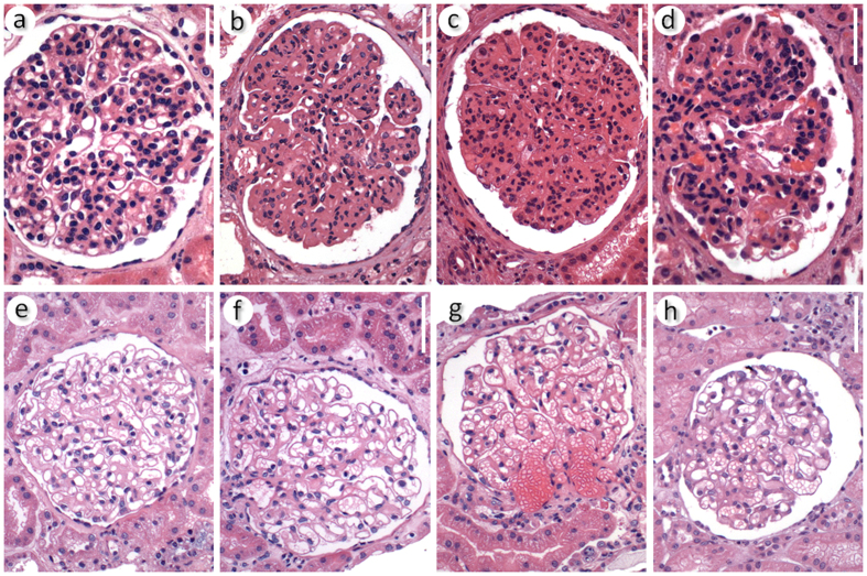
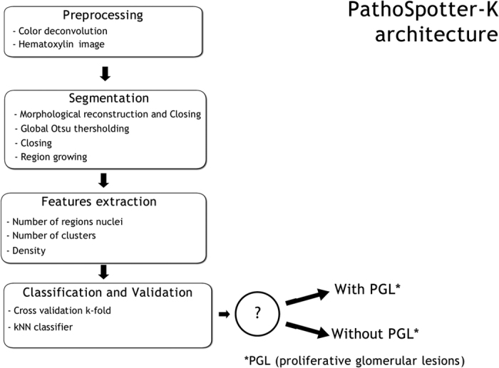
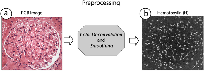
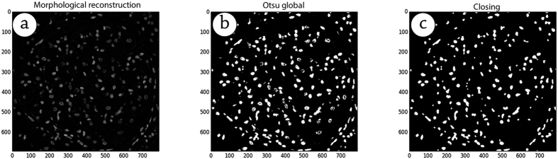
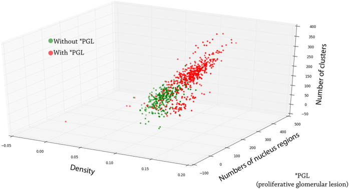
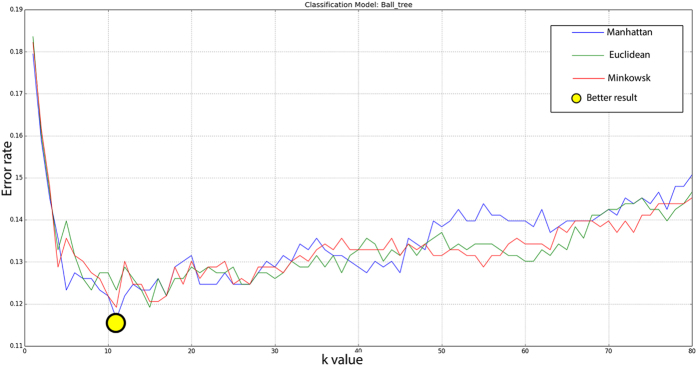
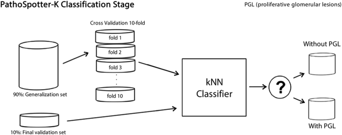
PathoSpotter is a computational system designed to assist pathologists in teaching about and researching kidney diseases. PathoSpotter-K is the version that was developed to detect nephrological lesions in digital images of kidneys. Here, we present the results obtained using the first version of PathoSpotter-K, which uses classical image processing and pattern recognition methods to detect proliferative glomerular lesions with an accuracy of 88.3 ± 3.6%. Such performance is only achieved by similar systems if they use images of cell in contexts that are much less complex than the glomerular structure. The results indicate that the approach can be applied to the development of systems designed to train pathology students and to assist pathologists in determining large-scale clinicopathological correlations in morphological research.
Conflict of interest statement
The authors declare no competing financial interests.
Figures
References
-
- Churg J., Bernstein J. & Glassock R. J. Renal disease: classification and atlas of glomerular diseases. 2 edn (Igaku-Shoin, 1995).
-
- Walker P. D., Cavallo T. & Bonsib S. M. Practice guidelines for the renal biopsy. Mod Pathol 17, 1555–1563 (2004). - PubMed
-
- Fogo A. B. Approach to renal biopsy. Am J Kidney Dis 42, 826–836, doi: S0272638603010540 (2003). - PubMed
-
- Tolles W. E. The cytoanalyzer-an example of physics in medical research. Trans N Y Acad Sci 17, 250–256 (1955). - PubMed
Publication types
MeSH terms
LinkOut - more resources
Full Text Sources
Other Literature Sources
Medical
