Review
. 2017 Aug;14(8):484-491.
doi: 10.1038/nrcardio.2017.57. Epub 2017 Apr 24.

Redefining the identity of cardiac fibroblasts

Affiliations
Review

Redefining the identity of cardiac fibroblasts

Michelle D Tallquist et al. Nat Rev Cardiol. 2017 Aug.

Abstract

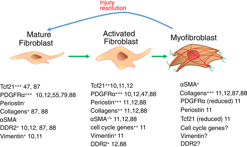
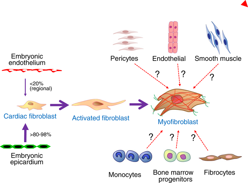
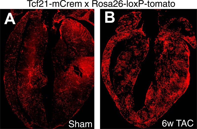
Cardiac fibroblasts deposit and maintain extracellular matrix during organogenesis and under physiological conditions. In the adult heart, activated cardiac fibroblasts also participate in the healing response after acute myocardial infarction and during chronic disease states characterized by augmented interstitial fibrosis and ventricular remodelling. However, delineation of the characteristics, plasticity, and origins of cardiac fibroblasts is an area of ongoing investigation and controversy. A set of genetic mouse models has been developed that specifically addresses the nature of these cells, in terms of both their origins and their response during cardiac disease and ventricular remodelling. As our understanding of cardiac fibroblasts becomes more defined and refined, so does the potential to develop new therapeutic strategies to control fibrosis and adverse ventricular remodelling.


Conflict of interest statement

Competing interests statement

The authors declare no competing interests.

Figures

Figure 1 |
Figure 1 |. Current categories of resident cardiac fibroblasts.
At least three gene-expression profiles can be used to describe fibroblasts in the adult heart−,,. Mature, resident fibroblasts are interspersed in the myomesial space throughout the heart and are proposed to maintain the extracellular matrix (ECM). They have a low level of proliferation and do not express smooth muscle α-actin (α-SMA) or periostin. After injury, a population of fibroblasts, often associated with inflammatory cell accumulation and cardiomyocyte death, rapidly proliferate and become activated to express many of the gene products shown. Later in the response, a smaller population of these activated fibroblasts further differentiate into presumed myofibroblasts, which express α-SMA and other genetic signatures shown. In the past, these myofibroblasts were reported to have increased ECM deposition and contractile capacities, although in vivo contractile activity has been predominantly documented in skin myofibroblasts. Myofibroblasts have also been shown to regress their activated gene-expression profile and morphology back to the more ‘quiescent’ and resident fibroblast state. Ddr2, discoidin domain-containing receptor 2; NA, not available; Pdgfr-α, platelet-derived growth factor receptor-α; Tcf21; transcription factor 21.
Figure 2 |
Figure 2 |. Developmental and alternative sources of fibroblasts.
There are two documented sources of tissue resident cardiac fibroblasts, the developmental epicardium and developmental endothelial cells. The epicardial-derived fibroblasts emerge by the process of epithelial-to-mesenchymal transition of the epicardium at embryonic day E13.5 in the mouse, and constitute a majority of the ventricular and atrial cardiac fibroblasts. How the endothelial-derived cardiac fibroblasts arise during development is uncertain, but they contribute tissue-resident fibroblasts to regions of the right ventricle and of the ventricular septum. Studies have suggested a number of nonfibroblast cellular sources, such as endothelial cells, smooth muscle cells, monocytes, fibrocytes, and bone-marrow progenitors (designated with question marks), as the primary origin for newly generated activated fibroblasts and myofibroblasts in the heart after injury. However, recent studies with more highly refined genetic markers have not confirmed these results, and instead have shown that tissue-resident cardiac fibroblasts of developmental origin generate all the activated fibroblasts and myofibroblasts after injury. Pericytes have been reported to have fibroblast-like qualities in the heart when activated, but further work is needed to understand the potential role of these cells.
Figure 3 |
Figure 3 |. Lineage tracing of resident cardiac fibroblasts.
Hearts from adult Tcf21mCrem mice containing a Cre-dependent ROSA26TdT indicator that was induced with tamoxifen perinatally. Tissue-resident fibroblasts are labelled red. Mice were subjected to either a | a sham surgical procedure (no injury) or b | 6 weeks of a thoracic aortic constriction surgical procedure before harvesting. The images show that Tcf21-lineage tissue-resident fibroblasts are uniformly distributed throughout the heart and that they expand with pressure-overload stimulation.

References

    1. Mozaffarian D et al. Heart disease and stroke statistics—2015 update: a report from the American Heart Association. Circulation 131, e29–322 (2015). - PubMed
    1. Gourdie RG, Dimmeler S & Kohl P Novel therapeutic strategies targeting fibroblasts and fibrosis in heart disease. Nat. Rev. DrugDiscov 15, 620–638 (2016). - PMC - PubMed
    1. Shinde AV & Frangogiannis NG Fibroblasts in myocardial infarction: a role in inflammation and repair. J. Mol. Cell. Cardiol 70, 74–82 (2014). - PMC - PubMed
    1. Davis J & Molkentin JD Myofibroblasts: trust your heart and let fate decide. J. Mol. Cell. Cardiol 70, 9–18 (2014). - PMC - PubMed
    1. Borthwick LA, Wynn TA & Fisher AJ Cytokine mediated tissue fibrosis. Biochim. Biophys. Acta 1832, 1049–1060 (2013). - PMC - PubMed