. 2017 Jul;35(7):1773-1785.
doi: 10.1002/stem.2627. Epub 2017 Apr 24.

Reduced Erg Dosage Impairs Survival of Hematopoietic Stem and Progenitor Cells

Affiliations

Reduced Erg Dosage Impairs Survival of Hematopoietic Stem and Progenitor Cells

Ying Xie et al. Stem Cells. 2017 Jul.

Abstract

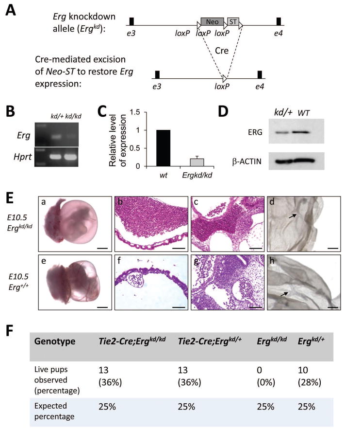
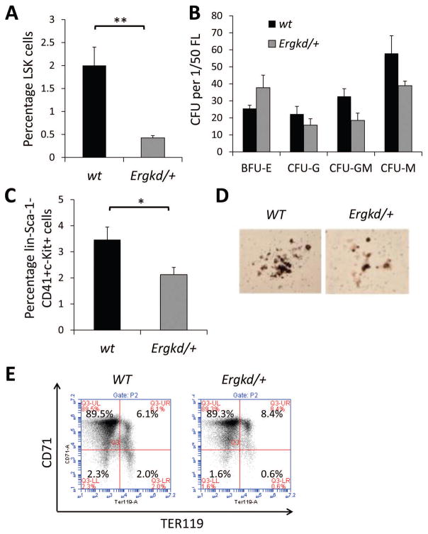
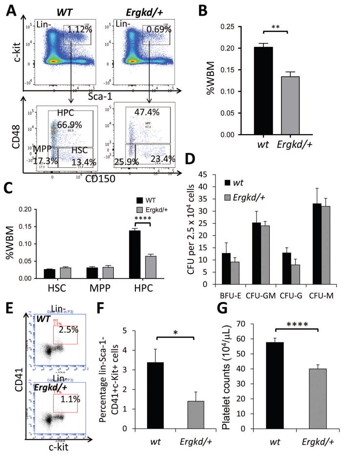
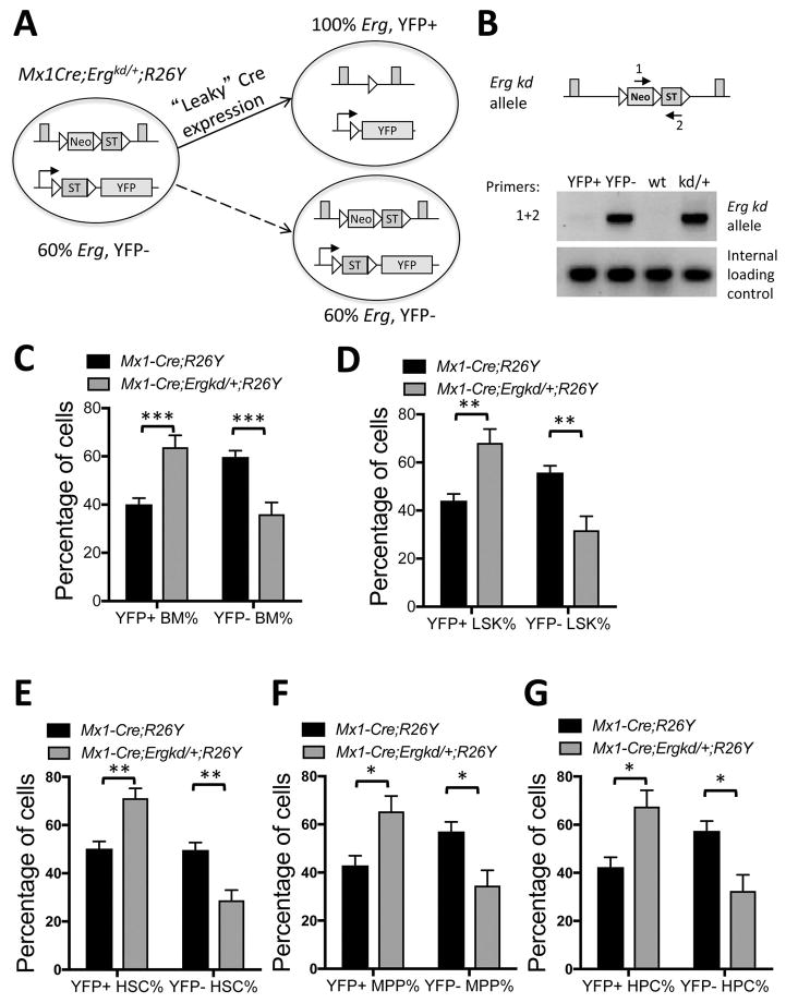
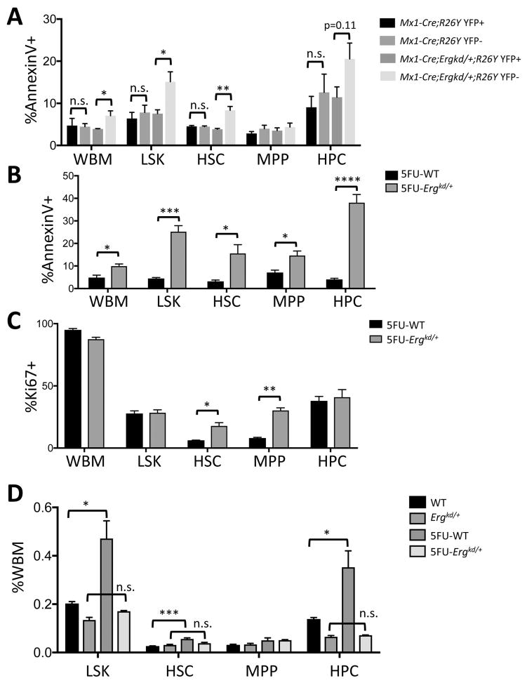
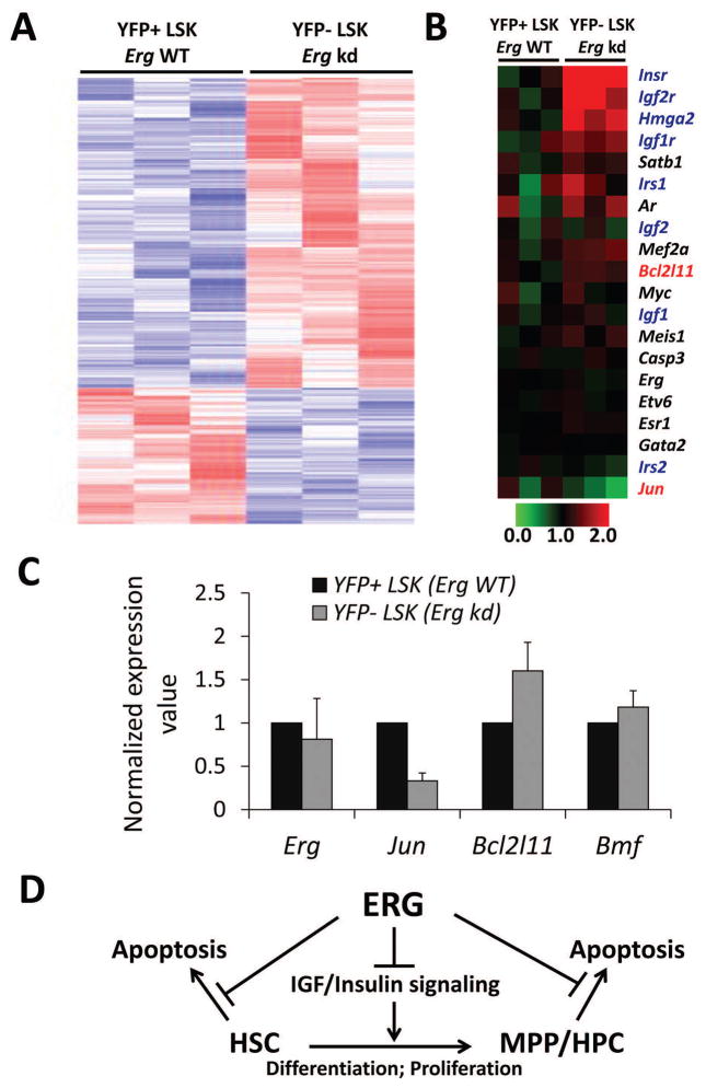
ERG, an ETS family transcription factor frequently overexpressed in human leukemia, has been implicated as a key regulator of hematopoietic stem cells. However, how ERG controls normal hematopoiesis, particularly at the stem and progenitor cell level, and how it contributes to leukemogenesis remain incompletely understood. Using homologous recombination, we generated an Erg knockdown allele (Ergkd ) in which Erg expression can be conditionally restored by Cre recombinase. Ergkd/kd animals die at E10.5-E11.5 due to defects in endothelial and hematopoietic cells, but can be completely rescued by Tie2-Cre-mediated restoration of Erg in these cells. In Ergkd/+ mice, ∼40% reduction in Erg dosage perturbs both fetal liver and bone marrow hematopoiesis by reducing the numbers of Lin- Sca-1+ c-Kit+ (LSK) hematopoietic stem and progenitor cells (HSPCs) and megakaryocytic progenitors. By genetic mosaic analysis, we find that Erg-restored HSPCs outcompete Ergkd/+ HSPCs for contribution to adult hematopoiesis in vivo. This defect is in part due to increased apoptosis of HSPCs with reduced Erg dosage, a phenotype that becomes more drastic during 5-FU-induced stress hematopoiesis. Expression analysis reveals that reduced Erg expression leads to changes in expression of a subset of ERG target genes involved in regulating survival of HSPCs, including increased expression of a pro-apoptotic regulator Bcl2l11 (Bim) and reduced expression of Jun. Collectively, our data demonstrate that ERG controls survival of HSPCs, a property that may be used by leukemic cells. Stem Cells 2017;35:1773-1785.

Keywords: Animal models; Apoptosis; Hematopoietic progenitors; Hematopoietic stem cells; Leukemia; Transcription factors.


Conflict of interest statement

Disclosure of Conflicts of Interest

The authors declare no competing financial interests.

Figures

Figure 1
Figure 1. ERG plays essential roles in hematopoietic and endothelial cells during development
(A) Schematic diagram showing design of the Ergkd allele. Cre-mediated excision of the floxed Neo-ST cassette (ST: transcriptional stopper) knocked-in to the Ergkd allele can lead to its restoration to WT Erg. (B) RT-PCR showing dramatic reduction in Erg transcripts in yolk sac-derived hematopoietic cells from Ergkd/kd embryos in comparison to Ergkd/+ embryos. Hprt expression level was used as the loading control. Note the Ergkd/kd (kd/kd) sample was slightly overloaded to show residual Erg transcript expressed from the Ergkd allele. (C) qRT-PCR showing ~80% reduction in Erg transcripts in day-10 EBs from Ergkd/kd ES cells compared to those from WT ES cells. (D) Western blot showing reduced ERG expression at the protein level in Ergkd/+ versus WT bone marrow cells; β-actin expression was used as the loading control. (E) E10.5 Ergkd/kd embryos (a) appeared much paler than WT embryos (e). Massive amount of blood cells leaked out from blood vessels within the yolk sac (b) and the embryo proper (c) of the Ergkd/kd animal, in comparison to the WT control (f–g), although its major vasculature appeared normal, as indicated by CD31 (PECAM-1) staining (d, compare to h, arrows indicate blood vessels). Scale bars = 1,000μm (a,e), 100μm (b,c,f,g), and 500μm (d,h), respectively. (F) Tie2-Cre leads to restoration of Erg expression in hematopoietic and endothelial cells and rescues the lethal phenotype of Ergkd/kd embryos; animals counted here were from crosses between Tie2-Cre;Ergkd/kd males and Ergkd/+ females (from 8 litters).
Figure 2
Figure 2. Fetal hematopoiesis is perturbed upon ~40% reduction in the Erg level
(A) FACS analysis showing a significant reduction in the percentage of HSPCs in the LSK gate in E12.5 Ergkd/+ fetal livers (FLs) compared to that of their WT littermates. (B) Hematopoietic colony analysis of E12.5 FLs showing slight reduction in myeloid colony formation [CFG-G (granulocyte), CFU-GM (granulocyte, macrophage), CFU-M (macrophage)], and no significant change in erythroid colony formation (BFU-E) in Ergkd/+ mice compared to WTs. (C) FACS analysis showing a reduction of Lin-Sca-1c-kit+CD41+ megakaryocytic progenitors (MPs) in Ergkd/+ E12.5 FLs compared to WTs. (D) Mega-Cult megakaryocytic colony-forming assay showing less acetylcholine+ megakaryocytes (large brown cells) in megakaryocytic colonies formed from Ergkd/+ E12.5 FL cells compared to WTs (magnification, 40x). (E) FACS analysis based on CD71 and TER119 staining in mid-gestation FLs showing largely normal erythropoiesis in Ergkd/+ mice compared to WT controls. Data represent mean ± SEM. P values: *p≤0.05; **p≤0.01.
Figure 3
Figure 3. Adult hematopoiesis is perturbed upon ~40% reduction in the Erg level
(A) FACS analysis showing changes in the percentage of LSK cells, as well as the HSC, MPP and HPC subsets of LSK cells in the bone marrow (BM) of Ergkd/+ mice compared to WTs. Percentages of LSK cells were shown as percentages of lineage-negative (Lin-) cells. (B) Quantification of BM LSK cells in Ergkd/+ animals compared to WTs. Percentages of LSK cells here were shown as percentages of whole bone marrow (WBM) cells. (C) Quantification of the percentage of HSC, MPP and HPC subsets within the WBM in Ergkd/+ animals compared to WTs. (D) Hematopoietic colony analysis of BM samples. (E) FACS analysis showing a reduction in the percentage of Lin-Sca-1c-kit+CD41+ MPs in the BM of Ergkd/+ mice compared to WTs. (F) Quantification of BM MPs in Ergkd/+ animals compared WTs for E. (G) CBC analysis demonstrated significantly reduced platelet counts in Ergkd/+ adult mice compared to WT controls. Data represent mean ± SEM. P values: *p≤0.05; **p≤0.01; ****p≤0.001.
Figure 4
Figure 4. Reduced Erg dosage affects survival and proliferation of HSPCs
(A–B) Summaries of FACS data showing percentages of Annexin V+ apoptotic cells (A) and Ki67+ proliferating cells (B) in each indicated population. (C) Summary of FACS-based cell cycle analysis data for each indicated BM hematopoietic cell subpopulation. Data represent mean ± SEM. P values: *p≤0.05; **p≤0.01; ***p≤0.005; ****p≤0.001.
Figure 5
Figure 5. Ergkd/+ HSPCs were outcompeted by Erg-restored HSPCs in the same animal
(A) Schematic diagram showing Cre-mediated restoration of Erg and simultaneous activation of the conditional YFP reporter in the same cell leading to formation of YFP+ Erg-restored cells from YFP Ergkd/+ cells. (B) PCR analysis (primers 1+2) of genomic DNAs of YFP+ and YFP BM cells sorted from the same Mx1-Cre;Ergkd/+;R26Y mouse showing deletion of the Neo-ST cassette in the YFP+ BM cells (thus Erg restored to WT). Genomic DNAs from WT and Ergkd/+ mice were used as controls for the PCR reaction. (C–G) Quantification of percentages of YFP+ (Erg-restored) and YFP (Erg-kd) WBM cells (C), LSK cells (D), HSCs (E), MPPs (F) and HPCs (G) from Mx1-Cre;R26Y control mice and Mx1-Cre;Ergkd/+;R26Y experimental mice. Data represent mean ± SEM. P values: *p≤0.05; **p≤0.01; ***p≤0.005.
Figure 6
Figure 6. Reduced Erg dosage impaired survival of proliferating HSPCs
(A) Quantification of percentages of Annexin V+ apoptotic cells in YFP+ and YFP WBM, LSK, HSC, MPP and HPC subsets from Mx1-Cre;R26Y control mice and Mx1-Cre;Ergkd/+;R26Y experimental mice. (B–C) Quantification of percentages of Annexin V+ apoptotic cells (B) and Ki67+ proliferating cells (C) in WBM, LSK, HSC, MPP and HPC subsets from Ergkd/+ and WT mice 15 days after 5-FU treatment. (D) Quantification of percentages of LSK, HSC, MPP and HPC subsets in WBM from Ergkd/+ and WT mice 15 days after 5-FU treatment. Data represent mean ± SEM. P values: *p≤0.05; **p≤0.01; ***p≤0.005; ****p≤0.001.
Figure 7
Figure 7. Microarray analysis and validation of select target genes of ERG
(A) Heatmap (red to white to blue indicate highest to intermediate to lowest expression level) showing differential expression of potential target genes of ERG (based on ChIP-seq from ) in YFP+ and YFP LSK cells sorted from the same Mx1-Cre;Ergkd/+;R26Y mouse. (B) Heatmap showing differential expression of select ERG target genes as in A. Genes related to apoptosis are marked in red; genes involved in IGF/insulin signaling are marked in dark blue. Expression values were normalized to the mean of those of Erg-WT samples (=1). (C) qRT-PCR validation showing reduced expression of Jun and increased expression of Bcl2l11 and Bmf in YFP LSK cells, compared to YFP+ LSK cells (=1) from the same Mx1-Cre;Ergkd/+;R26Y mouse. (D) Schematic diagram showing multiple roles of ERG in regulating HSPCs.

Similar articles

Cited by

References

    1. Martens JH. Acute myeloid leukemia: a central role for the ETS factor ERG. Int J Biochem Cell Biol. 2011;43:1413–1416. - PubMed
    1. Tomlins SA, Rhodes DR, Perner S, et al. Recurrent fusion of TMPRSS2 and ETS transcription factor genes in prostate cancer. Science. 2005;310:644–648. - PubMed
    1. Zucman J, Melot T, Desmaze C, et al. Combinatorial generation of variable fusion proteins in the Ewing family of tumours. Embo J. 1993;12:4481–4487. - PMC - PubMed
    1. Ginsberg JP, de Alava E, Ladanyi M, et al. EWS-FLI1 and EWS-ERG gene fusions are associated with similar clinical phenotypes in Ewing’s sarcoma. J Clin Oncol. 1999;17:1809–1814. - PubMed
    1. Shimizu K, Ichikawa H, Tojo A, et al. An ets-related gene, ERG, is rearranged in human myeloid leukemia with t(16;21) chromosomal translocation. Proc Natl Acad Sci U S A. 1993;90:10280–10284. - PMC - PubMed

Publication types

MeSH terms