. 2018 May;38(5):755-766.
doi: 10.1177/0271678X17705567. Epub 2017 Apr 24.

Pharmacologically increasing collateral perfusion during acute stroke using a carboxyhemoglobin gas transfer agent (Sanguinate™) in spontaneously hypertensive rats

Affiliations

Pharmacologically increasing collateral perfusion during acute stroke using a carboxyhemoglobin gas transfer agent (Sanguinate™) in spontaneously hypertensive rats

Marilyn J Cipolla et al. J Cereb Blood Flow Metab. 2018 May.

Abstract

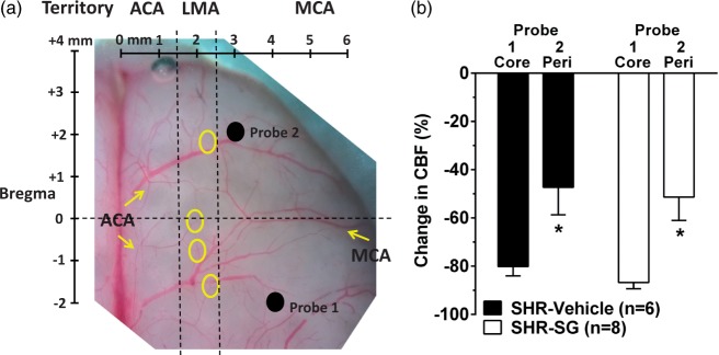
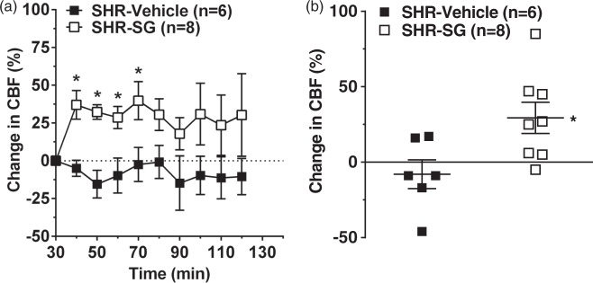
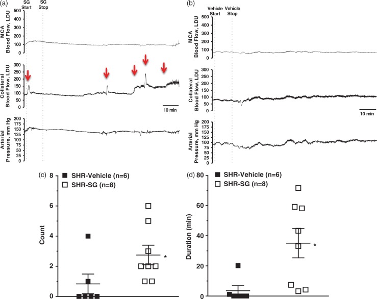
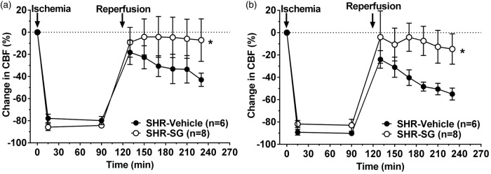
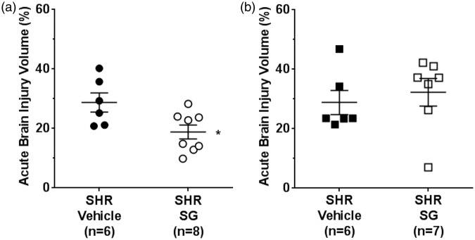
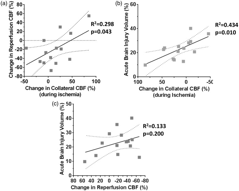
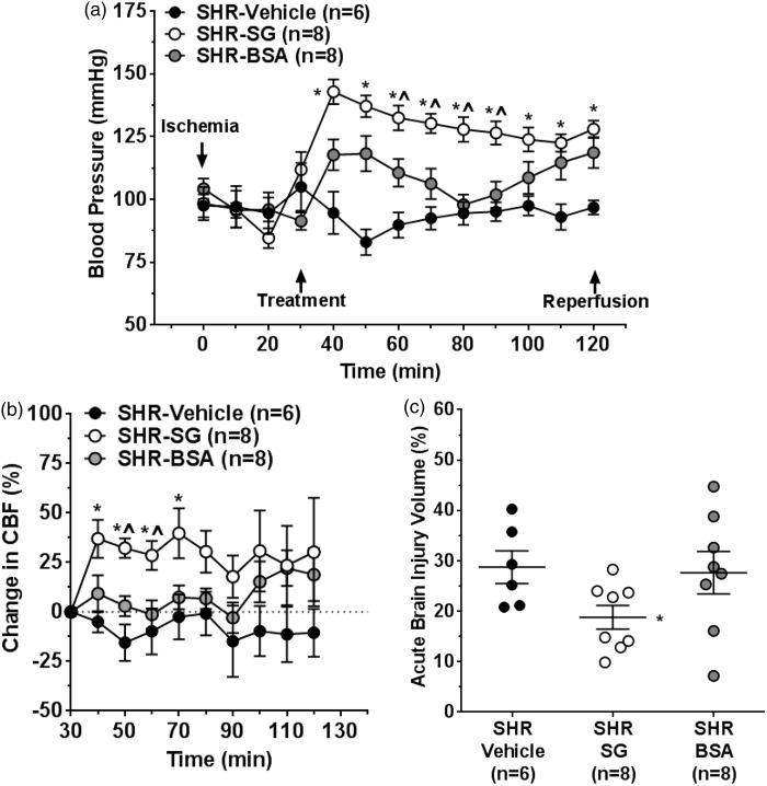
Similar to patients with chronic hypertension, spontaneously hypertensive rats (SHR) develop fast core progression during middle cerebral artery occlusion (MCAO) resulting in large final infarct volumes. We investigated the effect of Sanguinate™ (SG), a PEGylated carboxyhemoglobin (COHb) gas transfer agent, on changes in collateral and reperfusion cerebral blood flow and brain injury in SHR during 2 h of MCAO. SG (8 mL/kg) or vehicle ( n = 6-8/group) was infused i.v. after 30 or 90 min of ischemia with 2 h reperfusion. Multi-site laser Doppler probes simultaneously measured changes in core MCA and collateral flow during ischemia and reperfusion using a validated method. Brain injury was measured using TTC. Animals were anesthetized with choral hydrate. Collateral flow changed little in vehicle-treated SHR during ischemia (-8 ± 9% vs. prior to infusion) whereas flow increased in SG-treated animals (29 ± 10%; p < 0.05). In addition, SG improved reperfusion regardless of time of treatment; however, brain injury was smaller only with early treatment in SHR vs. vehicle (28.8 ± 3.2% vs. 18.8 ± 2.3%; p < 0.05). Limited collateral flow in SHR during MCAO is consistent with small penumbra and large infarction. The ability to increase collateral flow in SHR with SG suggests that this compound may be useful as an adjunct to endovascular therapy and extend the time window for treatment.

Keywords: Collateral perfusion; hypertension; infarction; ischemic stroke; reperfusion.


Figures

Figure 1.
Figure 1.
Measurement of MCA core and collateral CBF using multi-site laser Doppler probes. (a) Photomicrograph of rat brain demonstrating coordinates of dual laser Doppler probe placement on the skull relative to Bregma. Probe 1 was placed within the middle cerebral artery (MCA) ischemic core territory. Probe 2 was placed within the MCA territory but lateral to leptomeningeal anastomoses (LMA) between the anterior cerebral artery (ACA) and MCA territories such that changes in flow corresponded to changes in LMA flow. The LMA territory was determined by connections between branches of MCA and ACA (circles). (b) Graph comparing initial drop in CBF from baseline before filament insertion between MCA (Core) and peri-infarct (Peri) collateral CBF. Drop in collateral CBF (probe 2) was significantly less than that of the core MCA territory (probe 1) demonstrating that each probe was measuring different hemodynamic areas. *p < 0.05 vs. Probe 1.
Figure 2.
Figure 2.
Effect of SG treatment on collateral CBF during ischemia. (a) Graph showing percent change in collateral CBF (probe 2) calculated as a percent change from prior to treatment and 30 min after filament occlusion. Vehicle-treated SHR had minimal changes in collateral flow during the entire ischemic duration. However, treatment with SG significantly increased collateral flow during the first 40 min after treatment and remained elevated throughout ischemia. (b) Graph showing the average change in collateral flow for 90 min in response to SG or vehicle. Vehicle-treated SHR had collateral flow that was below baseline versus prior to treatment whereas SG treatment significantly increased collateral flow. *p < 0.05 vs. SHR vehicle-treated.
Figure 3.
Figure 3.
Effect of SG treatment on discrete collateral perfusion events. Representative laser Doppler CBF tracings from SG-treated (a) or vehicle-treated (b) SHR for core MCA CBF (upper), collateral CBF (middle) and blood pressure (lower tracing) demonstrating discrete increases in collateral flow (arrows). The collateral CBF tracings were used to compare the number, duration and magnitude (AUC) of these discrete events by investigators blinded to group. Graph showing number (c) and duration (d) of increased collateral perfusion events in SHR with either vehicle or SG treatment given after 30 min of ischemia. SHR had discrete collateral perfusion events that were infrequent and short in duration. Treatment with SG significantly increased the number and duration of collateral perfusion events in SHR. *p < 0.05 vs. SHR-vehicle treatment.
Figure 4.
Figure 4.
Effect of early and delayed SG treatment on reperfusion CBF in SHR. Graphs showing changes in CBF in the MCA ischemic core territory (probe 1) during ischemia and reperfusion for vehicle- and SG-treated SHR for early (a) and delayed (b) treatment. Animals were excluded if the drop in CBF during ischemia was <66% of baseline and therefore there was no difference in ischemic CBF in any of the groups. Vehicle-treated SHR had reperfusion that was below baseline and became progressively worse over time. Both early and delayed SG treatment improved reperfusion compared to vehicle treatment in SHR. *p < 0.05 vs. CBF at baseline by repeated measures ANOVA.
Figure 5.
Figure 5.
Effect of early and delayed treatment with SG on acute brain injury volume in SHR. (a) Acute brain injury volume of SHR was significantly decreased after early treatment with SG compared to vehicle treatment. (b) There was no beneficial effect of SG in SHR on brain injury volume when SG treatment was delayed 90 min. *p < 0.05 vs. SHR vehicle treatment.
Figure 6.
Figure 6.
Relationship between changes in collateral flow, reperfusion CBF and acute brain injury. (a) Graph showing the relationship between collateral flow and reperfusion CBF in all animals regardless of treatment. There was a positive correlation between reperfusion CBF and collateral flow such that animals with better collateral flow had better reperfusion. (b) Graph showing the correlation between collateral flow and infarction in all groups of animals with early treatment. There was a significant correlation between collateral flow and brain injury volume such that the greater increase in flow the smaller the injury. (c) Graph showing correlation between reperfusion CBF and brain injury volume. There was no significant correlation found.
Figure 7.
Figure 7.
Effect of blood pressure on collateral flow and acute brain injury. (a) Graph showing changes in blood pressure during ischemia. Treatment with SG and BSA both increased blood pressure in SHR compared to vehicle treatment, but not to the same level. (b) Graph showing change in collateral flow from baseline after infusion of SG, BSA or vehicle treatment during filament insertion. SG infusion increased collateral perfusion during ischemia but BSA and vehicle treatment had no effect. (c) Graph showing the effect of SG, BSA and vehicle treatment on acute brain injury volume. SG, but not BSA, reduced brain injury volume compared to vehicle treatment. *p < 0.05 vs. SHR vehicle treatment; ^p < 0.05 vs. SHR-BSA.

References

    1. Ginsberg MD. Neuroprotection for ischemic stroke: past, present and future. Neuropharmacology 2008; 55: 363–389. - PMC - PubMed
    1. Chamorro Á, Dirnagl U, Urra X, et al. Neuroprotection in acute stroke: targeting excitotoxicity, oxidative and nitrosative stress, and inflammation. Lancet Neurol 2016; 15: 869–881. - PubMed
    1. Ginsberg MD. Expanding the concept of neuroprotection for acute ischemic stroke: The pivotal roles of reperfusion and the collateral circulation. Prog Neurobiol 2016; 145–146: 46–77. - PubMed
    1. Palomares SM, Cipolla MJ. Vascular protection following cerebral ischemia and reperfusion. J Neurol Neurophysiol 2011; 20: piiS1–004. - PMC - PubMed
    1. Linfante I, Starosciak AK, Walker GR, et al. Predictors of poor outcome despite recanalization: a multiple regression analysis of the NASA registry. J Neurointerv Surg 2016; 8: 224–229. - PubMed

Publication types

LinkOut - more resources