Case Reports
. 2017 Jun;23(3):307-312.
doi: 10.1177/1591019917694839. Epub 2017 Jan 1.

A hemorrhagic complication after Onyx embolization of a tentorial dural arteriovenous fistula: A caution about subdural extension with pial arterial supply

Affiliations
Case Reports

A hemorrhagic complication after Onyx embolization of a tentorial dural arteriovenous fistula: A caution about subdural extension with pial arterial supply

Kenichi Sato et al. Interv Neuroradiol. 2017 Jun.

Abstract

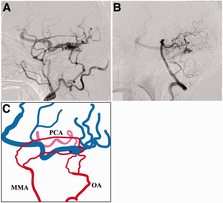
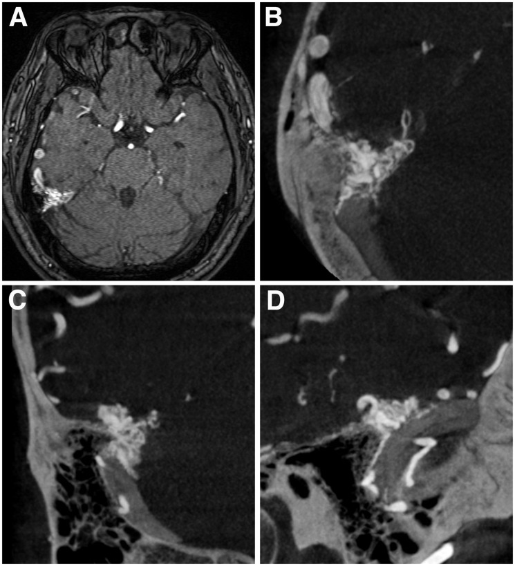
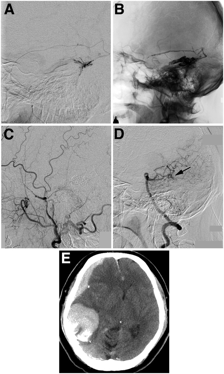
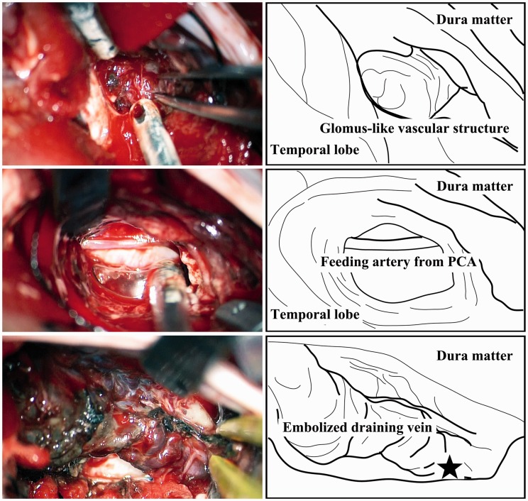
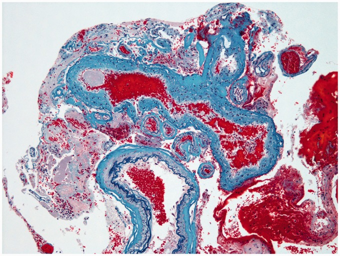
We report a case of tentorial dural arteriovenous fistula (DAVF) with a severe intracranial hemorrhage occurring after Onyx embolization. A 40-year-old man presented with an asymptomatic tentorial DAVF on angiography. Transarterial embolization with Onyx was performed via the middle meningeal artery, and the cast filled the fistula itself and its proximal draining vein. Postoperative angiography confirmed complete occlusion of the DAVF. A computed tomography scan performed immediately after the procedure demonstrated an acute subdural hematoma with the temporal hemorrhage. Emergency craniotomy revealed continuous arterial bleeding from a viable glomus-like vascular structure around the proximal part of the embolized draining vein, fed by a pial artery arising from the posterior cerebral artery. Pathologic findings suggested diagnosis of vascular malformation extending into the subdural space. Tentorial DAVFs can extend to the subdural space along their drainage route, and may be involved in severe hemorrhagic complications of curative endovascular treatment using Onyx, particularly those with pial arterial supply.

Keywords: Dural arteriovenous fistula; Onyx; endovascular treatment; hemorrhage.


Figures

Figure 1.
Figure 1.
a) The right external carotid artery angiogram demonstrating a tentorial dural arteriovenous fistula (DAVF). (b) The left vertebral artery angiogram. The DAVF is also fed by a pial branch originating from the right posterior cerebral artery (PCA). (c) Schematic drawing of the angioarchitecture showing the feeding arteries arising from the right external carotid artery (red) and from the PCA (pink), and the cortical venous drainage (blue). MMA: middle meningeal artery; OA: occipital artery; PCA: posterior cerebral artery.
Figure 2.
Figure 2.
a) Time-of-flight magnetic resonance image. No parenchymal vascular malformation is seen. (b)–(d) Cone-beam computed tomography scan images ((b) axial; (c) coronal; (d) sagittal) obtained by rotational angiography of the external carotid artery demonstrating a vessel cluster located lateral to the point where the tentorium joins the dura of the right middle cranial fossa and a dilated cortical vein in the right temporal lobe.
Figure 3.
Figure 3.
a) Superselective angiogram of the petrosquamous branch of the middle meningeal artery. (b) Cranial plain radiography showing Onyx cast. The right external carotid artery angiogram (c) and the left vertebral artery angiogram (d) demonstrating the complete disappearance of the dural arteriovenous fistula. The pial feeding artery seems to be occluded (arrow). (e) Computed tomography after endovascular treatment demonstrating a right acute subdural hematoma with a temporal hemorrhage.
Figure 4.
Figure 4.
Operative photographs (left) and corresponding illustrations (right) show a viable glomus-like vascular structure protruding from the inner surface of the dura into the subdural space (upper row), a pial feeding artery originating from the right posterior cerebral artery and traveling laterally along the superior surface of the tentorium (middle row), and the hypervascularized tentorium and vasa vasorum around the embolized draining vein (lower row). The asterisk indicates the location where the viable glomus-like vascular structure was found.
Figure 5.
Figure 5.
Histological images demonstrating an abnormal aggregation of vascular structures, including vessels that have an irregularly thickened wall caused by elastic fibrosis (corresponding to arterialized veins), and vessels with internal elastic lamina and regular smooth muscle (corresponding to normal arteries). Modified Verhoeff Elastic-Masson Trichrome Stain. Original magnification × 100.

Similar articles

Cited by

References

    1. Hiramatsu M, Sugiu K, Hishikawa T, et al. Epidemiology of dural arteriovenous fistula in Japan: Analysis of Japanese Registry of Neuroendovascular Therapy (JR-NET2). Neurol Med Chir (Tokyo) 2014; 54: 63–71. - PMC - PubMed
    1. Miller TR, Gandhi D. Intracranial dural arteriovenous fistulae: Clinical presentation and management strategies. Stroke 2015; 46: 2017–2025. - PubMed
    1. Bulters DO, Mathad N, Culliford D, et al. The natural history of cranial dural arteriovenous fistulae with cortical venous reflux—the significance of venous ectasia. Neurosurgery 2012; 70: 312–318. - PubMed
    1. Youssef PP, Schuette AJ, Cawley CM, et al. Advances in surgical approaches to dural fistulas. Neurosurgery 2014; 74: 532–541. - PubMed
    1. Cannizzaro D, Brinjikji W, Rammos S, et al. Changing clinical and therapeutic trends in tentorial dural arteriovenous fistulas: A systematic review. AJNR Am J Neuroradiol 2015; 36: 1905–1911. - PMC - PubMed

Publication types

MeSH terms

LinkOut - more resources