

Quantitative microvascular analysis of retinal venous occlusions by spectral domain optical coherence tomography angiography
- PMID: 28437483
- PMCID: PMC5402954
- DOI: 10.1371/journal.pone.0176404
Quantitative microvascular analysis of retinal venous occlusions by spectral domain optical coherence tomography angiography
Abstract
Purpose: To quantitatively evaluate the retinal microvasculature in human subjects with retinal venous occlusions (RVO) using optical coherence tomography angiography (OCTA).
Design: Retrospective, cross-sectional, observational case series.
Participants: Sixty subjects (84 eyes) were included (20 BRVO, 14 CRVO, 24 unaffected fellow eyes, and 26 controls).
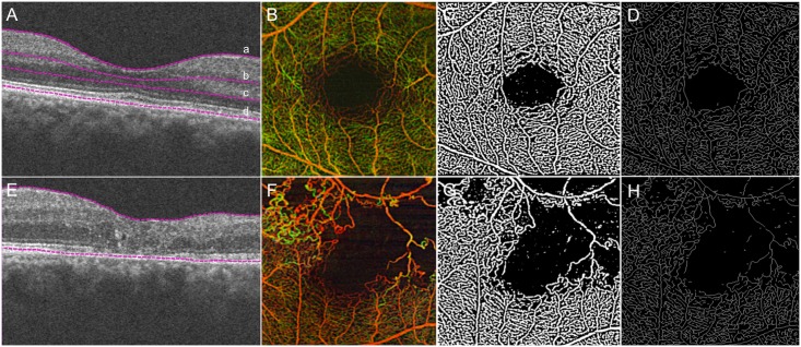
Methods: OCTA was performed on a prototype, spectral domain-OCTA system in the 3x3mm central macular region. Custom software was used to quantify morphology and density of retinal capillaries using four quantitative parameters. The vasculature of the segmented retinal layers and nonsegmented whole retina were analyzed.
Main outcome measures: Fractal dimension (FD), vessel density (VD), skeletal density (SD), and vessel diameter index (VDI) within the segmented retinal layers and nonsegmented whole retina vasculature.
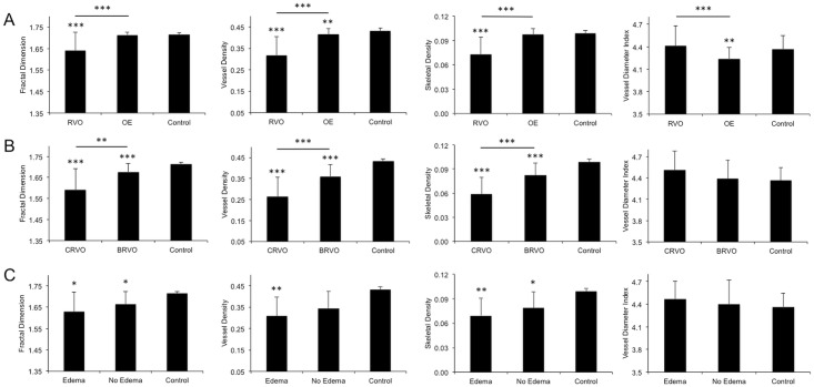
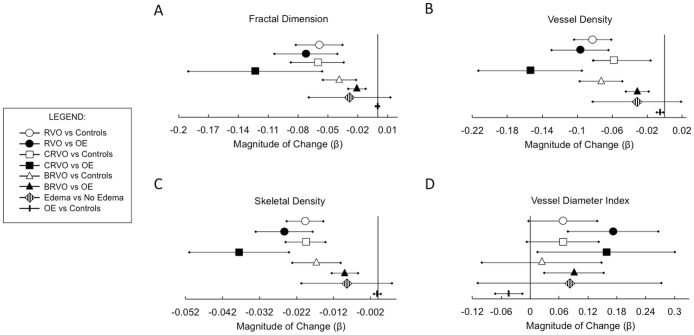
Results: Nonsegmented analysis of RVO eyes demonstrated significantly lower FD (1.64±0.01 vs 1.715±0.002; p<0.001), VD (0.32±0.01 vs 0.432±0.002; p<0.001), and SD (0.073±0.004 vs 0.099±0.001; p<0.001) compared to controls. Compared to the fellow eye, FD, VD and SD were lower (p<0.001), and VDI was higher (p<0.001). FD, VD, and SD progressively decreased as the extent (or type) of RVO increased (control vs BRVO vs CRVO; p<0.001). In the unaffected fellow eye FD, VD and SD showed significant differences when compared to control eyes or affected RVO eyes (p<0.001).
Conclusions: Quantitative OCTA of the central 3x3mm macular region demonstrates significant differences in capillary density and morphology among subjects with BRVO and CRVO compared to controls or unaffected fellow eyes in all vascular layers. The unaffected fellow eyes also demonstrate significant differences when compared to controls. OCTA allows for noninvasive, layer-specific, quantitative evaluation of RVO-associated microvascular changes.
Conflict of interest statement
Figures
Similar articles
-
Evaluation of microvascular network with optical coherence tomography angiography (OCTA) in branch retinal vein occlusion (BRVO).BMC Ophthalmol. 2020 Apr 19;20(1):154. doi: 10.1186/s12886-020-01405-0. BMC Ophthalmol. 2020. PMID: 32306978 Free PMC article.
-
Quantifying Retinal Microvascular Changes in Uveitis Using Spectral-Domain Optical Coherence Tomography Angiography.Am J Ophthalmol. 2016 Nov;171:101-112. doi: 10.1016/j.ajo.2016.08.035. Epub 2016 Sep 2. Am J Ophthalmol. 2016. PMID: 27594138 Free PMC article.
-
Retinal Capillary Network and Foveal Avascular Zone in Eyes with Vein Occlusion and Fellow Eyes Analyzed With Optical Coherence Tomography Angiography.Invest Ophthalmol Vis Sci. 2016 Jul 1;57(9):OCT486-94. doi: 10.1167/iovs.15-18907. Invest Ophthalmol Vis Sci. 2016. PMID: 27442342
-
Retinal oximetry and systemic arterial oxygen levels.Acta Ophthalmol. 2018 Nov;96 Suppl A113:1-44. doi: 10.1111/aos.13932. Acta Ophthalmol. 2018. PMID: 30460761 Review.
-
[A new approach for studying the retinal and choroidal circulation].Nippon Ganka Gakkai Zasshi. 2004 Dec;108(12):836-61; discussion 862. Nippon Ganka Gakkai Zasshi. 2004. PMID: 15656089 Review. Japanese.
Cited by
-
Maximum value projection produces better en face OCT angiograms than mean value projection.Biomed Opt Express. 2018 Nov 26;9(12):6412-6424. doi: 10.1364/BOE.9.006412. eCollection 2018 Dec 1. Biomed Opt Express. 2018. PMID: 31065439 Free PMC article.
-
Correlation Between Macular Microstructural Changes with Disease Staging and Visual Acuity in Diabetic Retinopathy.Int J Gen Med. 2025 May 20;18:2619-2628. doi: 10.2147/IJGM.S516938. eCollection 2025. Int J Gen Med. 2025. PMID: 40417418 Free PMC article.
-
OCT angiography features associated with macular edema recurrence after intravitreal bevacizumab treatment in branch retinal vein occlusion.Sci Rep. 2019 Oct 2;9(1):14153. doi: 10.1038/s41598-019-50637-8. Sci Rep. 2019. PMID: 31578437 Free PMC article.
-
Three-Dimensional Analysis of Choroidal Vessels in the Eyes of Patients With Unilateral BRVO.Front Med (Lausanne). 2022 Apr 5;9:854184. doi: 10.3389/fmed.2022.854184. eCollection 2022. Front Med (Lausanne). 2022. PMID: 35479961 Free PMC article.
-
Optical coherence tomography angiography: A comprehensive review of current methods and clinical applications.Prog Retin Eye Res. 2017 Sep;60:66-100. doi: 10.1016/j.preteyeres.2017.07.002. Epub 2017 Jul 29. Prog Retin Eye Res. 2017. PMID: 28760677 Free PMC article. Review.
References
-
- Yannuzzi LA, Rohrer KT, Tindel LJ, Sobel RS, Costanza MA, Shields W, et al. Fluorescein angiography complication survey. Ophthalmology. 1986;93(5):611–7. - PubMed
Publication types
MeSH terms
Grants and funding
LinkOut - more resources
Full Text Sources
Other Literature Sources
