Review
. 2017 May;19(5):529-540.
doi: 10.1177/1098612X17706466.

The feline blood film

Affiliations
Review

The feline blood film

John W Harvey. J Feline Med Surg. 2017 May.

Abstract

Practical relevance: Many veterinary practices have invested in quality automated hematology instruments for use in-house. However, regardless of the specific choice of analyzer, there are important hematology findings that can only be determined by microscopic examination of stained blood films. For this reason, and also for the purpose of quality control for the analyzer, a quick blood film review should be performed alongside every automated complete blood count. Even those practices that submit their blood samples to outside diagnostic laboratories for evaluation, still require the capability to examine stained blood films in emergency situations. Series outline: This is the first of a two-part article series that aims to familiarize the practitioner with normal findings on feline blood films, with a particular focus on unique features in the cat, as well as to assist with interpretation of common abnormalities. Part 1 focuses on how to prepare and examine blood films in order to maximize the reliability of the information they convey, and describes the morphology of feline erythrocytes in health and disease. Evidence base: The information and guidance offered is based on the published literature and the author's own extensive clinical pathology research.


Conflict of interest statement

The author declared no potential conflicts of interest with respect to the research, authorship, and/or publication of this article.

Figures

Figure 1
Figure 1
Blood film preparation. (a) A glass slide is placed on a flat surface and a small drop of well-mixed blood is placed on one end of the slide using a microhematocrit tube. (b) A second glass slide (spreader slide) is placed on the first slide at about a 30–45° angle in front of the drop of blood. The spreader slide is then backed into the drop of blood. (c) As soon as the blood flows along the back edge of the spreader slide, the spreader slide is rapidly pushed forward. (d) The blood film produced is thick at the back of the slide, where the drop of blood was placed, and thin at the front (feathered) edge of the slide. Reproduced from Harvey (2012), with permission
Figure 2
Figure 2
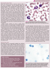
Neutrophil (bottom left), eosinophil (red granules) and a lymphocyte (round nucleus) are present in this blood film from a cat. Erythrocytes exhibit rouleaux, which is normal in cats. Wright-Giemsa stain
Figure 3
Figure 3
Reticulocytes in cat blood. Three whole aggregate reticulocytes (containing blue-staining aggregates of RNA) and one half of an aggregate reticulocyte (far right) in blood from a cat with a markedly regenerative anemia. The majority of the remaining cells are punctate reticulocytes containing discrete dot-like inclusions. New methylene blue reticulocyte stain. Reproduced from Harvey (2012), with permission
Figure 4
Figure 4
Aggregate of cells, which appears to contain a macrophage with phagocytized nucleated erythrocytes (black arrows), located at the feathered end of a blood film from a cat with Mycoplasma haemofelis infection. The large oval magenta structure near the bottom of the image (red arrow) is the nucleus of the macrophage. The nucleus contains a blue-staining nucleolus. Wright-Giemsa stain
Figure 5
Figure 5
Agglutination of polychromatophilic erythrocytes (aggregate reticulocytes) in blood from a cat with immune-mediated hemolytic anemia. Many mature erythrocytes appear to be spherocytes. Wright-Giemsa stain
Figure 6
Figure 6
Increased polychromasia and anisocytosis in a Mycoplasma haemofelis-infected cat with a regenerative anemia. Most polychromatophilic erythrocytes have a stomatocyte appearance. Wright-Giemsa stain
Figure 7
Figure 7
Increased anisocytosis without polychromasia in blood from a feline immunodeficiency virus-infected cat with a non-regenerative anemia. Wright-Giemsa stain
Figure 8
Figure 8
Marked anisocytosis in blood from a 6-week-old kitten after a blood transfusion. The kitten presented with severe iron deficiency anemia and marked lipemia. The small, hypochromic erythrocytes are from the kitten, and the larger erythrocytes are from the blood donor cat. A large polychromatophilic erythrocyte and two lysed erythrocytes (red smudges on the left side of the image) are present. The lipemia promoted erythrocyte lysis during blood film preparation. Wright-Giemsa stain
Figure 9
Figure 9
Poikilocytosis in blood from an anemic cat with a portosystemic shunt. Multiple elliptocytes (ovalocytes) and two keratocytes with ruptured vacuoles (black arrows) are present. A discocyte containing a Howell-Jolly body (red arrow) and a large platelet (green arrow) are also present. Wright-Giemsa stain
Figure 10
Figure 10
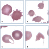
Abnormal erythrocyte shapes. (a) A normal discocyte (left) and an echinocyte (right) in blood from a cat with uremia. (b) Three acanthocytes in blood from a cat with hepatic lipidosis. (c) A keratocyte with intact vacuole (left) and a discocyte (right) in blood from a cat with a portosystemic shunt. (d) A schistocyte in blood from a cat with diabetes mellitus, interstitial lung disease and erythrocyte fragmentation. Wright-Giemsa stain
Figure 11
Figure 11
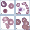
Abnormal erythrocyte shapes. (a) A polychromatophilic erythrocyte (top left), discocyte (top right) and spherocyte (bottom) in blood from a cat with immunemediated hemolytic anemia. (b) Three elliptocytes (right) in blood from a cat with a portosystemic shunt. A large activated platelet (center) with aggregated granules that may be confused with a nucleus is also present. (c) A tear dropshaped dacryocyte appears below two discocytes, each of which contains a pale, barely visible Heinz body in blood from a cat with a portosystemic shunt. (d) Stomatocytes in the thick portion of a feline blood film. This shape was not present in thinner areas of the film. Wright-Giemsa stain
Figure 12
Figure 12
Abnormal erythrocyte shapes. (a) Three eccentrocytes in blood from a cat with acetaminophen toxicity. Pale Heinz bodies are also visible in two of the eccentrocytes (left and top). (b) A hemoglobin crystal is present within an erythrocyte. The faintly stained cell membrane (top) is visible. Wright-Giemsa stain
Figure 13
Figure 13
Nucleated erythrocytes. (a) Polychromatophilic rubricyte and discocyte in blood from a cat. (b) Metarubricyte with nuclear extrusion (left), a polychromatophilic erythrocyte (right) and discocytes (top) in blood from a 3-week-old kitten with a regenerative response to anemia. Wright-Giemsa stain
Figure 14
Figure 14
Two metarubricytes (black arrows), a prorubricyte (red arrow) and a rubriblast (green arrow) in blood from a cat with erythroleukemia (AML-M6). Wright-Giemsa stain
Figure 15
Figure 15
Staining characteristics and appearance of Heinz bodies versus Howell-Jolly bodies in a cat with acetaminophen toxicity. (a) The erythrocyte on the top contains a barely visible, pale-pink staining Heinz body at the margin at the 7 o’clock position; in contrast, the erythrocyte below it contains a Howell-Jolly body that stains dark blue. Wright-Giemsa stain. (b) For comparison, erythrocytes from the same cat were stained with a new methylene blue reticulocyte stain. The erythrocyte on the left contains a dark-blue staining Howell-Jolly body and the other two cells each contain a lighter blue staining Heinz body. Reproduced from Harvey (2012), with permission
Figure 16
Figure 16
Heinz body hemolytic anemia in a cat with acetaminophen toxicity. The two erythrocytes on the left contain light-pink staining Heinz bodies, with prominent protrusion of the Heinz body in the top cell. The erythrocyte on the right is an eccentrocyte. Wright-Giemsa stain
Figure 17
Figure 17
Heinz body hemolytic anemia in a cat with acetaminophen toxicity. The cell at the bottom has lysed and a pale-pink Heinz body is visible in the erythrocyte ‘ghost’. The remaining intact erythrocytes all contain pale-pink staining Heinz bodies. Wright-Giemsa stain
Figure 18
Figure 18
Focal basophilic stippling in two erythrocytes from a cat with dyserythropoiesis and persistent mild anemia. These are called siderotic inclusions because they stained positive for iron using the Perl’s iron stain. In the human literature, siderotic inclusions have been referred to as Pappenheimer bodies. A platelet (left) is also present. Modified Wright’s stain. Photograph of a stained blood film from a 2012 ASVCP slide review submitted by P Cazzini, M Camus, J Messick, B Garner and K Latimer
Figure 19
Figure 19
Large numbers of small blue/purple-staining Mycoplasma haemofelis bacterial organism on the surface of erythrocytes from a cat with a natural infection. Wright-Giemsa stain
Figure 20
Figure 20
Large numbers of small blue/purple-staining Mycoplasma haemofelis organisms on the surface of erythrocytes from two cats with a natural infection. (a) Ring forms of the organism can be seen on the surface of an erythrocyte. A polychromatophilic erythrocyte (aggregate reticulocyte) without organisms is also present (bottom right). (b) Organisms appear on the periphery of erythrocytes in a thin area of the blood film. Wright-Giemsa stain
Figure 21
Figure 21
Protozoal parasites in feline erythrocytes. (a) Erythrocytes containing Cytauxzoon felis merozoites in blood from a domestic cat with a natural infection. Organisms have a magenta staining nucleus and pale-blue staining cytoplasm. Wright-Giemsa stain. (b) Erythrocytes containing Babesia felis in blood from a cat from South Africa. Wright stain. Reproduced from Harvey (2012), with permission
None
None
None
None
None
None
None

Similar articles

Cited by

References

    1. Harvey JW. Veterinary hematology. A diagnostic guide and color atlas. St Louis, Mo: Elsevier Saunders, 2012.
    1. Mitzner BT. Why automated differentials fall short. J Am Anim Hosp Assoc 2001; 37: 117–118. - PubMed
    1. Bain BJ. Diagnosis from the blood smear. N Engl J Med 2005; 353: 498–507. - PubMed
    1. Welles EG, Hall AS, Carpenter DM. Canine complete blood counts: a comparison of four in-office instruments with the ADVIA 120 and manual differential counts. Vet Clin Pathol 2009; 38: 20–29. - PubMed
    1. Fujino Y, Nakamura Y, Matsumoto H, et al. . Development and evaluation of a novel in-clinic automated hematology analyzer, ProCyte Dx, for canine erythrocyte indices, leukogram, platelet counts and reticulocyte counts. J Vet Med Sci 2013; 75: 1519–1524. - PMC - PubMed

MeSH terms

LinkOut - more resources