. 2017 Apr 24;15(1):32.
doi: 10.1186/s12951-017-0266-5.

Aggregation/dispersion transitions of T4 phage triggered by environmental ion availability

Affiliations

Aggregation/dispersion transitions of T4 phage triggered by environmental ion availability

Bożena Szermer-Olearnik et al. J Nanobiotechnology. .

Abstract

Background: Bacteriophage survives in at least two extremes of ionic environments: bacterial host (high ionic-cytosol) and that of soil (low ionic-environmental water). The impact of ionic composition in the micro- and macro-environments has not so far been addressed in phage biology.

Results: Here, we discovered a novel mechanism of aggregation/disaggregation transitions by phage virions. When normal sodium levels in phage media (150 mM) were lowered to 10 mM, advanced imaging by scanning electron microscopy, atomic force microscopy and dynamic light scattering all revealed formation of viral packages, each containing 20-100 virions. When ionic strength was returned from low to high, the aggregated state of phage reversed to a dispersed state, and the change in ionic strength did not substantially affect infectivity of the phage. By providing the direct evidence, that lowering of the sodium ion below the threshold of 20 mM causes rapid aggregation of phage while returning Na+ concentration to the values above this threshold causes dispersion of phage, we identified a biophysical mechanism of phage aggregation.

Conclusions: Our results implicate operation of group behavior in phage and suggest a new kind of quorum sensing among its virions that is mediated by ions. Loss of ionic strength may act as a trigger in an evolutionary mechanism to improve the survival of bacteriophage by stimulating aggregation of phage when outside a bacterial host. Reversal of phage aggregation is also a promising breakthrough in biotechnological applications, since we demonstrated here the ability to retain viable virion aggregates on standard micro-filters.


Figures
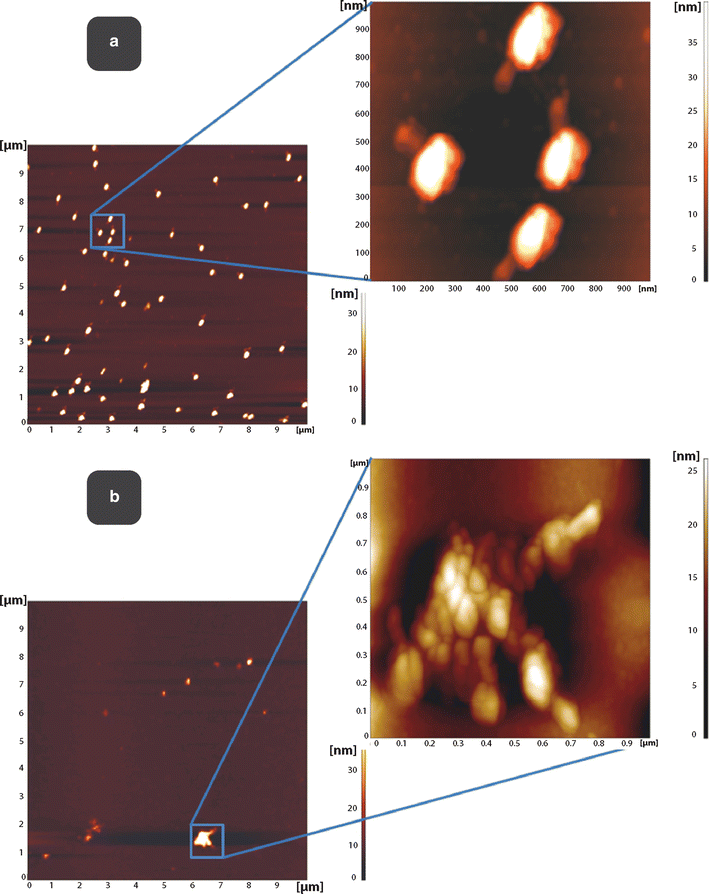
Fig. 1
Fig. 1
Aggregation of bacteriophage T4 visualized by atomic force microscopy, AFM. a. AFM images of T4 bacteriophages on PEI (polyethylene imine) modified mica surface deposited as separate objects from 150 mM NaCl solutions. Scan area 10 × 10 μm. b. AFM images of T4 bacteriophages on PEI (polyethylene imine) modified mica surface deposited in clusters from low ionic strength solution (10 mM NaHCO3) after 100 min of incubation at room temperature. Scan area 1 × 1 μm
Fig. 2
Fig. 2
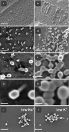
Aggregation of bacteriophage T4 visualized by scanning electron microscopy, SEM. In high-ionic strength 150 mM NaCl bacteriophage particles distributed uniformly on a silicon surface, as separate objects (a, c, e, g), while in contrast, in low-ionic strength (10 mM) phage particles get organized in clusters (aggregates) (b, d, f, hj). Images represent the typical forms of phage aggregates. Distribution of phage particles depended on solute, namely physiologic 150 mM NaCl (a, c, e, g) compared with low ionic strength 10 mM NaHCO3 (b, d, f, h, i). Visible phage particles, deposited on silicon substrate. In-lens SE1 detection (1.2 kV). Note the dispersed phenotype at higher salt concentrations (left panel), while aggregation of phages at low salt concentration (right panel). g Set of representative phage particles at high magnification, with high dispersion, under high (physiologic 150 mM NaCl) solute concentration. SEM scanned at low beam accelerating voltages with SE detection at 1.2 kV acceleration voltage of primary beam. h Set of representative phage particles at high magnification, clustered, under low (10 mM NaHCO3) solute concentration. In-lens SE1 detection at 1.2 kV acceleration voltage of primary beam. i, j SEM images of T4 bacteriophages on silicon (100) crystal surface deposited in clusters from low ionic strength solution with cation of sodium as 10 mM NaHCO3, (i) or with cation of potassium as 10 mM KHCO3, (j). Please notice a similar morphology of aggregates in both cases, when at low Na+ or at low K+. Scale bars a, b 1 µm; c, d 250 nm; e, f 100 nm; g, h 50 nm; i, j 250 nm
Fig. 3
Fig. 3
Kinetics of phage aggregation—dependence on ionic strength. a Under low ionic strength, the particles of phage rapidly aggregate to form clusters. The initial high rate of aggregation gradually slows down, with plateau in the late phase. Measured values fit to square-root function curve, shown in green. b The curves demonstrate DLS analysis where the particles of phage do not cluster when in high ionic concentration (red curve) while, upon ionic strength switched to the low range, the aggregation followed (green curve). These two contrasting behavior modes correspond to the clustering of nanoparticles, dynamic under liquid suspension conditions, and correlate well with observations of the static methods SEM and AFM
Fig. 4
Fig. 4
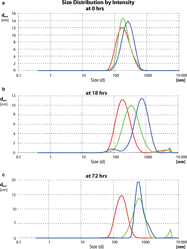
Dependence of aggregation on pH shown at time points a 0 h, b 18 h, c 72 h. Phage aggregation triggered by low ionic strength showed variable dynamics depending on pH. Under lower pH (5.8) we observed the slowest rate of aggregation (red line), intermediate rate at neutral pH (7.0) (green line) while the fastest under alkaline conditions (pH 8.6) with almost complete contribution of large objects already after 18 h (blue line). Aggregation reached the high yield level at the neutral pH only after 72 while at alkaline pH already 18 h were sufficient
Fig. 5
Fig. 5
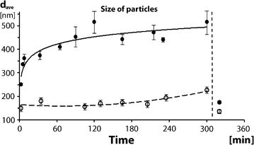
Aggregation of phage in dependence on temperature. Inhibition of aggregation at low temperature. Average effective particle diameter of bacteriophages in 37 °C (full points) in comparison to 4 °C (empty circles). The curve of a square-root function–the best fit for data measured at 37 °C—suggests a diffusion process being involved in the phage-aggregation progression. Dashed curve, fitting the measured particle dimensions under an inhibitory temperature, shows low starting value of particles’ size and much slower increase throughout timescale of the experiment (empty circles). Vertical line after time point ‘300 min’, indicates the addition of concentrated salt to previously formed aggregates to test reversibility of the aggregated state, triggered by high ionic strength. Please note a dramatic drop of average particle size at high salt (right part of the image, between time-points 300 and 360 min). Dispersion was noticed not only population of phage aggregated at 37 °C, but also in the less aggregated population at 4 °C, speaking for dispersion of clusters into individual phage particles in high ionic strength thus demonstrated the reversibility of the whole process, at both temperatures
Fig. 6
Fig. 6
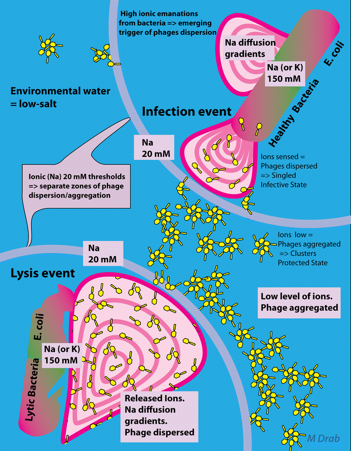
Ion availability as a trigger for aggregation/dispersion of bacteriophage T4—proposed mechanism in phage survival/infectivity cycles in (micro)-environment—the locally increased ions in proximity of bacteria serve as a cue, sensed by the phage, and converts into a quorum signal for re-organization. Infected (lytic phase) bacterium concomitantly releases cytosolic ions at high concentrations and phage in its dispersed form (left lower panel). Upon crossing the threshold gradient (range of 20 mM Na+, depicted as magenta circles) of monovalent cation, in low-ionic strength (water in soil) phage particles get clustered (aggregates), presumably to prolong bacteriophage survival (middle image panel). In contrast, in higher ionic concentration, like when immersed in zone of ionic fluxes contributed by live bacteria, bacteriophage particles disperse from clusters, as separate objects, to broaden the spread area and increase invasion events in proximity of the sensed host (right upper panel)

Similar articles

Cited by

References

    1. Partensky F, Garczarek L. Microbiology: Arms race in a drop of sea water. Nature. 2011;474(7353):582–583. doi: 10.1038/474582a. - DOI - PubMed
    1. Desnues C, Rodriguez-Brito B, Rayhawk S, Kelley S, Tran T, Haynes M, Liu H, Furlan M, Wegley L, Chau B, et al. Biodiversity and biogeography of phages in modern stromatolites and thrombolites. Nature. 2008;452(7185):340–343. doi: 10.1038/nature06735. - DOI - PubMed
    1. Bergh O, Borsheim KY, Bratbak G, Heldal M. High abundance of viruses found in aquatic environments. Nature. 1989;340(6233):467–468. doi: 10.1038/340467a0. - DOI - PubMed
    1. Dutilh BE, Cassman N, McNair K, Sanchez SE, Silva GG, Boling L, Barr JJ, Speth DR, Seguritan V, Aziz RK, et al. A highly abundant bacteriophage discovered in the unknown sequences of human faecal metagenomes. Nat Commun. 2014;5:4498. doi: 10.1038/ncomms5498. - DOI - PMC - PubMed
    1. Brussow H. The not so universal tree of life or the place of viruses in the living world. Philos Trans R Soc Lond B Biol Sci. 2009;364(1527):2263–2274. doi: 10.1098/rstb.2009.0036. - DOI - PMC - PubMed

MeSH terms

LinkOut - more resources