

Immunolocalization of steroidogenic enzymes in the vaginal mucous of Galea spixii during the estrous cycle
- PMID: 28438170
- PMCID: PMC5404681
- DOI: 10.1186/s12958-017-0248-3
Immunolocalization of steroidogenic enzymes in the vaginal mucous of Galea spixii during the estrous cycle
Abstract
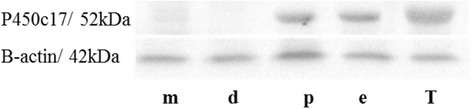
Background: The synthesis of sex steroids is controlled by several enzymes such as17α-hydroxylase cytochrome P450 (P450c17) catalyzing androgen synthesis and aromatase cytochrome P450 (P450arom) catalyzing estrogen synthesis, both of which must complex with the redox partner NADPH-cytochrome P450 oxidoreductase (CPR) for activity. Previous studies have identified expression of steroidogenic enzymes in vaginal tissue, suggesting local sex steroid synthesis. The current studies investigate P450c17, P450aromatase and CPR expression in vaginal mucosa of Galea spixii (Spix cavy) by immuno-histochemical and western immunoblot analyses.
Methods: Stages of estrous cyclicity were monitored by vaginal exfoliative cytology. After euthanasia, vaginal tissues were retrieved, fixed and frozen at diestrus, proestrus, estrus and metestrus. The ovaries and testis were used as positive control tissues for immunohistochemistry.
Results: Data from cytological study allowed identification of different estrous cycle phases. Immunohistochemical analysis showed different sites of expression of steroidogenic enzymes along with tissue response throughout different phases of the estrous cycle. However, further studies are needed to characterize the derived hormones synthesized by, and the enzymes activities associated with, vaginal tissues.
Conclusion: Current results not only support the expression of enzymes involved in sex steroid synthesis in the wall of the vagina, they also indicate that expression changes with the stage of the cycle, both the levels and types of cells exhibiting expression. Thus, changes in proliferation of vaginal epithelial cells and the differentiation of the mucosa may be influenced by local steroid synthesis as well as circulating androgens and estrogens.
Keywords: Androgens; Cytochrome P450; Estrogens; Estrous cycle; Vaginal epithelium.
Figures


Similar articles
-
Follicular development and morphological changes in the vaginal epithelium during the estrous cycle of Galea spixii.Microsc Res Tech. 2017 Feb;80(2):167-176. doi: 10.1002/jemt.22784. Epub 2016 Oct 7. Microsc Res Tech. 2017. PMID: 27717109
-
Steroidogenesis during postnatal testicular development of Galea spixii.Reproduction. 2017 Nov;154(5):645-652. doi: 10.1530/REP-17-0075. Epub 2017 Aug 8. Reproduction. 2017. PMID: 28982933
-
Vaginal fold histology reduces the variability introduced by vaginal exfoliative cytology in the classification of mouse estrous cycle stages.Toxicol Pathol. 2014 Dec;42(8):1212-20. doi: 10.1177/0192623314526321. Epub 2014 Apr 3. Toxicol Pathol. 2014. PMID: 24705880 Free PMC article.
-
Vaginal Cytology of the Laboratory Rat and Mouse: Review and Criteria for the Staging of the Estrous Cycle Using Stained Vaginal Smears.Toxicol Pathol. 2015 Aug;43(6):776-93. doi: 10.1177/0192623315570339. Epub 2015 Mar 3. Toxicol Pathol. 2015. PMID: 25739587 Free PMC article. Review.
-
Anterior pituitary cell renewal during the estrous cycle.Front Horm Res. 2006;35:9-21. doi: 10.1159/000094260. Front Horm Res. 2006. PMID: 16809919 Review.
Cited by
-
Letrozole administration effects on the P450aromatase expression and the reproductive parameters in male sheep (Ovis aries).Anim Reprod. 2023 Dec 1;20(4):e20230099. doi: 10.1590/1984-3143-AR2023-0099. eCollection 2023. Anim Reprod. 2023. PMID: 38074943 Free PMC article.
-
Galea spixii embryos have potential to produce steroid hormones.Anim Reprod. 2023 Jan 16;19(4):e20220091. doi: 10.1590/1984-3143-AR2022-0091. eCollection 2022. Anim Reprod. 2023. PMID: 36686856 Free PMC article.
References
-
- Adrian O, Sachser N. Diversity of social and mating systems in cavies: a review. J Mammal. 2011;92:39–53. doi: 10.1644/09-MAMM-S-405.1. - DOI
-
- Santos AC, Bertassoli BM, Viana DC, Vasconcelos BG, Oliveira MA, Miglino MA, et al. The morphology of female genitalia in Galea spixii (Caviidae, Caviinae) Biosc J. 2014;34:1793–1802.
-
- Santos AC, Viana DC, Bertassoli BM, Oliveira GB, Oliveira DM, Oliveira MF, et al. Characterization of the estrous cycle in Galea spixii (Wagler, 1831) Pesq Vet Bras. 2015;35:89–94. doi: 10.1590/S0100-736X2015000100017. - DOI
-
- IUCN. International Union of Conservation of Nature. Red List of Threatened Species. version 2014.1 Available at www.iucnredlist.org. Access at Nov 2014.
MeSH terms
Substances
LinkOut - more resources
Full Text Sources
Other Literature Sources
