. 2017 Apr 24;12(1):32.
doi: 10.1186/s13024-017-0174-z.

The PINK1 p.I368N mutation affects protein stability and ubiquitin kinase activity

Affiliations

The PINK1 p.I368N mutation affects protein stability and ubiquitin kinase activity

Maya Ando et al. Mol Neurodegener. .

Abstract

Background: Mutations in PINK1 and PARKIN are the most common causes of recessive early-onset Parkinson's disease (EOPD). Together, the mitochondrial ubiquitin (Ub) kinase PINK1 and the cytosolic E3 Ub ligase PARKIN direct a complex regulated, sequential mitochondrial quality control. Thereby, damaged mitochondria are identified and targeted to degradation in order to prevent their accumulation and eventually cell death. Homozygous or compound heterozygous loss of either gene function disrupts this protective pathway, though at different steps and by distinct mechanisms. While structure and function of PARKIN variants have been well studied, PINK1 mutations remain poorly characterized, in particular under endogenous conditions. A better understanding of the exact molecular pathogenic mechanisms underlying the pathogenicity is crucial for rational drug design in the future.

Methods: Here, we characterized the pathogenicity of the PINK1 p.I368N mutation on the clinical and genetic as well as on the structural and functional level in patients' fibroblasts and in cell-based, biochemical assays.

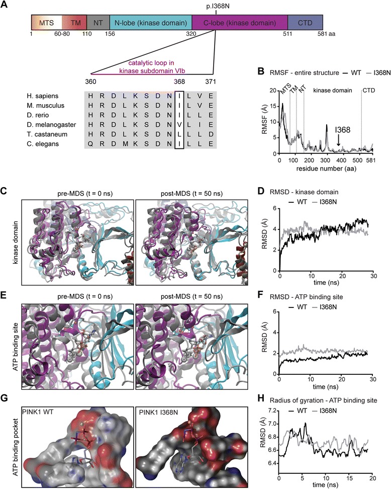
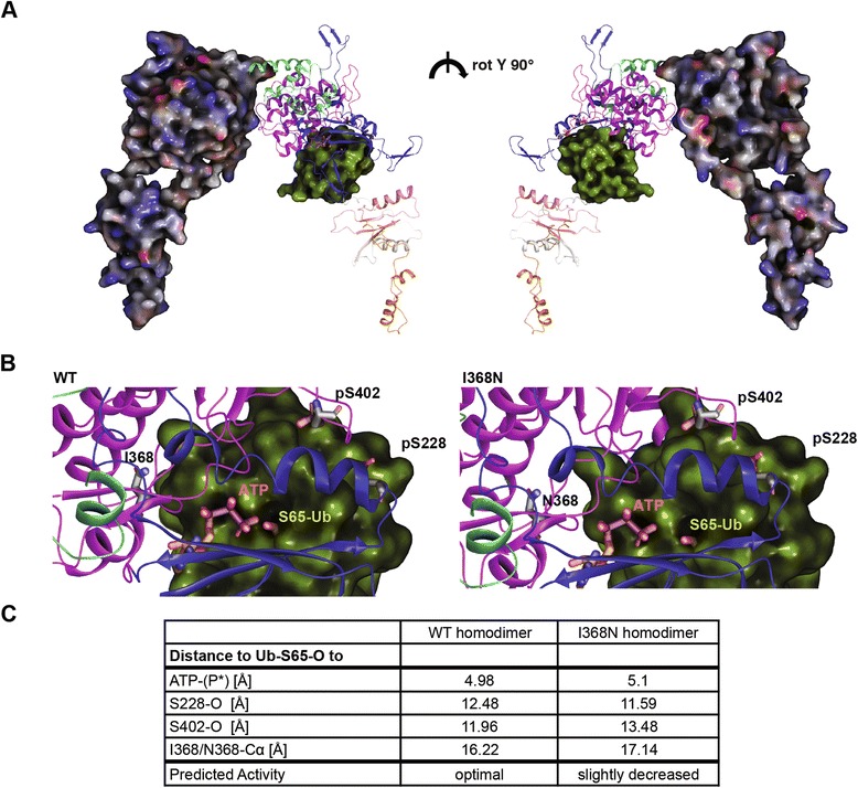
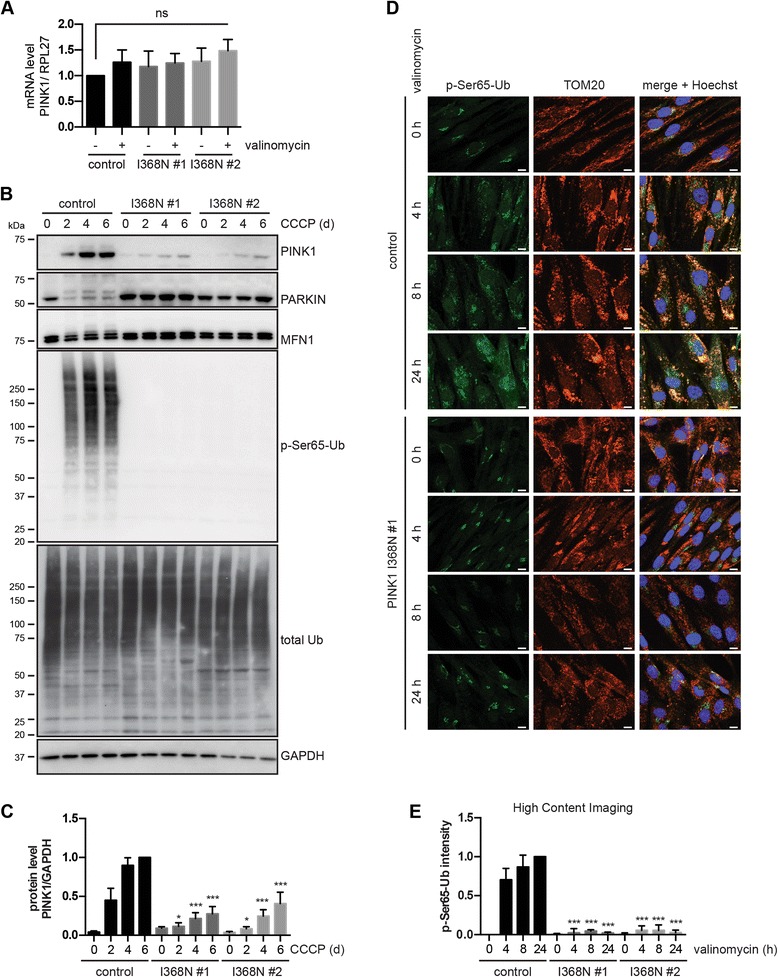
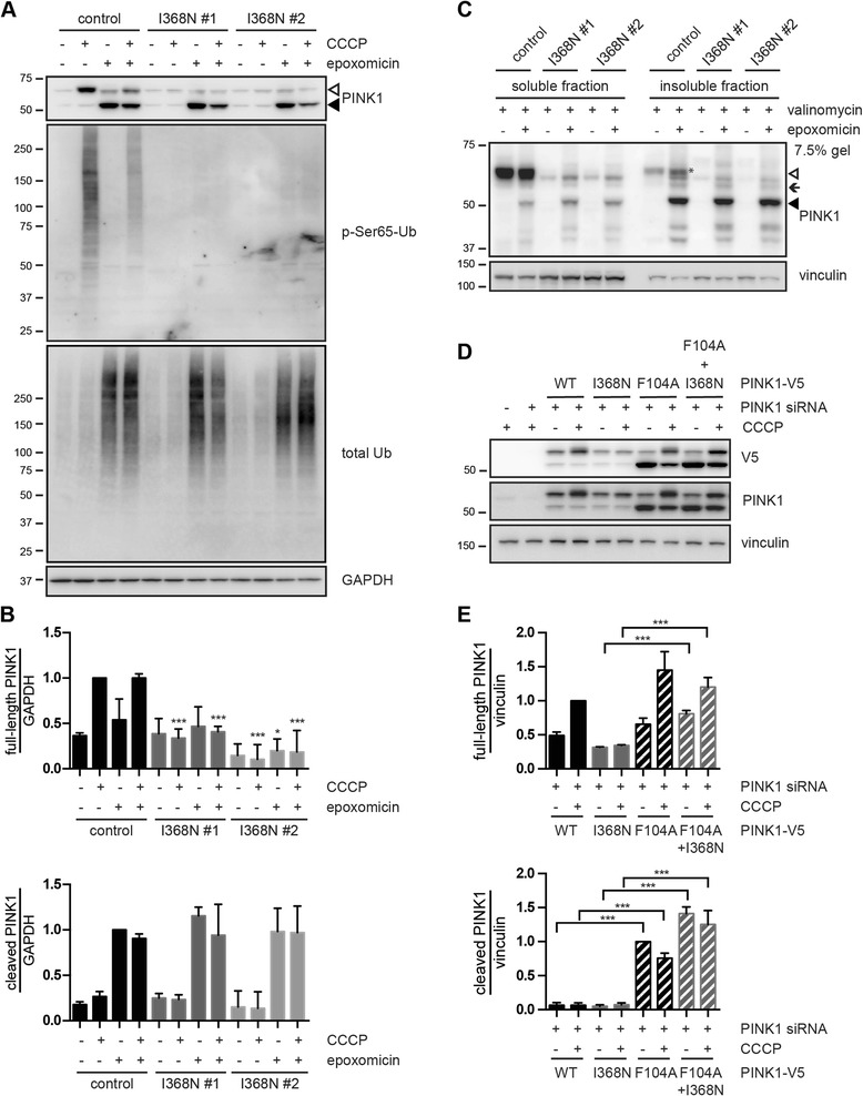
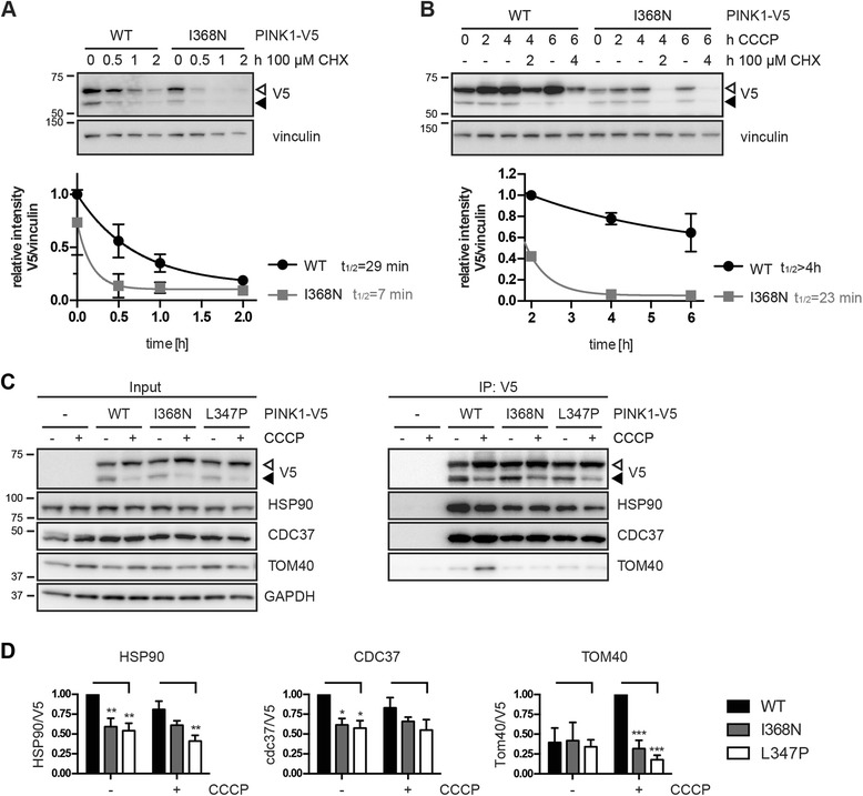
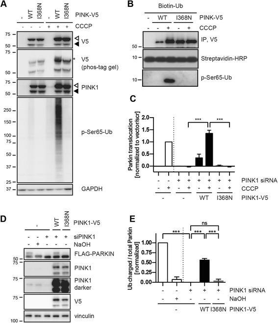
Results: Under endogenous conditions, PINK1 p.I368N is expressed, imported, and N-terminally processed in healthy mitochondria similar to PINK1 wild type (WT). Upon mitochondrial damage, however, full-length PINK1 p.I368N is not sufficiently stabilized on the outer mitochondrial membrane (OMM) resulting in loss of mitochondrial quality control. We found that binding of PINK1 p.I368N to the co-chaperone complex HSP90/CDC37 is reduced and stress-induced interaction with TOM40 of the mitochondrial protein import machinery is abolished. Analysis of a structural PINK1 p.I368N model additionally suggested impairments of Ub kinase activity as the ATP-binding pocket was found deformed and the substrate Ub was slightly misaligned within the active site of the kinase. Functional assays confirmed the lack of Ub kinase activity.

Conclusions: Here we demonstrated that mutant PINK1 p.I368N can not be stabilized on the OMM upon mitochondrial stress and due to conformational changes in the active site does not exert kinase activity towards Ub. In patients' fibroblasts, biochemical assays and by structural analyses, we unraveled two pathomechanisms that lead to loss of function upon mutation of p.I368N and highlight potential strategies for future drug development.

Keywords: Autophagy; E3 ubiquitin ligase; Mitochondria; Mitophagy; PARK2; PARKIN; PINK1; Parkinson’s disease; Phospho-ubiquitin; Ubiquitin.


Figures

Fig. 1
Fig. 1
The PINK1 p.I368N mutation is positioned in the kinase domain and alters the ATP-binding site. a Domain structure of human PINK1 protein. Individual domains are color-coded and labeled: mitochondrial targeting sequence (MTS); transmembrane domain (TM); N-terminal, regulatory domain (NT); N-lobe and C-lobe of the kinase domain; C-terminal domain (CTD). Residues surrounding Ile368 were aligned with corresponding sequences of PINK1 orthologs from different species. Horizontal bar denotes the catalytic loop in kinase subdomain VIb. b Root mean square fluctuations (RMSF) of the entire PINK1 structures. Dotted lines separate individual domains of PINK1 that include MTS, TM, NT, kinase domain and CTD. An arrow highlights the position of the p.I368N mutation. c Superposition overlay of the kinase domains before and after 50 ns of MDS. Structures are shown in cartoon ribbons with PINK1 WT in gray and p.I368N mutant in color according to the domain structure in (a). Residues Ile368 and Asn368 are shown in licorice rendering using gray or standard atom coloring, respectively. The Mg2+ ions are gray colored Van der Waals (VdW) spheres for both and the ATP-analogs in the active sites are shown in gray for WT and Goodsell coloring for p.I368N. d Root mean square deviation (RMSD) of the kinase domain of PINK1 WT and p.I368N mutant, shows only minor structural differences. e Zoom into the ATP-binding sites of PINK1 WT and p.I368N mutant in superposition overlay before and after 50 ns of MDS as described for (c). f RMSD of the ATP-binding pocket in the kinase domains of WT and mutant PINK1. Pocket residues include positions: 161,163, 217, 218, 219, 237, 240, 275, 316, 318, 319, 320, 321, 322, 325, 326, 327, 362, 363, 365, 366, 367, 368, 369, 383, 384, 385, 386, and 387. g The surface of the ATP-binding pocket of WT and p.I368N mutant PINK1 is shown for comparison in standard atom coloring. The Mg2+ ion (VdW sphere) and the ATP-analog (sticks colored by atom type) in the active site are given. h Radius of gyration measurements on the ATP-binding pocket from WT and mutant PINK1 provides the extent to which an object is extended from its center of mass
Fig. 2
Fig. 2
Protein docking of the kinase PINK1 and the substrate Ub. a Structural model of a PINK1 p.I368N homodimer docked with Ub in the kinase domain (between the N- and C-lobe). One molecule of the PINK1 dimer is shown in electrostatic surface rendering. The other one is shown in ribbons colored according to the individual domains of PINK1. The p-Ser228, p-Ser402, and Asn368 residues are shown in licorice sticks with the carbons colored in gray. ATP is shown in licorice rendering with orange carbons. The substrate Ub is shown in yellow-green surface culled to reveal Ser65 residue in VdW. Both sides of the dimer are rotated 180°. b Zoom into the active site region. Left panel: PINK1 WT (residues Ile368, p-Ser228, and p-Ser402 are highlighted) docked with ATP (terminal phosphate is highlighted), and Ub (Ser-65 is highlighted). Right panel: PINK1 p.I368N mutant with the same residues highlighted for comparison. c Metric for enzymatic comparison of the of PINK1 WT and p.I368N mutant is given in the table. Atomic distances between the docked Ub (Ser65-oxygen), the terminal phosphate of ATP (P*), and PINK1 residues (p-Ser228-oxygen, p-Ser402-oxygen, I368-Calpha or N368-Calpha) were determined
Fig. 3
Fig. 3
Full-length PINK1 protein and kinase activity are decreased in p.I368N mutant fibroblasts. a qRT-PCR was used to measure mRNA levels of PINK1 and housekeeping gene RPL27 in untreated cells and cells treated with 1 μM valinomycin for 24 h. Values of the PINK1/RPL27 mRNA ratio were normalized to untreated control cells. Error bars indicate mean ± SEM from six independent experiments (one-way ANOVA with Tukey’s post hoc; ns, not significant). b WT control and two PINK1 p.I368N fibroblasts were treated with 10 μM CCCP for 0, 2, 4 or 6 days and total lysates were analyzed by WB with the indicated antibodies. GAPDH served as a loading control. Slower migrating PINK1, PARKIN, and Mitofusin1 (MFN1) protein species indicate activated and/or modified forms and were only observed in control cells. p-Ser65-Ub signal increased in control cells over time but was undetectable in PINK1 p.I368N mutant cells. c Densitometric analysis of full-length PINK1 protein from (b). Values were normalized to controls cells treated for 6 days with CCCP. Error bars indicate mean ± SEM from four independent experiments. Values were tested for statistical significance compared to control cells (two-way ANOVA with Tukey’s posthoc test, *, p < 0.05, ***, p < 0.0005). d Representative images of fibroblasts upon treatment with 1 μM valinomycin. Cells were stained with anti p-Ser65-Ub (green) and TOM20 (red). Nuclei are shown in blue (Hoechst). Scale bars indicate 10 μm. Upon treatment, p-Ser65-Ub is induced only in control, but not in PINK1 p.I368N fibroblasts. e p-Ser65-Ub signal upon valinomycin treatment was quantified by HCI. Control and PINK1 p.I368N fibroblasts were incubated with 1 μM valinomycin for 0, 4, 8 and 24 h. Cells were fixed and stained with p-Ser65-Ub antibodies and a nuclear counterstain (Hoechst). Mean intensity of p-Ser65-Ub in a cytoplasmic ring around the nucleus was measured and normalized to values of untreated and 24 h valinomycin treated control cells. Error bars indicate mean ± SEM from three independent experiments (two-way ANOVA with Tukey’s post hoc; ***, p < 0.0005)
Fig. 4
Fig. 4
PARL-cleaved forms of PINK1 WT and p.I368N are similarly stabilized upon proteasome inhibition. a Control and PINK1 p.I368N fibroblasts were pretreated with 500 nM epoxomicin for 1 h followed by 10 μM CCCP treatment for additional 24 h, as indicated. Only PARL-cleaved PINK1 p.I368N (~52 kDa, black arrowhead), but not the full-length form (~63 kDa, white arrowhead) accumulated with epoxomicin ± CCCP in mutant fibroblasts. Anti-p-Ser65-Ub and total Ub antibodies were used as controls for CCCP and epoxomicin treatment, respectively. GAPDH served as a loading control. b WB quantification of full-length (top) and PARL-cleaved (bottom) PINK1 species from control and p.I368N mutant fibroblasts as performed in (a). Values of the full-length PINK1/GAPDH ratio were normalized to control cells treated only with CCCP. Values of the cleaved PINK1/GAPDH ratios were normalized to control cells treated only with epoxomicin. Error bars indicate mean ± SEM from three independent experiments (two-way ANOVA with Tukey’s post hoc; *, p < 0.05; ***, p < 0.0005). c Fibroblasts were pretreated with 500 nM epoxomicin for 1 h followed by 1 μM valinomycin treatment for additional 24 h, as indicated. Cells were homogenized in fractionation buffer and proteins were sequentially extracted into soluble (RIPA) and insoluble (urea) fractions and were analyzed by WB using PINK1 antibody. Vinculin served as a loading control. White and black arrowheads indicate PINK1 full-length (mostly in soluble fraction) and the PARL-cleaved form (mostly in insoluble fraction), respectively. Black arrow indicates an intermediate (probably MPP-cleaved) form of PINK1 (~60 kDa), which could be detected in the “insoluble fraction” after epoxomicin treatment. d HeLa cells were transfected with control or PINK1 siRNA together with constructs encoding for PINK1-V5 (WT, p.I368N, p.F104A, or the double mutant p.F104A + p.I368N). Cells were left untreated or treated with 10 μM CCCP for 2 h. Levels of full-length and cleaved PINK1 were analyzed by WB using anti-V5 and anti-PINK1 antibodies. Vinculin served as a loading control. e Densitometric analysis of the full-length (top) and PARL-cleaved (bottom) PINK1 species from (d). Values of the full-length PINK1-V5/vinculin ratio were normalized to CCCP-treated cells expressing WT PINK1-V5. Values of the cleaved PINK1-V5/vinculin ratio were normalized to untreated cells expressing PINK1-V5 p.F104A mutant. Error bars indicate mean ± SEM from three independent experiments (two-way ANOVA with Tukey’s post hoc; ***, p < 0.0005)
Fig. 5
Fig. 5
The half-life of the p.I368N mutant PINK1 protein is reduced. a-b HeLa cells were simultaneously transfected with PINK1 siRNA and DNA constructs encoding for PINK1-V5 WT or p.I368N mutant and incubated with 100 μg/ml cycloheximide (CHX) alone (a) or pretreated with 10 μM CCCP for 2 h before incubation with cycloheximide (b). Cell lysates were analyzed by WB and V5/vinculin ratios were normalized to PINK1-V5 WT. Shown is the mean ± SEM from three independent experiments. Half-life (t1/2) of PINK1-V5 proteins was determined by curve fitting (one phase exponential decay). c Co-immunoprecipitation (IP) of the HSP90/CDC37 chaperone complex and the import channel of the mitochondrial outer membrane TOM40 with PINK1-V5. HeLa cells were transfected with PINK1-V5 WT, p.I368N or another PD-mutant PINK1 p.L347P as a control and left untreated or treated with 10 μM CCCP for 2 h. PINK1-V5 protein complexes were immunoprecipitated from cell lysates using V5 antibody conjugated to agarose. Representative WB is shown for input and IP samples probed with antibodies against V5 (PINK1), HSP90, CDC37, and TOM40. GAPDH served as a loading control. d Quantification of the co-immunoprecipitated proteins as performed in (c) is shown as the protein/V5 ratio. Data represent mean ± SEM from six independent experiments. Statistical significance was assessed by two-way ANOVA with Tukey’s post hoc; ***, p < 0.0005; **, p < 0.005; *, p < 0.05
Fig. 6
Fig. 6
PINK1 p.I368N is a kinase dead mutant that fails to activate PARKIN and the mitochondrial quality control. a HeLa cells were simultaneously transfected with PINK1 siRNA and V5 empty vector, PINK1 WT or p.I368N mutant. Cells were left untreated or incubated with 10 μM CCCP for 4 h. Cell lysates were analyzed by WB, probed with anti-V5, PINK1 and p-Ser65-Ub antibodies. GAPDH was used as a loading control. Auto-phosphorylated PINK1 (anti-V5) (asterisk) was detected on phos-tag gels only in lysates from PINK1-V5 WT transfected, CCCP treated cells. In PINK1-V5 p.I368N transfected cells, CCCP-induced phosphorylation of Ub was largely abolished. b IP of PINK1-V5 using V5 antibody followed by in vitro kinase assay. HeLa cells were transfected with the V5 empty vector, PINK1-V5 WT or p.I368N and treated with or without CCCP for 2 h. Washed immunoprecipitates were incubated with N-terminally biotinylated mono-Ub and 500 μM ATP in phosphorylation buffer at 37 °C for 24 h. Total and phosphorylated Ub were analyzed by WB using streptavidin-HRP and p-Ser65-Ub antibody, respectively. c PARKIN translocation to damaged mitochondria measured by HCI. HeLa cells stably overexpressing GFP-tagged PARKIN were simultaneously transfected with PINK1 (or control) siRNA and with V5 empty vector, PINK1-V5 WT or p.I368N mutant, as indicated, and with mCherry as a selection marker. Cells were left untreated or treated with CCCP for 2 h and GFP-PARKIN translocation was measured in mCherry-positive cells. Data represents the mean of two independent experiments run with each six replicate wells. Statistical significance was assessed by two-way ANOVA with Tukey’s post hoc; **, p < 0.005; ***, p < 0.0005). d Ub-charging of PARKIN C431S as a measure of its enzymatic activation. HeLa cells stably expressing 3xFLAG-tagged PARKIN C431S were pretreated with control or PINK1 siRNA and then transfected with V5 empty vector, PINK1-V5 WT or p.I368N. After treatment with CCCP for 4 h, cell lysates were analyzed by WB with the indicated antibodies. PARKIN C431S traps Ub in an oxyester-bond (resulting in an 8 kDa band shift) that is cleavable by NaOH treatment. e Densitometric analysis of Ub-charged 3xFLAG-PARKIN that was normalized to total PARKIN levels. Error bars indicate mean ± SEM from three independent experiments (one-way ANOVA with Tukey’s post hoc; ***, p < 0.0005; ns, not significant)

References

    1. Corti O, Lesage S, Brice A. What Genetics Tells Us About the Causes and Mechanisms of Parkinson's Disease. Physiol Rev. 2011;91:1161–1218. doi: 10.1152/physrev.00022.2010. - DOI - PubMed
    1. Dawson TM, Ko HS, Dawson VL. Genetic Animal Models of Parkinson's Disease. Neuron. 2010;66:646–661. doi: 10.1016/j.neuron.2010.04.034. - DOI - PMC - PubMed
    1. Valente EM, Abou-Sleiman PM, Caputo V, Muqit MM, Harvey K, Gispert S, Ali Z, Del Turco D, Bentivoglio AR, Healy DG, et al. Hereditary early-onset Parkinson's disease caused by mutations in PINK1. Science. 2004;304:1158–1160. doi: 10.1126/science.1096284. - DOI - PubMed
    1. Kitada T, Asakawa S, Hattori N, Matsumine H, Yamamura Y, Minoshima S, Yokochi M, Mizuno Y, Shimizu N. Mutations in the parkin gene cause autosomal recessive juvenile parkinsonism. Nature. 1998;392:605–608. doi: 10.1038/33416. - DOI - PubMed
    1. Geisler S, Holmstrom KM, Skujat D, Fiesel FC, Rothfuss OC, Kahle PJ, Springer W. PINK1/Parkin-mediated mitophagy is dependent on VDAC1 and p62/SQSTM1. Nat Cell Biol. 2010;12:119–131. doi: 10.1038/ncb2012. - DOI - PubMed