. 2017 Apr 24;12(1):6.
doi: 10.1186/s13064-017-0083-4.

Semaphorin-Plexin signaling influences early ventral telencephalic development and thalamocortical axon guidance

Affiliations

Semaphorin-Plexin signaling influences early ventral telencephalic development and thalamocortical axon guidance

Manuela D Mitsogiannis et al. Neural Dev. .

Abstract

Background: Sensory processing relies on projections from the thalamus to the neocortex being established during development. Information from different sensory modalities reaching the thalamus is segregated into specialized nuclei, whose neurons then send inputs to cognate cortical areas through topographically defined axonal connections. Developing thalamocortical axons (TCAs) normally approach the cortex by extending through the subpallium; here, axonal navigation is aided by distributed guidance cues and discrete cell populations, such as the corridor neurons and the internal capsule (IC) guidepost cells. In mice lacking Semaphorin-6A, axons from the dorsal lateral geniculate nucleus (dLGN) bypass the IC and extend aberrantly in the ventral subpallium. The functions normally mediated by Semaphorin-6A in this system remain unknown, but might depend on interactions with Plexin-A2 and Plexin-A4, which have been implicated in other neurodevelopmental processes.

Methods: We performed immunohistochemical and neuroanatomical analyses of thalamocortical wiring and subpallial development in Sema6a and Plxna2; Plxna4 null mutant mice and analyzed the expression of these genes in relevant structures.

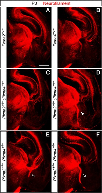
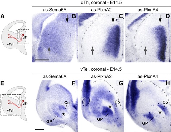
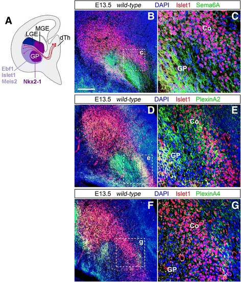
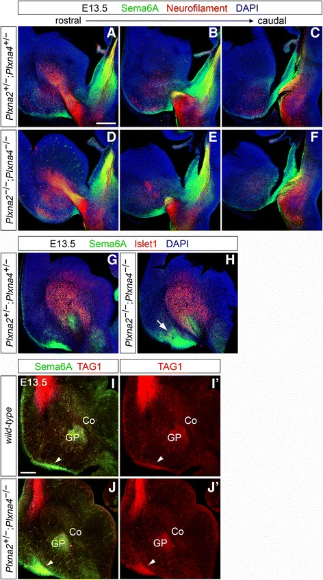
Results: In Plxna2; Plxna4 double mutants we discovered TCA pathfinding defects that mirrored those observed in Sema6a mutants, suggesting that Semaphorin-6A - Plexin-A2/Plexin-A4 signaling might mediate dLGN axon guidance at subpallial level. In order to understand where and when Semaphorin-6A, Plexin-A2 and Plexin-A4 may be required for proper subpallial TCA guidance, we then characterized their spatiotemporal expression dynamics during early TCA development. We observed that the thalamic neurons whose axons are misrouted in these mutants normally express Semaphorin-6A but not Plexin-A2 or Plexin-A4. By contrast, all three proteins are expressed in corridor cells and other structures in the developing basal ganglia. This finding could be consistent with an hypothetical action of Plexins as guidance signals through Sema6A as a receptor on dLGN axons, and/or with their indirect effect on TCA guidance due to functions in the morphogenesis of subpallial intermediate targets. In support of the latter possibility, we observed that in both Plxna2; Plxna4 and Sema6a mutants some IC guidepost cells abnormally localize in correspondence of the ventral path misrouted TCAs elongate into.

Conclusions: These findings implicate Semaphorin-6A - Plexin-A2/Plexin-A4 interactions in dLGN axon guidance and in the spatiotemporal organization of guidepost cell populations in the mammalian subpallium.

Keywords: Guidepost cells; Plexin-A2; Plexin-A4; Semaphorin-6A; Subpallium; Thalamocortical connectivity.


Figures

Fig. 1
Fig. 1
Immunohistochemical analysis of TCA development in Plxna2; Plxna4 single and double mutant P0 brains. Immunostaining for neurofilament (red) confirms the presence of a Sema6a-mutant-like TCA defect in Plxna2; Plxna4 double mutant postnatal brains (d; filled arrowhead), while no abnormal thalamic projections in the vTel are observed in either Plxna2 or Plxna4 single mutants (a, b). Additionally, misrouted TCAs are not present in Plxna2; Plxna4 double heterozygous brains (c). Some TCA guidance defects, similar to those observed in double mutant brains but restricted to only a few thalamic fibers, also characterize Plxna2 +/− ; Plxna4 −/− and Plxna2 −/− ; Plxna4 +/− postnatal brains (e, f; empty arrowheads). Scale: 500 μm
Fig. 2
Fig. 2
Neuroanatomical tracing experiments reveal a Sema6a mutant-like TCA phenotype in Plxna2; Plxna4 double mutant brains. ac Retrograde labeling with carbocyanine dyes from the vTel in wild-type (a) versus Plxna2 −/− ; Plxna4 −/− P0 brains (b, c). Insertion of DiI crystals in the vTel (asterisk) results in no back-labeling of dTh neurons in wild-type brains; on the other hand, in Plxna2 −/− ; Plxna4 −/− brains DiI back-labels thalamic axons and cell somas located in the dLGN (b), a finding that coincides with data obtained from Sema6a −/− brains. In addition, dye-labeled neurons are also found in the VB, indicating the extension of guidance defects to a subset of thalamic axons normally directed to somatosensory cortical areas. di Back-labeling of thalamic neurons with two distinct carbocyanine dyes from the visual (occipital) cortex and the somatosensory (parietal) cortex in P0 wild-type (wt) and Plxna2; Plxna4 double mutant (dKO) brains. c Schematic representation of the cortical sites of dye placement in P0 brain hemispheres (OB: olfactory bulb). DiA (green) and DiA (red) crystals are placed respectively on parietal (Par) and occipital (Occ) regions of the cortex. eh Insertion of DiI crystals in visual cortical areas of Plxna2 −/− ; Plxna4 −/− brains results in the back-labeling of some thalamic neurons of the dorso-lateral VB (h), rather than the dLGN (as instead observed in wild-type brains (f)), suggesting a miswiring of somatosensory TCAs to the visual cortex similar to that present in Sema6a mutants. Normal connectivity between ventro-medial VB neurons and the somatosensory cortex is preserved, as indicated by back-labeling of these cells by DiA. Scale: a, b: 250 μm; eh: 500 μm
Fig. 3
Fig. 3
Expression of Sema6a, Plxna2 and Plxna4 mRNA in the E14.5 mouse forebrain. Levels of mRNA expression were detected by in situ hybridization with antisense (as) RNA probes. ad mRNA expression in the dTh of E14.5 mouse brains. a Schematic representation of a coronal E14.5 section indicating the approximate region of interest (boxed area); TCAs are represented in red. bd Sema6a is strongly expressed in both lateral (grey arrows) and medial (black arrows) dTh (b); in contrast, Plxna2 and Plxna4 are highly transcribed only in the medial dTh (c, d). eh mRNA expression in the subpallium (vTel) of E14.5 mouse brains. e Schematic representation of a coronal E14.5 section indicating the approximate region of interest (boxed area); TCAs are represented in red. (fh) Sema6a is highly expressed in areas surrounding the IC (asterisks), in the pial surface of the vTel, and the ventricular zone; it is also moderately transcribed in subventricular subpallial regions (f). Plxna2 shows maximum expression in the globus pallidus, ventral to the IC, and strong expression levels in subventricular and mantle layers (g), while Plxna4 is highly transcribed in two discrete bands located dorsal and ventral to the IC (h). Scale: ad, 500 μm; eh, 500 μm. Coronal section schemes adapted from López-Bendito et al. [22]
Fig. 4
Fig. 4
Expression of Sema6A and PlxnA4 on thalamic neurons and TCAs during axonal growth into the subpallium. ab’ Double immunohistochemistry for Sema6A (red) and neurofilament (green) on wild-type E13.5 coronal brain sections indicates expression of Sema6A in thalamic neurons, in particularly in dorso-lateral populations (b, b’), as well as on extending TCAs; the protein can also be found in vTel areas ventral to the IC (a, a’). c–d’ Double immunohistochemistry for Sema6A (red) and neurofilament (green) on coronal sections of wild-type E14.5 brains shows expression of Sema6A extending to all thalamic nuclei (d, d’); the protein is furthermore expressed along TCAs positioned more caudally along the rostro-caudal axis (d, d’), while it is not found on TCAs projecting more rostrally (c, c’). e–f’ Double immunohistochemistry for PlxnA4 (red) and neurofilament (green) in wild-type E13.5 coronal brain sections reveals that expression of PlxnA4 is mostly concentrated in medial thalamic neural populations, and is present on TCAs. Immunostaining can also be observed on some fibers in the IC contacting the axon bundle dorso-medially and ventro-laterally, and in mantle and pial surface areas of the caudal vTel (e, e’). g–h’ Double immunohistochemistry for PlxnA4 (red) and neurofilament (green) on coronal sections of wild-type E14.5 brains demonstrates localization of PlxnA4 in medial thalamic neural populations (h, h’), as well as along TCAs projecting rostrally (g, g’). PlexinA4 seems to be further localized in some caudally-located TCAs (h, h’). i–j Immunohistochemistry for PlxnA4 (red) on coronal sections of Sema6a −/− E15.5 brains shows the presence of PlxnA4 on caudally-located TCAs extending within the IC (empty arrowhead), which presumably correspond to VB-originated axons. On the other hand, no immunostaining can be detected on misrouted projections corresponding to dLGN-originated fibers (filled arrowheads). Scale: a–b’, 300 μm; c–d’, 300 μm; e–f’, 300 μm; g–h’, 300 μm; i–j, 300 μm
Fig. 5
Fig. 5
Expression of Sema6A, PlxnA2 and PlxnA4 in corridor cells and other subpallial structures at E13.5. a Diagram illustrating the spatial expression patterns of LGE- and MGE-derived neural population markers. The transcription factors Ebf1, Islet1, and Meis2 are detected in striatal and corridor regions of the vTel (light purple), both derivatives of the LGE, but not in the GP and the ventricular/subventricular zone of the MGE (dark purple). These territories in turn express a transcription factor, Nkx2-1, not present in LGE-derived territories. (Adapted from López-Bendito et al. [22].). b, c Double immunohistochemistry for the corridor cell marker Islet1 (red) and Sema6A (green) on coronal wild-type brain sections demonstrates the expression of Sema6A on corridor cells (Co) during the growth of TCAs into subpallial populations. Sema6A is also highly expressed in globus pallidus (GP) cells (c). d–g Double immunohistochemistry for PlxnA2 (green) and Islet1 (red) (d, e), or PlxnA4 (green) and Islet1 (red) (f, g) on coronal wild-type brain sections indicates a strong presence of PlxnA2 within the corridor and in the globus pallidus (e); PlxnA4 is also moderately present on most dorso-medial corridor domains (g), and in the lateral half of the globus pallidus area (f). Both molecules are additionally lightly expressed in the vTel subventricular zone and pial surface, in an area close to the IC, and in a discrete band at the ventral edge of the striatum (PlxnA4 is particularly present here) (d, f). Scale: 150 μm
Fig. 6
Fig. 6
Normal overall expression of Islet1 in the vTel of Sema6a mutants and Plxna2; Plxna4 double mutants at E13.5. Double immunohistochemistry for neurofilament (green) and Islet1 (red) on E13.5 coronal brain sections indicates the preserved organization, at this stage, of Islet1-positive cell domains in the developing subpallium of Sema6a −/− (e, f), Plxna2 +/− ; Plxna4 +/− (c, d), Plxna2 −/− ; Plxna4 −/− (g, h) mouse brains, as compared to wild-type (a, b). Islet1-positive neurons are present in a narrow band situated immediately dorsal to extending TCAs, between the vTel subventricular zone and the globus pallidus (characterized by the absence of Islet1 immunostaining), and throughout the striatum, where neurofilament-expressing thalamocortical fibers can be observed to segregate (a, c, e, g). Caudally, a slight reduction and disorganization of the most posteriorly-located subset of Islet1-positive cells can be observed in the Plxna2 −/− ; Plxna4 −/− mouse vTel (h). Scale: 200 μm
Fig. 7
Fig. 7
A subset of IC guidepost cells is misplaced in Sema6a mutant and Plxna2; Plxna4 double mutant E13.5 brains. a Schematic representation of the dye tracing experiments performed. DiI crystals were inserted into the dTh of E13.5 Plxna2 +/− ; Plxna4 +/− and Plxna2 −/− ; Plxna4 −/− mouse brains; from this position, the dye diffuses along TCAs in an anterograde fashion, and on guidepost cell projections reaching the dTh. (Adapted from Garel and López-Bendito [21].). b, c Coronal sections of E13.5 brains illustrating the labeled IC (b) and the exact location of dye placement in the dTh (asterisk in c). ds DiI labels growing TCAs as well as guidepost cell bodies in the IC area, along the dorso-lateral path which TCAs will follow to proceed further into the subpallium (solid arrowheads), in late E13.5 wild-type (d–g), E13.5 Plxna2 +/− ; Plxna4 +/− (hk), E13.5 Plxna2 −/− ; Plxna4 −/− (lo), and E13.5 Sema6a −/− (ps) mouse brains. In Plxna2 −/− ; Plxna4 −/− and Sema6a −/− sections, however, back-labeling identifies a group of cells projecting to the dTh in an abnormal caudo-ventral position in the vTel, close to the pial surface, corresponding to presumptive amygdala territories (ls, empty arrowheads). No dye can be detected in this domain in either wild-type or Plxna2 +/− ; Plxna4 +/− brains. Scale: b, c: 250 μm; ds: 100 μm. t Quantification and comparison of DiI signal intensities in the caudo-ventral subpallium, expressed as ventral vTel/overall (DTB and vTel) integrated intensity ratios (IRRs), between wild-type, Plxna2 +/− ; Plxna4 +/−, Plxna2 −/− ; Plxna4 −/−, and Sema6a −/− E13.5 brains. A significantly higher DiI signal intensity compared to both wild-type (n = 10) and Plxna2 +/− ; Plxna4 +/− (n = 7) measurements was detected in case of Plxna2 −/− ; Plxna4 −/− (n = 6) as well as Sema6a −/− (n = 11) specimens. IRRwild-type = 0.09 ± 0.01; IRRPlxna2+/−; Plxna4+/− = 0.10 ± 0.02; IRRPlxna2−/−; Plxna4−/− = 0.23 ± 0.06; IRRSema6a−/− = 0.20 ± 0.02; p < 0.01
Fig. 8
Fig. 8
Sema6A expression in the dorsal thalamus, prethalamus, developing basal ganglia and lateral olfactory tract in Plxna2; Plxna4 double mutant E13.5 brains. a–f Double immunohistochemistry for neurofilament (red) and Sema6A (green) on E13.5 coronal brain sections reveals the normal fasciculation and spatial navigation of Sema6A-positive caudally-projecting TCAs at dorsal thalamic and prethalamic level in Plxna2 −/− ; Plxna4 −/− (ac) mouse brains, as compared to Plxna2 +/− ; Plxna4 +/− specimens (d–f). g, h Double immunohistochemistry for Islet1 (red) and Sema6A (green) on E13.5 coronal Plxna2 +/− ; Plxna4 +/− and Plxna2 −/− ; Plxna4 −/− brain sections demonstrates the preserved corridor region co-expression of the two proteins in the absence of PlxnA2/PlnxA4 function, as well as the normal presence of Sema6A in the globus pallidus and other subpallial areas. However, a Sema6A-positive axonal bundle, which normally elongates within the ventral vTel surface, can be observed to invade the subpallium in a ventro-dorsal direction in Plxna2 −/− ; Plxna4 −/− brains (arrow in h). i–j’ Double immunohistochemistry for TAG1 (red) and Sema6A (green) on E13.5 coronal wild-type and Plxna2 +/− ; Plxna4 −/− brain sections indicates that the Sema6A-positive axonal bundle invading the subpallium in mice lacking PlxnA2/PlxnA4 (arrowheads) co-expresses TAG1, and thus corresponds to the lateral olfactory tract. Moreover, immunohistochemical data confirms the normal expression of Sema6A in developing basal ganglia structures in Plxna2; Plxna4 null mutants. Co: corridor cells; GP: globus pallidus. Scale: a–g: 250 μm; i–j’: 200 μm

Similar articles

Cited by

References

    1. Van Battum EY, Brignani S, Pasterkamp RJ. Axon guidance proteins in neurological disorders. Lancet Neurol. 2015;14(5):532–46. doi: 10.1016/S1474-4422(14)70257-1. - DOI - PubMed
    1. Amaral DG, Schumann CM, Nordahl CW. Neuroanatomy of autism. Trends Neurosci. 2008;31(3):137–45. doi: 10.1016/j.tins.2007.12.005. - DOI - PubMed
    1. Ameis SH, Catani M. Altered white matter connectivity as a neural substrate for social impairment in Autism Spectrum Disorder. Cortex. 2015;62:158–81. doi: 10.1016/j.cortex.2014.10.014. - DOI - PubMed
    1. Belmonte MK, et al. Autism and abnormal development of brain connectivity. J Neurosci. 2004;24(42):9228–31. doi: 10.1523/JNEUROSCI.3340-04.2004. - DOI - PMC - PubMed
    1. Geschwind DH, Levitt P. Autism spectrum disorders: developmental disconnection syndromes. Curr Opin Neurobiol. 2007;17(1):103–11. doi: 10.1016/j.conb.2007.01.009. - DOI - PubMed

MeSH terms

LinkOut - more resources