. 2017 Apr 24;10(1):29.
doi: 10.1186/s13048-017-0324-5.

Very small embryonic-like stem cells (VSELs) in adult mouse uterine perimetrium and myometrium

Affiliations

Very small embryonic-like stem cells (VSELs) in adult mouse uterine perimetrium and myometrium

Deepa Bhartiya et al. J Ovarian Res. .

Abstract

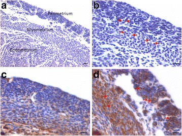
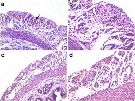
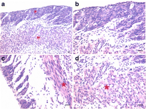
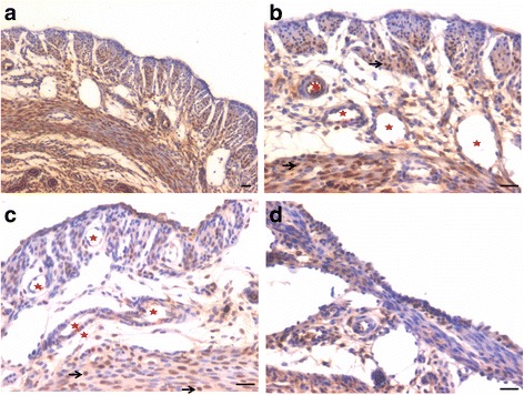
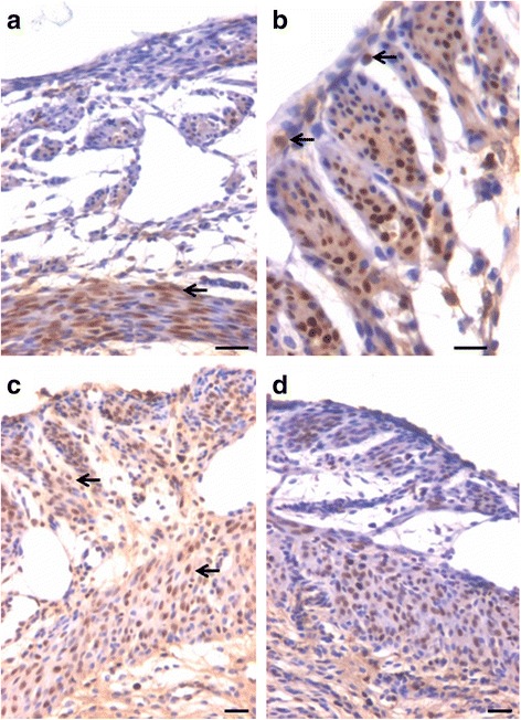
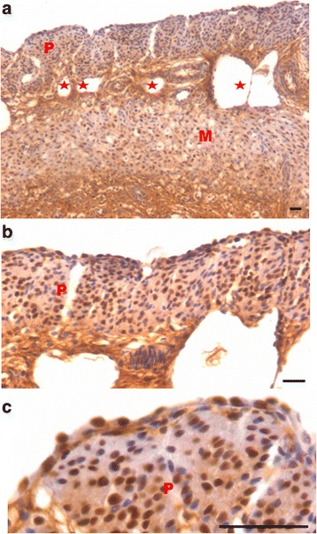
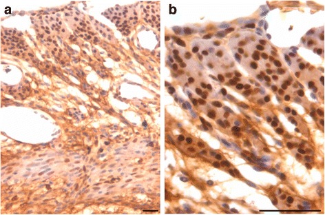
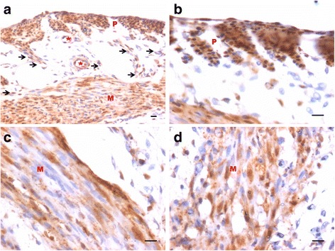
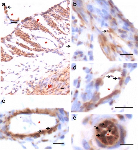
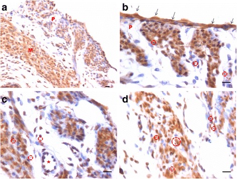
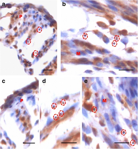
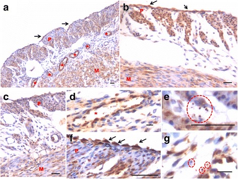
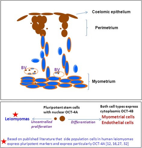
We have earlier reported the presence of very small embryonic-like stem cells (VSELs) in adult mouse uterus along with slightly bigger progenitors termed endometrial stem cells (EnSCs) and their regulation by ovarian hormones thus demonstrating a crucial role played by them during proliferation, differentiation and remodeling of the endometrium. Present study is a brief communication wherein we have examined the effect of higher dose of estrogen (E, 2 μg/day), progesterone (P, 1 mg/day) and follicle stimulating hormone (FSH, 5 IU/day for 5 days) specifically on the myometrium and perimetrium surrounding the endometrium in bilaterally ovariectomized mice. Similar treatment with E & P was recently used in a study published in the journal Nature to study the effect of steroid hormones on hematopoietic stem cells and this treatment regimen helps achieve hormone levels observed during pregnancy. Quiescent spherical stem cells (lacking PCNA expression) with high nucleo-cytoplasmic ratio and nuclear OCT-4A were detected in the perimetrium of atrophied (bilaterally ovariectomized) uterus. PCNA expression was observed after treatment and cells with cytoplasmic OCT-4B were invariably observed in the myometrium. VSELs were clearly visualized after treatment and the effect of P and FSH was more prominent compared to E on the development of myometrium. It is speculated that stem cells with nuclear OCT-4A located in the perimetrium differentiate to give rise to endothelial and myometrial cells with cytoplasmic OCT-4B. Based on the results of present study and published reports showing the presence of pluripotent markers (OCT-4, NANOG and SOX2) in human myometrial side population and expression of particularly OCT-4A in human leiomyomas, we speculate that these nuclear OCT-4 positive stem cells located in the perimetrium are the possible tumor initiating cells leading to the development of leiomyomas rather than the mesenchymal cells which express cytoplasmic OCT-4B.

Keywords: Hormones; Leiomyomas; Myometrium; Uterus; VSELs.


Figures

Fig. 1
Fig. 1
Bilaterally ovariectomized mouse uterine sections after H&E staining and expression of PCNA and OCT-4. a Myometrium and perimetrium surrounding the inner endometrium were clearly visualized. They comprised of two distinct layers including outer perimetrium and the inner myometrium. b At higher magnification, myometrium comprised rounded cells (arrow) as well as sparsely distributed stromal cells. c No expression of PCNA was observed whereas (d) Nuclear OCT-4A expression (arrow) was clearly evident in both perimetrium and myometrium in atrophied uterine sections. Scale bar represents 20 μm
Fig. 2
Fig. 2
Effect of progesterone and follicle stimulating hormone treatment on mouse uterine myometrium (M) and perimetrium (P). Cellularity was greatly increased after both progesterone (ab) and follicle stimulating hormone (cd) treatment compared to atrophied uterus shown in Fig. 1. Perimetrial cells were observed to be organized as small islands (arrow) and the myometrium was very conspicuous and comprised of tightly woven myoblasts. Note that the spherical cells observed in atrophied uterine sections (Fig. 1) were not observed after treatment with P and FSH. Scale bar represents 20 μm
Fig. 3
Fig. 3
Effect of estrogen treatment on mouse uterine myometrium (M) and perimetrium (P). Estrogen treatment also induced growth of the myometrium and perimetrium but (ab) small islands in the perimetrium observed after P and FSH treatment were not prominent after estrogen treatment. Histological appearance of the cells comprising the perimetrium was very different (appeared more undifferentiated) after E treatment compared to those after P and FSH treatment shown in Fig. 2. (c & d) Myometrium was also poorly organized comprising loosely arranged cells (asterix) compared to that observed after P and FSH treatment (Fig. 2). Scale bar represents 20 μm
Fig. 4
Fig. 4
Immuno-localization of PCNA after treatment with progesterone (ab) and (cd) follicle stimulating hormone. PCNA expression was observed in both P and FSH treated mouse perimetrium but was more in P treated sections compared to after FSH treatment. Cells in both perimetrium and myometrium showed positive expression. Note the development of small blood vessels (asterix) lined by a single layer of endothelial cells at peri- and myometrium junction. Excessive non-specific brown color in A is because of endogenous mouse Ig staining since PCNA antibody is monoclonal antibody being used on mouse sections. Scale bar represents 20 μm
Fig. 5
Fig. 5
Immuno-localization of PCNA after treatment with progesterone (ab) and (cd) follicle stimulating hormone. At higher magnification, nuclear PCNA expression was clearly visualized in cells lodged in the perimetrium. Few single cells along the outer lining were also PCNA positive (arrow). Progesterone treatment resulted in higher expression of PCNA compared to FSH treatment. Whereas cells in the myometrium showed nuclear to cytoplasmic expression of PCNA. Scale bar represents 20 μm
Fig. 6
Fig. 6
Immuno-localization of proliferation marker PCNA after treatment with estradiol (a-c). PCNA expression was also noted after estrogen treatment in cells present in both the perimetrium and myometrium. Single cells lining the perimetrium were prominent and expressed PCNA. Small blood capillaries were observed at the junction of peri- and myometrium (lacking in atrophied endometrium, Fig. 1). Excessive non-specific brown color in A is because of endogenous mouse Ig staining since PCNA antibody is monoclonal antibody being used on mouse sections. Scale bar represents 20 μm
Fig. 7
Fig. 7
Immuno-localization of PCNA after treatment with estradiol (a-b). At higher magnification, cells expressing PCNA were visualized in the perimetrium. Only few cells in the myometrium were positive for PCNA. Spindle shaped cells in the myometrium (a) were negative for PCNA expression. Scale bar represents 20 μm
Fig. 8
Fig. 8
Immuno-localization of OCT-4 in uterine myometrium (M) and perimetrium (P) after treatment with progesterone. OCT-4 expression was clearly observed in the perimetrium as well as myometrium. OCT-4 antibody used in the study detects both cells expressing nuclear OCT-4 (a) as well as cytoplasmic OCT-4 (b). It is intriguing to note that cells in the perimetrium expressed nuclear OCT-4 (b) whereas in the myometrium (cd) OCT-4 expression was predominantly cytoplasmic. These observations suggest that the pluripotent stem cells in the perimetrium possibly differentiate into the cells of myometrium. Please note the cells migrating from the perimetrium to the myometrium (arrow). Few blood vessels were also observed (asterix). Scale bar represents 20 μm
Fig 9
Fig 9
Immuno-localization of OCT-4 in uterine myometrium and perimetrium after treatment with P (a-e). At higher magnification, few small spherical cells with nuclear OCT-4 (a) were clearly visualized (arrow). Blood vessels (asterix) were visualized in the perimetrium which had spherical cells in their lumen expressing nuclear OCT-4 (arrow). Scale bar represents 20 μm
Fig. 10
Fig. 10
Immuno-localization of OCT-4 in uterine myometrium and perimetrium after treatment with FSH (a-d). Similar pattern of nuclear and cytoplasmic OCT-4 positive cells in the perimetrium and myometrium were observed as after P treatment. The spherical cells expressing nuclear OCT-4A were clearly visualized at higher magnification (encircled). Also cells lining the perimetrium were positive for OCT-4 (arrow). Scale bar represents 20 μm
Fig. 11
Fig. 11
Immuno-localization of OCT-4 in uterine myometrium (M) and perimetrium (P) after treatment with FSH (a-e). At higher magnification, OCT-4 expression was clearly observed in the perimetrium and myometrium after FSH treatment. Cells in the perimetrium showed cells expressing nuclear OCT-4. In the myometrium, most of cells showed cytoplasmic expression. Interestingly few spherical cells of small size were observed positive for OCT-4 expression (encircled). Scale bar represents 20 μm
Fig. 12
Fig. 12
Immuno-localization of OCT-4 in uterine myometrium (M) and perimetrium (P) after treatment with estradiol. With the help of OCT-4 antibody that detects both nuclear and cytoplasmic OCT-4, this figure provides evidence suggestive of pluripotent VSELs differentiate into both endothelial and myometrial cells. a At low magnification, OCT-4 expression was noted in cells in the perimetrium, myometrium, coelomic epithelium as well as in the cells lining the blood vessels (asterix). b Similar distinct pattern of OCT-4 expression including nuclear OCT-4 positive cells in the coelomic epithelium and perimetrium and cytoplasmic OCT-4 in the myometrium was clearly visualized after estradiol treatment. c This representative field is suggestive of how nuclear OCT-4 positive cells in the coelomic epithelium/perimetrium differentiate into cells with cytoplasmic OCT-4 that migrate downwards and align as the myometrial cells. Nuclear OCT-4 positive stem cells also differentiate into endothelial cells lining the blood vessels that appear after treatment compared to atrophied uterus (Fig. 1). d High magnification of a blood vessel in section with small spherical VSELs expressing nuclear OCT-4 in the lumen. Note that the endothelial cells express cytoplasmic OCT-4. (e and g) Higher magnification showing small, spherical cells with minimal cytoplasm expressing nuclear OCT-4. f High magnification showing coelomic epithelial cells lining the perimetrium expressing nuclear OCT-4 positive cells. Scale bar represents 20 μm
Fig. 13
Fig. 13
Schematic depicting stem cell biology in uterine myometrium and perimetrium based on our results shown in Figs. 10–12. Brown color depicts OCT-4 immuno-expression. Blue color nuclei depict Hematoxylin counterstained nuclei with absent nuclear OCT-4 expression. Cells lining the surface and in perimetrium express nuclear OCT-4. They are interspersed by small size cells which also express nuclear OCT-4. From the base of the perimetrium, spindle shaped mesenchymal cells emerge which express cytoplasmic OCT-4 (suggestive of differentiation of nuclear OCT-4 positive cells). They migrate downwards and align as myometrium. Few cells with absent OCT-4 expression possibly represent more differentiated state. This stem cells activity in the uterine myometrium and perimetrium became evident only as a result of treatment with higher dose of E, P or FSH to bilaterally ovariectomized mice. Otherwise under normal conditions, these stem cells act in a very subtle manner and have remained elusive so far in the published literature

Similar articles

Cited by

References

    1. Ratajczak M, Ratajczak J, Suszynska M, et al. A novel view of the adult stem cell compartment from the perspective of a quiescent population of very small embryonic-like stem cells. Circ Res. 2017;120:166–178. doi: 10.1161/CIRCRESAHA.116.309362. - DOI - PMC - PubMed
    1. Bhartiya D, Shaikh A, Anand S, Patel H, Kapoor S, Sriraman K, Parte S, Unni S. Endogenous, very small embryonic-like stem cells: critical review, therapeutic potential and a look ahead. Hum Reprod Update. 2016;23(1):41–76. doi: 10.1093/humupd/dmw030. - DOI - PubMed
    1. Shaikh A, Anand S, Kapoor S, Ganguly R, Bhartiya D. Mouse bone marrow VSELs exhibit differentiation into three embryonic germ lineages and germ & hematopoietic cells in culture. Stem Cell Rev. 2017. doi: 10.1007/s12015-016-9714-0. - PubMed
    1. Shaikh A, Bhartiya D, Kapoor S, et al. Delineating the effects of 5-fluorouracil and follicle-stimulating hormone on mouse bone marrow stem/progenitor cells. Stem Cell Res Ther. 2016;7(1):59. doi: 10.1186/s13287-016-0311-6. - DOI - PMC - PubMed
    1. Nakada D, Oguro H, Levi BP, Ryan N, Kitano A, Saitoh Y, Takeichi M, Wendt GR, Morrison SJ. Oestrogen increases haematopoietic stem-cell self-renewal in females and during pregnancy. Nature. 2014;505(7484):555–558. doi: 10.1038/nature12932. - DOI - PMC - PubMed

MeSH terms