Review
. 2017 Oct:139:153-160.
doi: 10.1016/j.visres.2017.03.006. Epub 2017 Aug 2.

A proposal for early and personalized treatment of diabetic retinopathy based on clinical pathophysiology and molecular phenotyping

Affiliations
Review

A proposal for early and personalized treatment of diabetic retinopathy based on clinical pathophysiology and molecular phenotyping

Thomas W Gardner et al. Vision Res. 2017 Oct.

Abstract

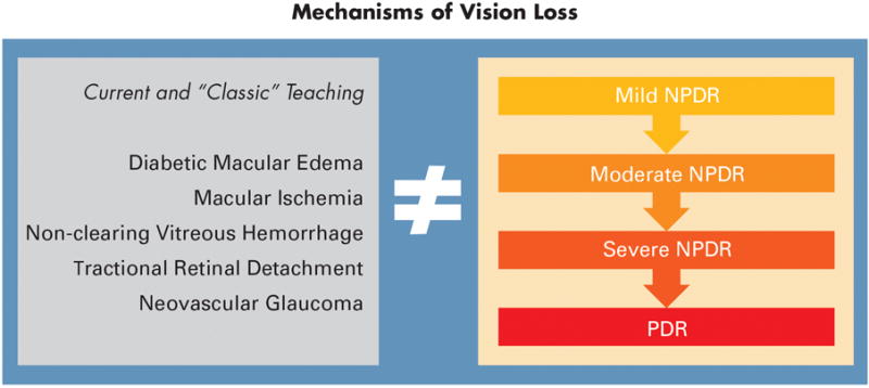
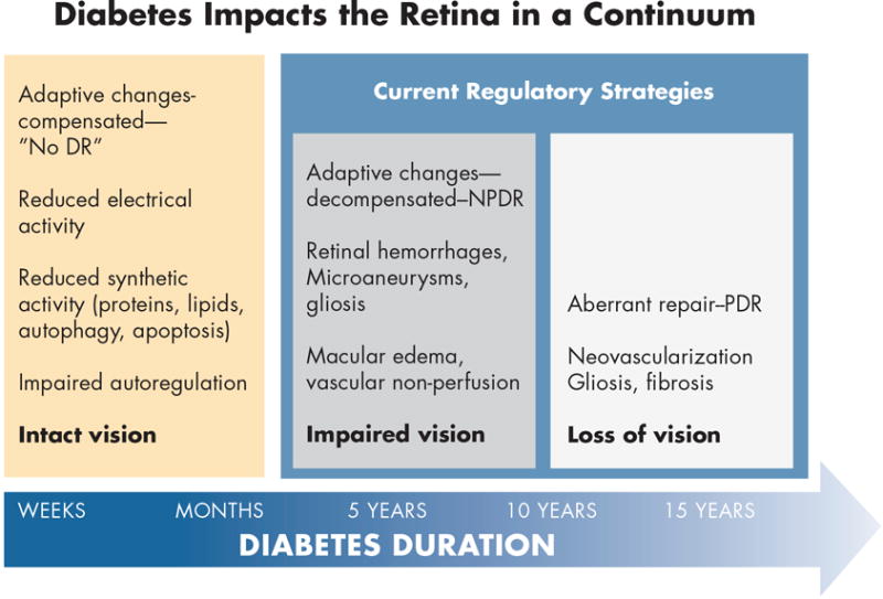
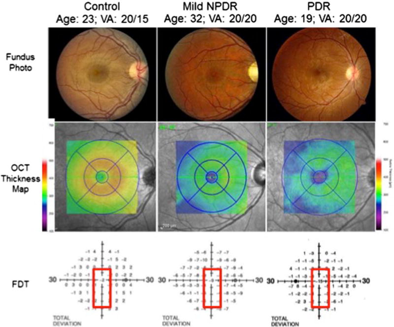
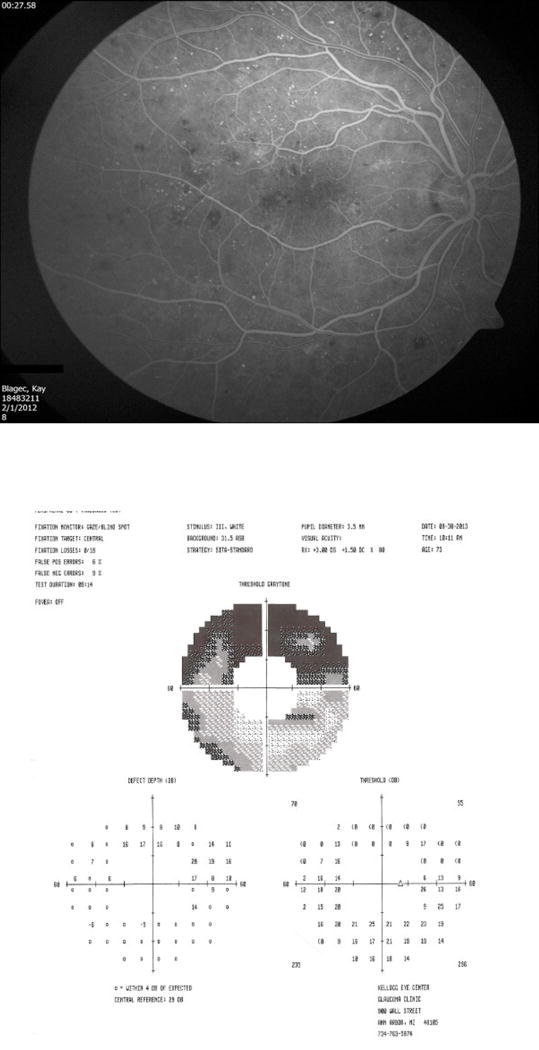
This paper presents a new approach to the prevention and treatment of early stage diabetic retinopathy before vision is severely impaired. This approach includes two major steps. The first step is to understand the mechanisms of vision impairment and classify diabetic retinopathy on the basis of pathophysiologic adaptations, rather than on the presence of advanced pathologic lesions, as defined by current clinical practice conventions. The second step is to develop patient-specific molecular diagnoses of diabetic retinopathy so that patients can be treated based on their individual characteristics, a process analogous to the individualized diagnosis and treatment of cancer patients. This step is illustrated by proteomic analysis of vitreous fluid that reveals evidence of neuroretinal degeneration and inflammation, as well as vascular proliferation. Together, these steps may lead to improved means to preserve vision in the ever-increasing number of patients with diabetes worldwide.

Keywords: Diabetic retinopathy; Molecular diagnosis; Neurovascular unit; Retinal failure; Vitreous proteomics.


Figures

Figure 1
Figure 1
Attendees at the 1968 Airlie House Symposium with Professor Eva Kohner front and center (44).
Figure 2
Figure 2. Classification of mechanisms of vision loss in diabetic retinopathy
The clinical features of diabetic retinopathy that are associated with vision loss (left) do not coincide directly with the severity scale (right).
Figure 3
Figure 3. A model of adaptive and maladaptive stages of diabetic retinopathy development
Within the first weeks to months of diabetes, the retina adapts to a lower metabolic steady-state with reduced electrical and biosynthetic activity, adaptive autophagy and apoptosis, and impaired autoregulation. Vision remains intact and there is no clinical evidence of diabetic retinopathy. After 5 - 10 years of diabetes, adaptive mechanisms begin to decompensate with the appearance of mild nonproliferative retinopathy, and early impairment of vision. Aberrant repair can ensue with the onset of proliferative retinopathy and loss of vision. Regulatory strategies are currently limited to the latter two stages (3). Artwork by David Murrel, MFA.
Figure 4
Figure 4
Color fundus photographs (top panel) of a healthy person (left), a person with mild nonproliferative diabetic retinopathy (NPDR; middle), and a person with proliferative diabetic retinopathy (PDR; right) demonstrate preserved macular anatomy without obvious vascular compromise and visual acuities 20/20 or better. Spectral-domain optical coherence tomography (SD-OCT) macular thickness mapping (central panel) demonstrates foveal and parafoveal thicknesses reduced by between 50 and 75 μm comparing healthy eyes to those with mild NPDR or PDR. The lower panel demonstrates a concomitant decline in foveal sensitivities in the diabetic patients as measured by frequency-doubling perimetry, with a 9 decibel (dB) visual field sensitivity loss in the patient with mild NPDR and a 15 dB sensitivity loss in the patient with PDR.
Figure 5
Figure 5. Peripheral visual field sensitivity loss in a patient with moderate NPDR
This 76 year old female had 20/25 visual acuity and moderate reduction of capillary perfusion, especially in the inferior retina. Her 60-4 Humphrey visual field showed marked (17 - 18 dB) sensitivity loss in the superior hemifield, corresponding to the inferior vascular nonperfusion.

Similar articles

Cited by

References

    1. Goldberg MF, Fine SL. Symposium on the treatment of diabetic retinopathy. Washington, DC: US Govt Printing Office; 1969.
    1. Diabetic retinopathy study. Report Number 6. Design, methods, and baseline results. Report Number 7. A modification of the Airlie House classification of diabetic retinopathy. Prepared by the Diabetic Retinopathy. Investigative ophthalmology & visual science. 1981;21(1 Pt 2):1–226. - PubMed
    1. Anonymous. Grading diabetic retinopathy from stereoscopic color fundus photographs–an extension of the modified Airlie House classification. ETDRS report number 10. Early Treatment Diabetic Retinopathy Study Research Group. Ophthalmology. 1991;98(5 Suppl):786–806. - PubMed
    1. Gardner TW, Davila JR. The neurovascular unit and the pathophysiologic basis of diabetic retinopathy. Graefes Arch Clin Exp Ophthalmol. 2017;255(1):1–6. - PMC - PubMed
    1. Fort PE, Losiewicz MK, Pennathur S, Jefferson LS, Kimball SR, Abcouwer SF, Gardner TW. mTORC1-independent reduction of retinal protein synthesis in type 1 diabetes. Diabetes. 2014;63(9):3077–90. - PMC - PubMed