. 2017 Jun 20;85(7):e00218-17.
doi: 10.1128/IAI.00218-17. Print 2017 Jul.

Host and Bacterial Factors Control Susceptibility of Drosophila melanogaster to Coxiella burnetii Infection

Affiliations

Host and Bacterial Factors Control Susceptibility of Drosophila melanogaster to Coxiella burnetii Infection

Reginaldo G Bastos et al. Infect Immun. .

Abstract

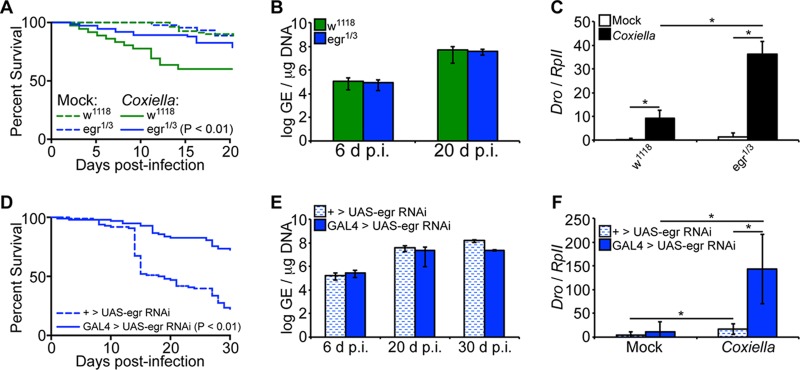
Coxiella burnetii is the causative agent of Q fever, a zoonotic disease that threatens both human and animal health. Due to the paucity of experimental animal models, little is known about how host factors interface with bacterial components and affect pathogenesis. Here, we used Drosophila melanogaster, in conjunction with the biosafety level 2 (BSL2) Nine Mile phase II (NMII) clone 4 strain of C. burnetii, as a model to investigate host and bacterial components implicated in infection. We demonstrate that adult Drosophila flies are susceptible to C. burnetii NMII infection and that this bacterial strain, which activates the immune deficiency (IMD) pathway, is able to replicate and cause mortality in the animals. We show that in the absence of Eiger, the only known tumor necrosis factor (TNF) superfamily homolog in Drosophila, Coxiella-infected flies exhibit reduced mortality from infection. We also demonstrate that the Coxiella type 4 secretion system (T4SS) is critical for the formation of the Coxiella-containing vacuole and establishment of infection in Drosophila Altogether, our data reveal that the Drosophila TNF homolog Eiger and the Coxiella T4SS are implicated in the pathogenesis of C. burnetii in flies. The Drosophila/NMII model mimics relevant aspects of the infection in mammals, such as a critical role of host TNF and the bacterial T4SS in pathogenesis. Our work also demonstrates the usefulness of this BSL2 model to investigate both host and Coxiella components implicated in infection.

Keywords: Eiger; IMD; NMII; Q fever; T4SS; TNF; innate immunity; pathogenesis; tumor necrosis factor.


Figures

FIG 1
FIG 1
C. burnetii replicates in Drosophila hemocyte-derived S2 cells. (A) Cells were infected (MOI = 100 GE/cell), and comparative growth kinetics of C. burnetii in insect and mammalian cells were determined by qPCR. The results are presented as log GE per microgram of DNA. (B) Immunoblotting detection of C. burnetii antigens in Drosophila S2 cells at 1, 6, and 12 days postinfection (dpi) using a rabbit polyclonal antibody against Coxiella. Nonspecific (n.s.) banding in S2 cell lysates is denoted by the arrow. (C and D) Drosophila hemocyte-derived S2 cells were infected with C. burnetii expressing mCherry and prepared for confocal microscopy at 4 dpi. Nuclei were stained with DAPI and actin (C) or LAMP1 (D). (E to G) A gentamicin protection assay was performed to evaluate the growth of mCherry-expressing C. burnetii in Drosophila S2 cells. (E and F) At the indicated times postinfection, total DNA was collected to determine GE levels (E) or mCherry intensity was measured at five different locations of three independent wells at the indicated time points postinfection (F). (G) Representative images for each condition at 2, 6, and 10 days postinfection. (H) mCherry-expressing Coxiella was isolated from infected S2 cells and used to infect HeLa cells at an MOI of 100 GE/cell. The intensity of mCherry was measured over the course of 10 days, and representative images are shown. The asterisks denote statistical significance (*, P < 0.05). The error bars indicate standard deviations.
FIG 2
FIG 2
C. burnetii induces the expression of AMPs in S2 cells. (A to C) A luciferase reporter assay was performed to investigate the activation of the Drosocin (Dro) (A), CecropinA1 (CecA1) (B), and Defensin (Def) (C) AMP promoters in S2 cells following infection. At 24 h posttransfection, the cells were infected with C. burnetii (MOI = 100 GE/cell), and luciferase (luc) activity was assessed at different times postinfection. The firefly luciferase activity of each sample was normalized to Actin5C-driven Renilla luciferase activity to correct for transfection efficiency. (D to I) Drosophila S2 cells were infected with C. burnetii (MOI = 100 GE/cell), and total RNA was collected at 4 h and 24 h postinfection to examine AMP expression. Gene expression levels for Drosocin (D), CecropinA1 (E), Defensin (F), Diptericin (G), AttacinA (H), and Drosomycin (I) were determined by qRT-PCR. The relative expression of AMP was normalized to Drosophila RpII. The asterisks denote statistical significance (*, P < 0.05). The error bars indicate standard deviations.
FIG 3
FIG 3
Adult Drosophila flies are susceptible to C. burnetii and elicit a host immune response. (A and B) Four-day-old Oregon-R female (A) and male (B) Drosophila flies were infected with live (102 or 105 GE/fly) or HK (105 GE/fly) C. burnetii, and survival was evaluated for 30 days. (C) Four-day-old adult Oregon-R flies were infected with C. burnetii (100 GE/fly), and bacterial levels were determined at 6 and 30 days postinfection by quantitative real-time PCR. (D) C. burnetii antigens were detected in infected flies at 6 and 30 days postinfection, as shown by immunoblotting using a rabbit polyclonal antibody against Coxiella. Biological duplicates are shown. Nonspecific (n.s.) banding from fly homogenates is denoted by the arrow. (E and F) Antimicrobial peptide levels of Drosocin (E) and Defensin (F) were determined in Oregon-R adults infected with C. burnetii (100 GE/fly) at 12 days postinfection. (G and H) Confocal microscopy showing mCherry-Coxiella invasion of hemocytes (white arrows) derived from 3rd-instar larvae infected in vivo (G) or ex vivo (H). The hemocytes expressed GFP, and the nuclei were stained with DAPI. Bars = 2 μm. Numbers by arrows designate the same Coxiella-mCherry signal among images in the same panel. Dotted lines in insets represent where the cross-section is made. The asterisks denote statistical significance (*, P < 0.05). The error bars indicate standard deviations.
FIG 4
FIG 4
Drosophila PGRP-LC7454 and RelE20 mutants are more susceptible to C. burnetii NMII clone 4. (A to F) Adult w1118, PGRP-LC7454, and RelE20 male flies, 4 days of age, were mock infected or infected with C. burnetii (100 GE/fly). Percent survival was evaluated for a period of 20 days, comparing infected flies to one another (A) or mock- and Coxiella-infected flies for each genotype (B to D). (E) Bacterial loads were determined at 6 and 20 days postinfection by qPCR. (F) Expression of Drosocin in w1118, PGRP-LC7454, and RelE20 flies was determined at 12 days postinfection by reverse transcriptase quantitative real-time PCR, and the results were normalized to the Drosophila RpII transcripts. (G to I) Four-day-old sibling adult flies carrying a UAS-induced dsRNA cassette targeting Relish (TRiP.HMS00070) or PGRP-LC (TRiP.HMS00259) with an Actin5C-driven GAL4 element (GAL4 > UAS) or lacking the GAL4 element (+ > UAS) were infected with C. burnetii (100 GE/fly). (G) Percent survival was evaluated for a period of 30 days. (H) The bacterial loads were determined at 6, 20, and 30 dpi by qPCR. (I) Expression of Drosocin was determined at 12 dpi. The asterisks denote statistical significance (*, P < 0.05). The error bars indicate standard deviations.
FIG 5
FIG 5
Eiger mutant Drosophila flies display tolerance for C. burnetii. (A to C) Adult w1118 and Eiger mutant (egr1/3) male flies, 4 days of age, were mock infected or infected with C. burnetii (100 GE/fly). (A) Mortality was significantly increased (P < 0.01) in w1118 flies compared to Eiger mutant flies. (B) Coxiella GE was quantified at 6 and 20 days postinfection by qPCR. (C) Levels of Drosocin were measured in Eiger mutant flies and control w1118 flies at 12 days postinfection. (D to F) Four-day-old sibling adult flies carrying a UAS-induced dsRNA cassette targeting Eiger (TRiP.HMC03963) with an Actin5C-driven GAL4 element (GAL4 > UAS-egr RNAi) or lacking the GAL4 element (+ > UAS-egr RNAi) were infected with C. burnetii (100 GE/fly). (D) Percent survival was evaluated for a period of 30 days. (E) Bacterial loads were determined at 6, 20, and 30 days postinfection by qPCR. (F) Expression of Drosocin was determined at 12 days postinfection. The asterisks denote statistical significance (*, P < 0.05). The error bars indicate standard deviations.
FIG 6
FIG 6
The C. burnetii type 4 secretion system is essential for establishment of infection in Drosophila. (A) S2 cells were infected with NMII clone 4 or the ΔdotA or ΔpmrA mutant (MOI = 100 GE/cell), and bacterial growth was assessed by qPCR. (B) S2 cells were infected with NMII clone 4 or the ΔdotA and ΔpmrA mutant expressing GFP. CCV formation was observed by confocal microscopy at 6 days postinfection. (C) Coxiella antigens were examined in S2 cells by immunoblotting using an anti-Coxiella polyclonal antibody at 6 and 12 days following infection with NMII clone 4 or the ΔdotA and ΔpmrA mutants. Nonspecific (n.s.) banding in S2 cell lysates is denoted by the arrow. (D) Four-day-old adult Oregon-R flies were infected with 100 GE/fly of NMII clone 4 or the ΔdotA or ΔpmrA mutant, and mortality was monitored for 30 days. The asterisks denote statistical significance (*, P < 0.05). The error bars indicate standard deviations.

Similar articles

Cited by

References

    1. Maurin M, Raoult D. 1999. Q fever. Clin Microbiol Rev 12:518–553. - PMC - PubMed
    1. Schimmer B, Morroy G, Dijkstra F, Schneeberger PM, Weers-Pothoff G, Timen A, Wijkmans C, van der Hoek W. 2008. Large ongoing Q fever outbreak in the south of The Netherlands, 2008. Euro Surveill 13:18939. - PubMed
    1. Sondgeroth KS, Davis MA, Schlee SL, Allen AJ, Evermann JF, McElwain TF, Baszler TV. 2013. Seroprevalence of Coxiella burnetii in Washington State domestic goat herds. Vector Borne Zoonotic Dis 13:779–783. doi:10.1089/vbz.2013.1331. - DOI - PMC - PubMed
    1. Madariaga MG, Rezai K, Trenholme GM, Weinstein RA. 2003. Q fever: a biological weapon in your backyard. Lancet Infect Dis 3:709–721. doi:10.1016/S1473-3099(03)00804-1. - DOI - PubMed
    1. Hackstadt T, Peacock MG, Hitchcock PJ, Cole RL. 1985. Lipopolysaccharide variation in Coxiella burnetti: intrastrain heterogeneity in structure and antigenicity. Infect Immun 48:359–365. - PMC - PubMed

MeSH terms

LinkOut - more resources