. 2017 Apr;5(8):e13178.
doi: 10.14814/phy2.13178.

Acute inflammation reveals GABAA receptor-mediated nociception in mouse dorsal root ganglion neurons via PGE2 receptor 4 signaling

Affiliations

Acute inflammation reveals GABAA receptor-mediated nociception in mouse dorsal root ganglion neurons via PGE2 receptor 4 signaling

In Jeong Jang et al. Physiol Rep. 2017 Apr.

Abstract

Gamma-aminobutyric acid (GABA) depolarizes dorsal root ganglia (DRG) primary afferent neurons through activation of Cl- permeable GABAA receptors but the physiologic role of GABAA receptors in the peripheral terminals of DRG neurons remains unclear. In this study, we investigated the role of peripheral GABAA receptors in nociception using a mouse model of acute inflammation. In vivo, peripheral administration of the selective GABAA receptor agonist muscimol evoked spontaneous licking behavior, as well as spinal wide dynamic range (WDR) neuron firing, after pre-conditioning with formalin but had no effect in saline-treated mice. GABAA receptor-mediated pain behavior after acute formalin treatment was abolished by the GABAA receptor blocker picrotoxin and cyclooxygenase inhibitor indomethacin. In addition, treatment with prostaglandin E2 (PGE2) was sufficient to reveal muscimol-induced licking behavior. In vitro, GABA induced sub-threshold depolarization in DRG neurons through GABAA receptor activation. Both formalin and PGE2 potentiated GABA-induced Ca2+ transients and membrane depolarization in capsaicin-sensitive nociceptive DRG neurons; these effects were blocked by the prostaglandin E2 receptor 4 (EP4) antagonist AH23848 (10 μmol/L). Furthermore, potentiation of GABA responses by PGE2 was prevented by the selective Nav1.8 antagonist A887826 (100 nmol/L). Although the function of the Na+-K+-2Cl- co-transporter NKCC1 was required to maintain the Cl- ion gradient in isolated DRG neurons, NKCC1 was not required for GABAA receptor-mediated nociceptive behavior after acute inflammation. Taken together, these results demonstrate that GABAA receptors may contribute to the excitation of peripheral sensory neurons in inflammation through a combined effect involving PGE2-EP4 signaling and Na+ channel sensitization.

Keywords: EP4 receptors; Gamma‐aminobutyric acid; TTX‐resistant sodium channels; formalin test; peripheral sensitization; prostaglandin E2.


Figures

Figure 1
Figure 1
GABA induces Ca2+ transients via membrane depolarization in mouse dorsal root ganglion neurons. (A) Sequential application of GABA (300 μmol/L, 10 sec) induces Ca2+ transients in DRG neurons. (B) Mean relative amplitude of sequential GABA‐induced Ca2+ responses (n = 54 neurons, 6 coverslips from 4 mice; ns, not significant, one‐way ANOVA). (C) Ca2+ transients are elicited by GABA in a concentration‐dependent manner. (D) Concentration‐response curve of GABA in cultured DRG neurons (n = 22–32 neurons, 9 coverslips from 4 mice). (E) GABA‐induced Ca2+ responses are abolished by extracellular Ca2+‐free solution. (F) Normalized Ca2+ responses relative to peak amplitude of 50 mmol/L KCl response (n = 16 cells, coverslips from 3 mice; ***< 0.001, one‐way ANOVA with Bonferroni post‐test). (G) Thapsigargin (1 μmol/L) treatment to deplete intracellular Ca2+ stores had no effect on GABA‐induced Ca2+ transients (H) Quantification of normalized Ca2+ responses relative to peak amplitude of 50 mmol/L KCl response (n = 21 cells, 3 coverslips from 3 mice; one‐way ANOVA with Bonferroni post‐test). (I) Ca2+ transients evoked by supramaximal GABA (300 μmol/L, 10 sec) are blocked by picrotoxin (300 μmol/L). (J) Concentration‐response curve of GABA (300 μmol/L)‐induced Ca2+ responses inhibited by picrotoxin. (K) Gramicidin perforated patch‐clamp recording of adult mouse DRG neurons. Supramaximal GABA (300 μmol/L) application led to a fast, rapidly decaying depolarization of the membrane potential in small‐sized (12–20 μm diameter) DRG neurons. (L) Box and whisker plot showing the mean membrane potential at rest (−53.7 ± 0.8 mV) and peak (−35.3 ± 0.6 mV) during GABA (300 μmol/L) application (***< 0.001, paired t‐test; n = 12 neurons, 12 coverslips from 4 mice). Whiskers above and below represent max and min values, respectively. DRG, dorsal root ganglia.
Figure 2
Figure 2
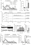
Activation of peripheral GABAA receptors induces pain‐like behavior after acute formalin inflammation but not in naïve mice. (A) Time course of hind paw licking behaviors during pre‐conditioning with formalin (0.8%, 20 μL) followed by injection of muscimol (1 mmol/L, 20 μL) (open circles, n = 5 mice) or 0.9% saline (black circles, n = 4 mice) (‘stimulation’); pre‐conditioning with 0.9% saline vehicle followed by injection of muscimol (open squares, n = 5 mice) or 0.9% saline (black squares, n = 5 mice) into the same dorsum hind paw area. (B) GABAA receptor agonist muscimol significantly increased hind paw licking behavior in formalin pre‐conditioning group. Bar graph represents accumulative licking time during 30 min stimulation phase (**< 0.01, one‐way ANOVA with Bonferroni post‐test compared with muscimol in saline pre‐conditioning group; (*< 0.05, one‐way ANOVA with Bonferroni post‐test compared with saline in formalin pre‐conditioning group). (C) Extracellular unit recording of spinal WDR neurons showing the pattern of action potential (AP) discharge frequency of three units (a, b, and c) in response to sequential injections of muscimol (1 mmol/L, 20 μL) and formalin (0.8%, 20 μL) into the hind paw. (D–F) Pre‐injection of formalin (0.8%) increases frequency of action potential (AP) discharge in spinal WDR neurons by injection of muscimol (1 mmol/L, 20 μL) into the hind paw. (D) Representative traces showing the AP discharge of two units induced by muscimol before and after formalin injection. (E) Frequency histogram of muscimol‐induced AP discharge (1 min bins). (F) Mean frequency of total muscimol‐induced AP discharge per min for 20 min, n = 12 units recorded from 6 animals, **< 0.01, Wilcoxon signed‐rank test. (G) Effect of GABAA antagonist picrotoxin (PTx) on time course of hind paw licking behaviors during pre‐conditioning with formalin (0.8%, 20 μL) followed by injection of muscimol (1 mmol/L, 20 μL) into the same dorsum hind paw area. Picrotoxin was given at 1 mmol/L (gray triangle, n = 5 mice), 5 mmol/L (black squares, n = 6 mice) or vehicle (0.9% saline, open circles, n = 12 mice). (H) Picrotoxin significantly inhibited the second phase of the formalin response (*P < 0.05, *** P < 0.001, one‐way ANOVA with Bonferroni post‐test) as well as muscimol‐induced pain behavior (**P < 0.01, P < 0.001, one‐way ANOVA with Bonferroni post‐test). WDR, wide dynamic range.
Figure 3
Figure 3
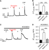
Formalin potentiates GABA‐induced Ca2+ transients in a TRPA1 independent manner. (A, B) Formalin potentiates GABA‐induced Ca2+ transients in nociceptive neurons. (A) Representative traces showing potentiated GABA (300 μmol/L)‐induced Ca2+ by formalin pre‐treatment (0.001%, 120 sec) in a capsaicin‐sensitive DRG neuron. (B) Mean amplitude of GABA‐induced Ca2+ transients (n = 18 cells, 6 coverslips from 3 mice; ***< 0.001, one‐way ANOVA). (C) Potentiated GABA‐induced Ca2+ transients by formalin (0.001%) are not blocked by TRPA1 receptor selective antagonist HC030031 (30 μmol/L). (D) Quantification of GABA‐induced Ca2+ transients in capsaicin‐sensitive DRG neurons (n = 16 cells, 3 coverslips from 2 mice; ***< 0.001, one‐way ANOVA with Bonferroni post‐test). DRG, dorsal root ganglia.
Figure 4
Figure 4
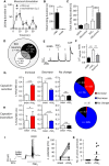
Prostaglandin E2 contributes to GABAA receptor‐mediated pain and neuronal excitability. (A) Effect of intraperitoneal injection of indomethacin (40 mg/kg) (open squares) or PBS vehicle (black circles) on muscimol (1 mmol/L, 20 μL)‐induced hind paw licking behavior in formalin (0.8%, 20 μL) preconditioned mice. (B) Cyclooxygenase inhibitor indomethacin abolishes muscimol‐induced hind paw licking behavior after formalin pre‐conditioning (n = 6 mice per group, **< 0.05, Student's t‐test). (C) PGE 2 potentiates muscimol‐induced hind paw licking behavior. Quantification represents total duration of hind paw licking behavior during 30 min after PGE 2 (10 nmol, 20 μL) or saline vehicle with or without muscimol (1 mmol/L, 20 μL) (n = 5–6 mice per group, *P < 0.05, ***< 0.001, one‐way ANOVA with Bonferroni post‐test). (D) Venn diagram of GABA (300 μmol/L) and capsaicin (CAP; 1 μmol/L) sensitivity among KCl‐responsive neuronal cells in acute (<24 h) cultures of adult mouse DRG (n = 960 cells, 19 coverslips from 3 mice). (E, F) PGE 2 potentiates GABA‐induced Ca2+ transients in nociceptive neurons. (E) Representative trace showing potentiated GABA (300 μmol/L)‐induced Ca2+ by PGE 2 pre‐treatment (10 μmol/L, 180 sec) in a capsaicin‐sensitive DRG neuron. (F) Mean amplitude of GABA‐induced GABA‐induced Ca2+ transients (n = 34 cells, 11 coverslips from 7 mice; ***< 0.001, one‐way ANOVA). (G, H) The effect of PGE 2 on the amplitude of GABA‐induced Ca2+ transients. GABA‐induced Ca2+ transients in capsaicin‐sensitive (G) and capsaicin‐insensitive (H) GABA‐responsive neurons were classified according to whether the amplitude (change in F340/380 ratio) was increased >0.03 (‘potentiated’), decreased >0.02, or unchanged (−0.02 to +0.03) during PGE 2 (10 μmol/L) compared to control responses (*P < 0.05, ***P < 0.001, paired two‐tailed t‐test). (I, J, K) PGE 2 increases GABA‐induced neuronal excitability. (I) Representative traces of GABA (100 μmol/L)‐induced depolarization before and after pre‐incubation with PGE 2 (10 μmol/L, 180 sec). (J) Quantification of GABA‐induced changes in membrane potentials and (K) number of action potentials by GABA before and after pre‐incubation with PGE 2 (10 μmol/L) (n = 21 cells). DRG, dorsal root ganglia.
Figure 5
Figure 5
Potentiation of GABA‐induced neuronal excitability is mediated by EP4 receptors. (A) Potentiation of GABA (300 μmol/L)‐induced Ca2+ transients in capsaicin‐sensitive DRG neurons by PGE 2 (10 μmol/L) is inhibited by the EP4 receptor antagonist AH23848 (10 μmol/L) (B) Quantification of GABA‐induced Ca2+ transients relative to peak amplitude of 1st GABA response in the presence of AH23848 (n = 19 cells, 12 coverslips from 4 mice; ***< 0.001, one‐way ANOVA with Bonferroni post‐test) (C) Potentiation of GABA (300 μmol/L)‐induced Ca2+ transients by PGE 2 (10 μmol/L) is not affected by EP1‐2 receptor antagonist AH6809 (50 μmol/L). (D) Quantification of GABA‐induced Ca2+ transients relative to peak amplitude of 1st GABA response in the presence of AH6809 (n = 15 cells, 5 coverslips from 3 mice). (E, F) The potentiating effect of PGE 2 on GABA‐induced changes in membrane potentials (E) and generation of action potentials (F) is abolished by EP4 receptor antagonist AH23848 (10 μmol/L) in small‐sized DRG neurons (n = 10 cells). (G) Potentiated GABA (300 μmol/L)‐induced Ca2+ transients by formalin (0.001%) is blocked by EP4 receptor antagonist AH23848 (10 μmol/L) in capsaicin‐sensitive DRG neurons. (H) Quantification of potentiated GABA‐induced Ca2+ transients by formalin (0.001%) in the presence of AH23848 relative to peak amplitude of first GABA response (n = 8 cells, 6 coverslips from 3 mice; ***< 0.001, repeat‐measures one‐way ANOVA with Bonferroni post‐test). DRG, dorsal root ganglia.
Figure 6
Figure 6
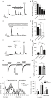
PGE 2 potentiates GABA –induced neuronal excitability through Nav1.8 channel modulation. (A) Potentiation of GABA (300 μmol/L)‐induced Ca2+ transients by PGE 2 (10 μmol/L) is inhibited by voltage‐gated sodium channel blocker lidocaine (300 μmol/L) in capsaicin‐sensitive DRG neurons. (B) Quantification of GABA‐induced Ca2+ transients in the presence and absence of lidocaine relative to peak amplitude of 1st GABA response (n = 12 cells, 9 coverslips from 4 mice; ***< 0.001, repeat‐measures one‐way ANOVA with Bonferroni post‐test). (C) GABA‐induced Ca2+ transients are unaffected by lidocaine (300 μmol/L, 360 sec). (D) Quantification of GABA‐induced Ca2+ transients amplitudes in the presence of lidocaine (n = 17 cells, 4 coverslips from 3 mice; one‐way ANOVA with Bonferroni post‐test). (E) PGE 2 (10 μmol/L, 180 sec) potentiates TTX‐resistant sodium currents in small‐sized DRG neurons. (F) Quantification of normalized TTX‐resistant sodium currents amplitude (n = 5 cells, ***< 0.001, paired Student's t‐test). (G) Normalized conductance‐voltage relationship of Nav1.8 currents in the presence and absence of PGE 2 (10 μmol/L) (n = 6 cells) fit with a Boltzmann function. Test pulses were preceded by a 500‐msec step to −50 mV to inactivate TTX‐resistant Nav1.9. (H) A887826 (100 nmol/L) inhibits the membrane depolarization caused by PGE 2 (10 μmol/L) application (n = 12–20 cells; *< 0.05, unpaired Student's t‐test). (I) Potentiation of GABA (300 μmol/L)‐induced Ca2+ transients by PGE 2 (10 μmol/L) is abolished by Nav1.8 channel blocker A887826 (100 nmol/L) in DRG neurons (J) Quantification of GABA‐induced Ca2+ transients in the presence and absence of A887826 relative to peak amplitude of 1st GABA response (n = 10 cells, 4 coverslips from 3 mice; **< 0.01, repeat‐ measures one‐way ANOVA with Bonferroni post‐test). (K, L) The potentiating effect of PGE 2 on GABA‐induced changes in membrane potentials (K) and generating action potentials (L) in small‐sized DRG neurons is blocked by Nav1.8 channel blocker A887826 (100 nmol/L) (n = 9 cells). DRG, dorsal root ganglia.
Figure 7
Figure 7
NKCC1 is not required for GABAA receptor‐mediated pain behavior in formalin inflammation. (A) GABA‐induced Ca2+ transients are decreased by sequential application of GABA (300 μmol/L) in the presence of NKCC1 co‐transporter inhibitor bumetanide (10 μmol/L). (B) Quantification of GABA‐induced Ca2+ transients normalized by 1st GABA response (n = 24 cells, 6 coverslips from 3 mice; **< 0.01, ***< 0.001, one‐way ANOVA with Bonferroni post‐test. (C) GABA (300 μmol/L)‐induced Ca2+ transient amplitudes vary with different concentrations of extracellular Cl in dissociated DRG neurons. (D) Quantification of GABA‐induced Ca2+ transients normalized by the amplitude of Ca2+ transients in control extracellular solution with 134 mmol/L Cl (n = 10 cells, 5 coverslips from 4 mice; **< 0.01, ***< 0.001, repeat‐measures ANOVA with Bonferroni post‐test). (E) Representative traces showing potentiation of GABA (300 μmol/L)‐induced Ca2+ transients by PGE 2 pre‐treatment (10 μmol/L, 180 sec) in NKCC1−/− DRG neurons. (F) Normalized amplitude of GABA‐induced Ca2+ transients in NKCC1−/− DRG neurons (n = 12 cells, 4 coverslips from 3 mice; **< 0.01, repeat‐measures one‐way ANOVA with Bonferroni post‐test). (G, H) PGE 2 (10 μmol/L) does not affect GABA (100 μmol/L)‐induced currents at a holding potential of −60 mV in small‐size DRG neurons (n = 11 cells, two‐tailed paired Student's t‐test). (I, J) Formalin‐evoked pain behavior and GABAA receptor agonist muscimol‐induced pain behavior after formalin test are not altered in NKCC1−/− mice. Formalin (0.8%, 20 μL)‐induced biphasic licking behavior (1st and 2nd phases) followed by the injection of muscimol (1 mmol/L, 20 μL) (GABAA receptor‐mediated pain) in wild type (open squares) and NKCC1−/− (black circles) mice. (F) Bar graph represents accumulative licking time during 1st (0–10 min) and 2nd phases (10–70 min) of formalin‐induced pain‐like behavior and muscimol‐induced pain‐like behavior (70–100 min) (n = 5 mice per genotype; two‐way ANOVA compared with WT mice). DRG, dorsal root ganglia.

Similar articles

Cited by

References

    1. Barry, P. H. 1994. JPCalc, a software package for calculating liquid junction potential corrections in patch‐clamp, intracellular, epithelial and bilayer measurements and for correcting junction potential measurements. J. Neurosci. Methods 51:107–116. - PubMed
    1. Berta, T. , Poirot O., Pertin M., Ji R. R., Kellenberger S., and Decosterd I.. 2008. Transcriptional and functional profiles of voltage‐gated Na(+) channels in injured and non‐injured DRG neurons in the SNI model of neuropathic pain. Mol. Cell Neurosci. 37:196–208. - PubMed
    1. Bravo‐Hernandez, M. , Feria‐Morales L. A., Torres‐Lopez J. E., Cervantes‐Duran C., Delgado‐Lezama R., Granados‐Soto V., et al. 2014. Evidence for the participation of peripheral alpha5 subunit‐containing GABAA receptors in GABAA agonists‐induced nociception in rats. Eur. J. Pharmacol. 734:91–97. - PubMed
    1. Carlton, S. M. , Zhou S., and Coggeshall R. E.. 1999. Peripheral GABA(A) receptors: evidence for peripheral primary afferent depolarization. Neuroscience 93:713–722. - PubMed
    1. Carr, R. W. , Sittl R., Fleckenstein J., and Grafe P.. 2010. GABA increases electrical excitability in a subset of human unmyelinated peripheral axons. PLoS ONE 5:e8780. - PMC - PubMed

MeSH terms

LinkOut - more resources