. 2017 Apr 24;7(1):1103.
doi: 10.1038/s41598-017-01304-3.

Translation, but not transfection limits clinically relevant, exogenous mRNA based induction of alpha-4 integrin expression on human mesenchymal stem cells

Affiliations

Translation, but not transfection limits clinically relevant, exogenous mRNA based induction of alpha-4 integrin expression on human mesenchymal stem cells

Adam Nowakowski et al. Sci Rep. .

Abstract

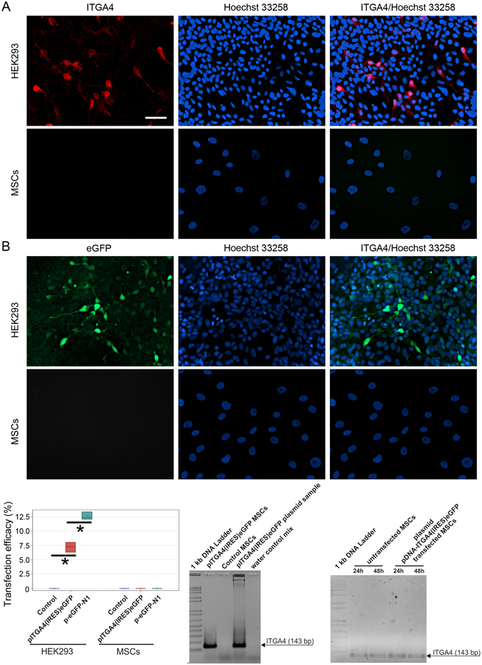
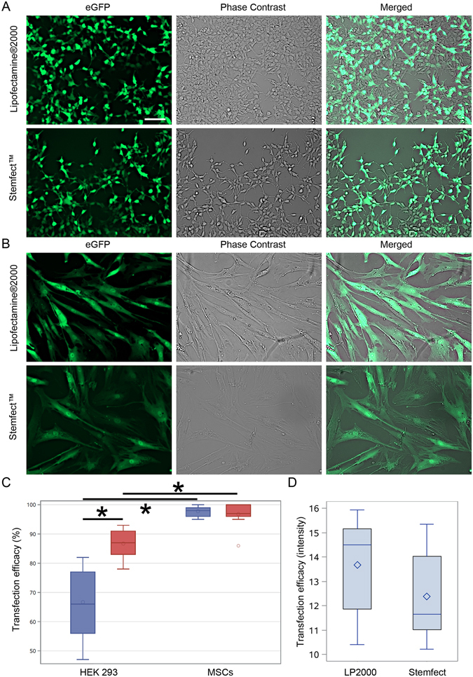
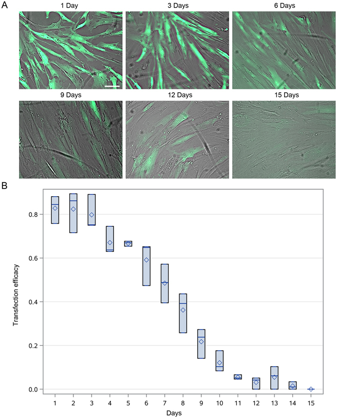
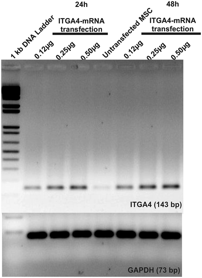
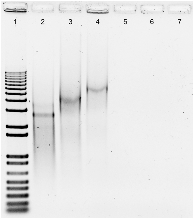
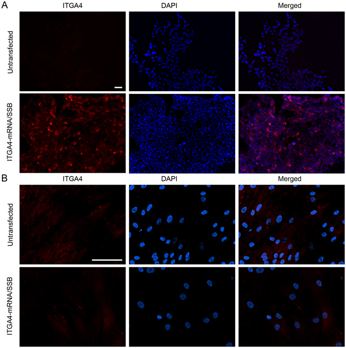
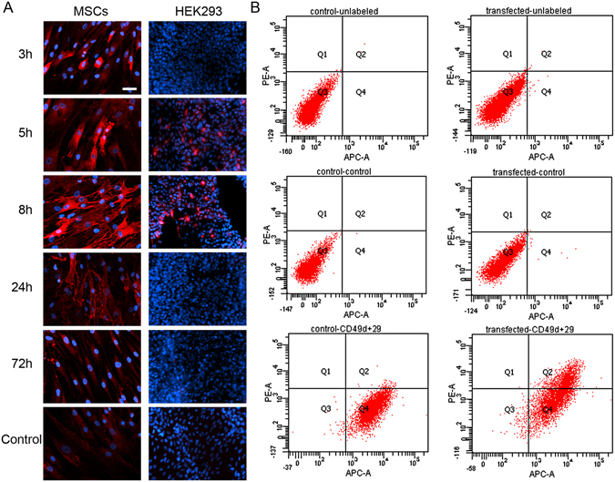
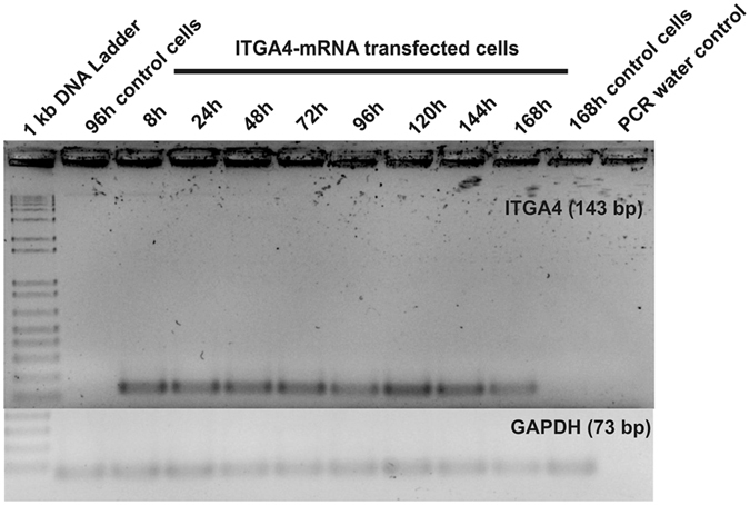
Mesenchymal stem cells (MSCs) represent promising resource of cells for regenerative medicine in neurological disorders. However, efficient and minimally invasive methods of MSCs delivery to the brain still have to be developed. Intra-arterial route is very promising, but MSCs are missing machinery for diapedesis through blood-brain barrier. Thus, here we have tested a mRNA-based method to induce transient expression of ITGA4, an adhesion molecule actively involved in cell extravasation. We observed that transfection with an ITGA4-mRNA construct bearing a conventional cap analogue (7-methylguanosine) failed to produce ITGA4 protein, but exogenous ITGA4-mRNA was detected in transfected MSCs. This indicates that not transfection, but rather translation being the major roadblock. Stabilization of ITGA4-mRNA with SSB proteins resulted in ITGA4 protein synthesis in HEK293 cells only, whereas in MSCs, satisfactory results were obtained only after using an anti-reverse-cap-analogue (ARCA). The presence of ITGA4 protein in MSCs was transient and lasted for up to 24 h after transfection. Membranous location was confirmed by flow cytometry of viable non-permeabilized cells using anti-ITGA4 antibody. The mRNA-based expression of itga4 transgene is potentially sufficient for diapedesis after intra-arterial delivery. To conclude, mRNA-based engineering of stem cells is a rapid and integration-free method and attractive from the perspective of potential future clinical application.


Conflict of interest statement

The authors declare that they have no competing interests.

Figures

Figure 1
Figure 1
Plasmid DNA-based transfections of MSCs and HEK293 cells. MSCs and HEK293 visualized 24 h post transfection by immunofluorescence after pITGA4(IRES)eGFP plasmid (A) and p-eGFP-N1 plasmid (B). Quantification of percentage of cells expressing transgenes [pITGA4(IRES)eGFP/red/, p-eGFP-N1/green/] (C). PCR for presence of pITGA4(IRES)eGFP plasmid in MSCs 24 h after transfection (D). PCR for presence of itga4 mRNA in MSCs transfected with pITGA4(IRES)eGFP (E). The arrows point on PCR-product corresponding itga4 gene (143 bp). Error bars are box-and-whiskers plots containing the mean, quartiles (box) and minimum and maximum observations (whiskers). Transfection efficiencies have been calculated from three independent experiments. Scale bar located in the top left image: 20 µm.
Figure 2
Figure 2
mRNA-based transfections of MSCs and HEK293 cells. The eGFP-mRNA transfection of HEK293 (A) and MSCs (B) with different transfection agents. Quantification of percentage of cells expressing transgenes (C). Quantification of pixel intensity in MSCs transfected with different agents (D). Green – eGFP protein. Error bars are box-and-whiskers plots containing the mean, quartiles (box) and minimum and maximum observations (whiskers). Transfection efficiencies have been calculated from three independent experiments. Scale bar located in the top left image: 20 µm.
Figure 3
Figure 3
Time-course eGFP protein synthesis after mRNA transfection in MSCs. Time-course of eGFP protein presence in MSCs after eGFP-mRNA transfection on intravital fluorescent images (A) with subsequent quantification (B). Green – eGFP protein. Error bars are box-and-whiskers plots containing the mean, quartiles (box) and minimum and maximum observations (whiskers). Transfection efficiencies have been calculated from three independent experiments. Scale bar located in the top left image: 20 µm.
Figure 4
Figure 4
Exogenous ITGA4-mRNA delivery to MSCs. RT-PCR of MSC lysates after ITGA4-mRNA transfection: The higher band refers to ITGA4-mRNA (143 bp) and lower band to housekeeping gene: GAPDH (73 bp).
Figure 5
Figure 5
SSB protein binding assay in vitro. Electrophoresis after SSB protein to ITGA4-mRNA in vitro binding assay: 1) 1 kb DNA ladder, 2) ITGA4-mRNA, 3) ITGA4-mRNA/SSB, 4) ITGA4-mRNA/SSB/Lipofectamine®2000, 5) SSB/Lipofectamine®2000, 6) SSB, 7) Lipofectamine®2000.
Figure 6
Figure 6
ITGA4 protein synthesis after ITGA4-mRNA/SSB transfection. Immunocytochemistry study for assessment of the ITGA4 protein presence after ITGA4-mRNA/SSB transfection using Lipofectamine®2000. Scale bars separate for HEK293 and MSC cells located in the respective left upper images of the panel indicate 100 µm.
Figure 7
Figure 7
ITGA4 protein distribution in (ARCA)-ITGA4-mRNA transfected MSCs. Time-course of ITGA4 protein production – note its translocation from perinuclear area in 3 and 5 hour time-points toward the cell membrane in 8 and 24 hour time points (A). The presence of ITGA4 protein on the cell membrane was confirmed in living cells by flow cytometry performed 20 h after transfection (B). red – ITGA4 protein, blue – Hoechst 33258. Scale bar: 20 µm.
Figure 8
Figure 8
Longitudinal study of ITGA4-mRNA presence in transfected MSCs. RT-PCR results from MSCs transfected with (ARCA)-ITGA4-mRNA confirmed that delivered (ARCA)-ITGA4-mRNA was present in transfected cells at least for 168 h.

References

    1. Janowski M, Wagner DC, Boltze J. Stem Cell-Based Tissue Replacement After Stroke: Factual Necessity or Notorious Fiction? Stroke. 2015;46(8):2354–2363. doi: 10.1161/STROKEAHA.114.007803. - DOI - PMC - PubMed
    1. Kabadi SV, Faden AI. Neuroprotective strategies for traumatic brain injury: improving clinical translation. Int J Mol Sci. 2014;15(1):1216–1236. doi: 10.3390/ijms15011216. - DOI - PMC - PubMed
    1. Savitz SI, Cramer SC, Wechsler L. STEPS 3 Consortium. Stem cells as an emerging paradigm in stroke 3: enhancing the development of clinical trials. Stroke. 2014;45(2):634–639. doi: 10.1161/STROKEAHA.113.003379. - DOI - PMC - PubMed
    1. Nowakowski A, Walczak P, Janowski M, Lukomska B. Genetic Engineering of Mesenchymal Stem Cells for Regenerative Medicine. Stem Cells Dev. 2015;24(19):2219–2242. doi: 10.1089/scd.2015.0062. - DOI - PubMed
    1. Huang B, Tabata Y, Gao JQ. Mesenchymal stem cells as therapeutic agents and potential targeted gene delivery vehicle for brain diseases. J Control Release. 2012;162(2):464–473. doi: 10.1016/j.jconrel.2012.07.034. - DOI - PubMed

Publication types

LinkOut - more resources