. 2017 Apr 24;7(1):1109.
doi: 10.1038/s41598-017-01187-4.

ETS Related Gene mediated Androgen Receptor Aggregation and Endoplasmic Reticulum Stress in Prostate Cancer Development

Affiliations

ETS Related Gene mediated Androgen Receptor Aggregation and Endoplasmic Reticulum Stress in Prostate Cancer Development

Taduru L Sreenath et al. Sci Rep. .

Erratum in

Abstract

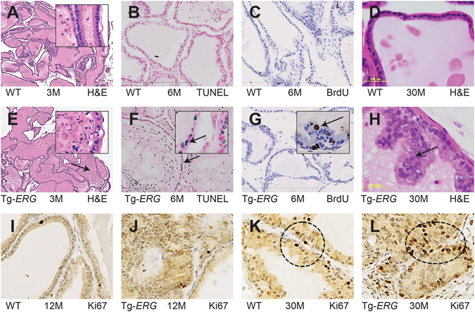
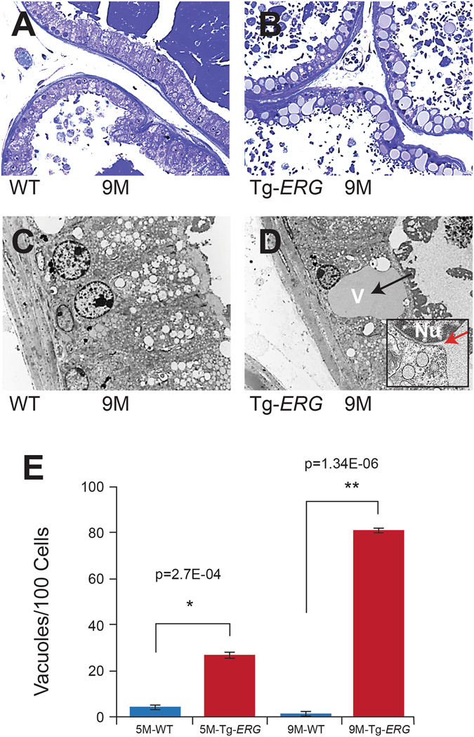
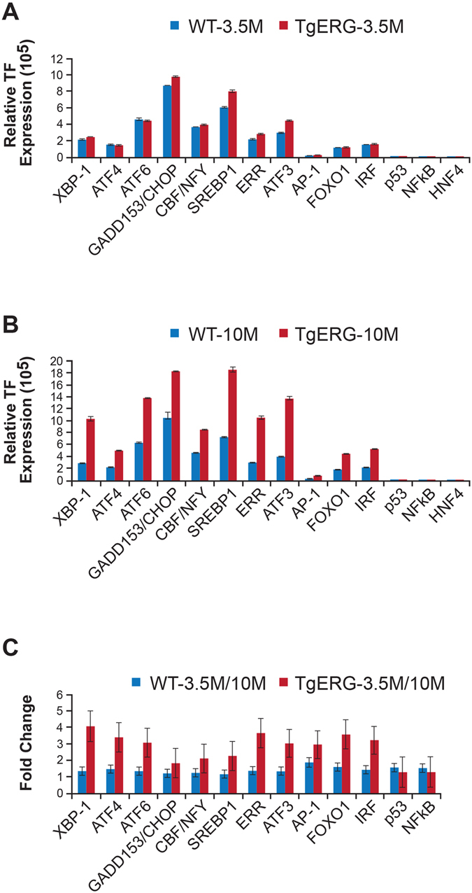
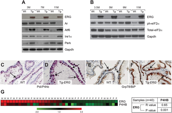
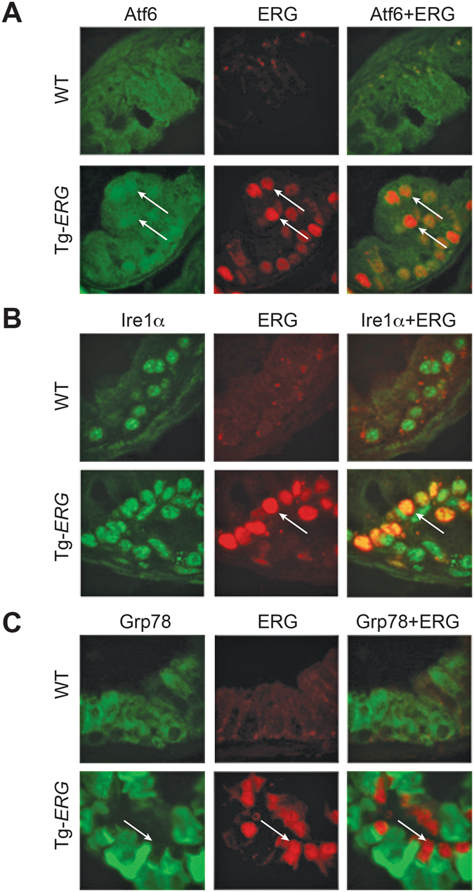
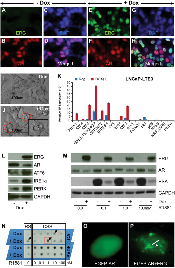
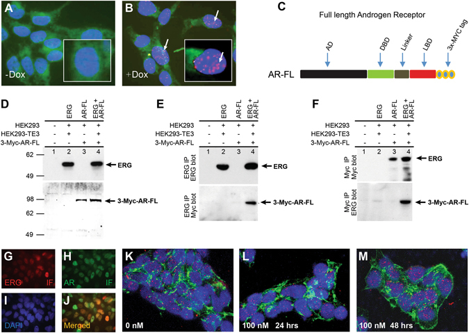
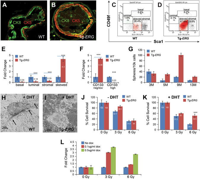
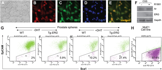
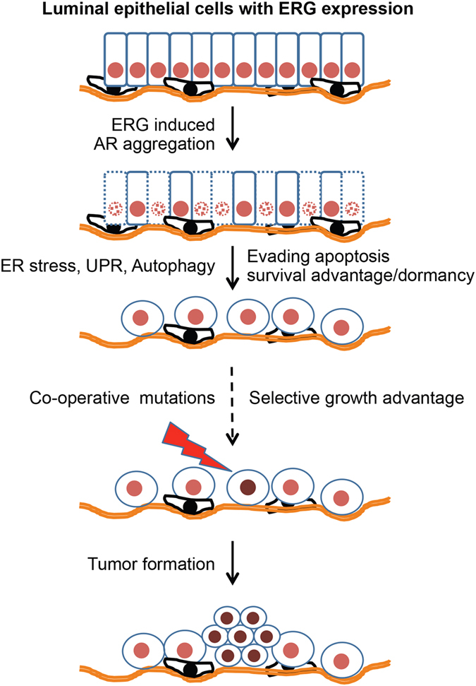
Mechanistic studies of deregulated ERG in prostate cancer and other cancers continue to enhance its role in cancer biology and its utility as a biomarker and therapeutic target. Here, we show that ERG, through its physical interaction with androgen receptor, induces AR aggregation and endoplasmic reticulum stress in the prostate glands of ERG transgenic mice. Histomorphological alterations and the expression of ER stress sensors Atf6, Ire1α, Perk, their downstream effectors Grp78/BiP and eIF2α in ERG transgenic mouse prostate glands indicate the presence of chronic ER stress. Transient activation of apoptotic cell death during early age correlated well with the differential regulation of ER stress sensors, in particular Perk. Epithelial cells derived from ERG transgenic mouse prostates have increased prostasphere formation with resistance to radiation induced cell death. Continued activation of cell survival factors, Atf6 and Ire1α during chronic ER stress due to presence of ERG in prostate epithelium induces survival pathways and provides a selection pressure in the continuum of ERG dependent neoplastic process. These novel insights will enhance the understanding of the mechanistic functions of ERG in prostate tumor biology and towards development of early targeted therapeutic strategies for prostate cancer.


Conflict of interest statement

The authors declare that they have no competing interests.

Figures

Figure 1
Figure 1
Morphological and histological differences in the ventral prostates of Tg-ERG mice. Hematoxylin and eosin staining of prostate glands from 3–30 months-old wild-type (A,C,G) and Tg-ERG mouse (B,D,H) prostate glands. Increased dell death is seen in 3 and 6 months-old display significant cell death (B,D). Inserts show nuclear fragmentation and sparsely organized and morphologically distinct luminal epithelial cell layer (note the arrows). TUNEL staining of wild-type (C) and Tg-ERG (D) prostates confirm the cell death due to apoptosis. Short arrow (insert) points to an intact luminal cell undergoing apoptotic cell death while long arrow points to fragmented nuclei of dead cells in the lumen. Cell proliferation analysis has shown an increase in the number of BrdU-positive cells in Tg ERG mouse (F) than wild-type (E). Hematoxylin and eosin staining of 30 month-old transgenic mice display clustered luminal cells resembling high grade PIN lesions (H). Similarly, Ki67 immunostaining was also increased in Tg-ERG mouse (J,L) than wild-type (I,K). There is an increase in the Ki67 staining pattern in 30 month-old mouse prostates than 12 month-old prostates (I–L). Total number of mice used for each analysis is 5 (n = 5).
Figure 2
Figure 2
ERG induces histo-morphological phenotype in mouse prostate glands. Toluidine blue stained plastic sections of wild-type mouse prostate glands (A) display well-organized, terminally differentiated, tall columnar luminal epithelial cells with basally positioned nuclei. Tg-ERG mouse ventral prostate (B) luminal epithelial cells displayed cuboidal cells with irregularly bordered nuclei and increased vacuolization. TEM analysis of the wild-type (C) ventral prostates displayed tall columnar luminal cells, basal nuclei and large number of clear secretory vesicles in the apical cytoplasm. Tg-ERG mouse prostate epithelial cells (D) displayed an abnormally large vacuole adjacent to nuclei (red arrow showing the extension of nuclear envelop into ER). The quantitative analysis of morphological changes (appearance of vacuoles in the Tg-ERG mouse prostates) in the luminal epithelial cells was performed on Toluidine blue stained plastic sections of Tg-ERG and wild-type prostate glands of 5 and 9 months-old mice (E).
Figure 3
Figure 3
ERG deregulates ER stress transcription factors. ER Stress (UPR) TF Activation Profiling Plate Array was used to analyze transcription factors (TFs) in the prostates of 3.5 and 10 month-old Tg-ERG mice compared to wild-type mice (A,B). Activity of ER transcription factors Xbp1, Gadd153/Chop, Cbf/Nfy, Serbp1, Err, Atf3 were slightly up regulated in 3.5 month-old prostates (A). However, by 10 months of age, Xbp1, Atf4, Atf6, Gadd153/Chop, Cbf/Nfy, Srebp1, Err, Atf3, Foxo1, Irf were up regulated significantly (B). Expression of ER stress transcription factors in 10 month-old prostates from Tg-ERG showed significant increase compared to 3 months old prostates from Tg-ERG (C).
Figure 4
Figure 4
Regulation of ER stress sensors by ERG in mouse prostates. Western blot analysis  (A) of ERG, Ar and ER stress sensors Atf6, Ire1α and Perk show increased expression in 3, 7 and 11 month-old Tg-ERG mouse prostates. Ar expression appears to decrease in the Tg-ERG prostates and is more significant in older mice. Similarly, there is an increase in the phosphorylation of eIF2α, a down stream target for Perk, in the transgenic mouse prostate glands (B). Immunohistochemical analysis of and P4hb/Pdi (C,D) and Grp78/BiP (E,F) in wild-type (C,E) and Tg-ERG (D,F) mouse prostates show elevated expression. Analysis of ERG and P4HB expression (G) in human prostate has shown significant correlation (n) (r = 0.65; P = 0.001). Expression data obtained from an 80GeneChip microarray show up-regulation as red and down-regulation as green.
Figure 5
Figure 5
Colocalization of ERG with ER stress sensors Atf6, Ire1α and ER chaperone protein Grp78/BiP by indirect immunofluorescence in 9 month-old Tg-ERG mouse prostate glands. Immunolocalization of Atf6 reveals the presence of activated Atf6 in the nucleus of Tg-ERG prostate luminal epithelial cells along with colocalized ERG (white arrows in the lower panel of A). Ire1α expression was increased in the Tg-ERG mouse prostates, correlating well with the colocalized expression of ERG in the luminal epithelial cells (white arrows in the lower panel of B). ER resident chaperone protein Grp78/BiP,  localized mainly in the cytoplasm of the luminal epithelial cells was increased in Tg-ERG mouse prostates (white arrows in the lower panel of C).
Figure 6
Figure 6
ERG induces ER stress and AR aggregation in LNCaP cells. Stable and doxycycline inducible ERG expressing LNCaP-LnTE3 cells were generated by lentiviral expression vector and characterized by immunofluorescence using mouse monoclonal ERG antibodies (9FY) and anti-AR antibodies (A–H). ERG expression was detected upon induction with 1 ug/ml of doxycycline treatment for 24 hrs. Note the ERG expression in un-induced (A) and induced (B). Increased numbers of vacuole formations (red dotted circles) were observed with doxycycline induction of ERG (J and Supplementary Figure S7). Analysis of the ER stress transcription factors was analyzed in LNCaP-LnTE3 cell lysates prepared from without and with induction of ERG by doxycycline. Levels of XBP1, ATF4, ATF6, GADD153/CHOP, CBF/NFY, SREBP1, ERR, ATF3 were increased with induced ERG (K). Western blot analysis showing enhancement of critical ER sensors such as ATF6, IRE1α and PERK (L). Induced expression of ERG in the presence of synthetic androgen, R1881 showing altered regulation of AR target genes PSA (M). AR aggregation was observed in cells treated with 10% FBS in the presence of induced ERG (N; shown by black small arrow). PVDF membrane, which traps monomers shows inverse AR signal with doxycycline induced ERG at 1.0 nm concentration of R1881 (N; red solid box). However, higher AR aggregation at 10.0 nM of R1881in the absence of ERG induction was (red dotted box) observed. AR aggregation is shown in LNCaP-LnTE3 cells transfected with pEGFP-C1-AR (O,P). pEGFP-C1-AR transfected LNCaP-LnTE3 cells were grown without (O) and with (P) doxycycline induction showing aggregates.
Figure 7
Figure 7
ERG physically interacts with Androgen Receptor. Proximity ligation assay (PLA) on LNCaP-LnTE3 cells without (A) or with (B) induction of ERG shows a positive interaction with AR protein. Graphical representation of 3x-MYC tagged AR full length and AR deletion construct (C) used to transfect HEK293 and HEK293-TE3 cells. HEK-293 and ERG expressing HEK 293-LTE3 cells grown in serum-starved conditions for 20 hours were transfected with full length AR and ERG constructs and stimulated with 1 nM R1881 for another 24 hrs. Immunoblot of cell lysates used in co-IP experiment were tested with anti-ERG MAb and anti-MYC-tag antibody as controls (D). ERG interactions with AR were detected by immunoprecipitation of ERG followed by westernblot with anti-ERG MAb (9FY) with mouse polyclonal IgG as control and immunoblotted with rabbit polyclonal anti-MYC-tag antibody (E). As a second approach, AR was immunoprecipitated with anti-MYC-tag antibody with rabbit polyclonal IgG as controll and immunoblotted with anti-ERG MAb (9FY) antibody (F). To further analyze if the interactions of ERG with AR are dependent on AR dimers or monomers, VCaP cells were grown on cover glasses in the presence of 100 mM resverotrol for 48 hours and PLA was performed to detect changes in physical interactions. Immunoflurescence analysis of ERG and AR in VCaP cells grown for 48 hrs showed colocalization (G–J). ERG-AR interactions by PLA showed the presence of interactions at 48 hrs resveratrol treated VCaP cells (K–M).
Figure 8
Figure 8
ERG alters molecular and morphological characteristics of luminal epithelial cells. Expression of Ck5 (basal) and Ck8 (luminal) cell markers in wild-type and Tg-ERG mouse prostates was performed by immuno-fluorescence on frozen sections. Wild-type mouse prostate glands express Ck5 (red) and Ck8 (green) (A). Tg-ERG prostate glands express increased number of cells double positive for both Ck5 and Ck8 (B). FACS analysis of isolated luminal cells from wild-type (C) and Tg-ERG mice (D) showed a significant increase in CD49f (low) and Sca-1 (med) in Tg-ERG mice. Normalized fold induction/reduction in frequencies of basal, luminal, stromal, skewed populations show about 4-fold increase of skewed population in Tg-ERG prostates (E). The frequencies of CD133 expression in percentages within skewed (red), stromal (grey) populations in wild-type and Tg-ERG mice show increase in CD133negative/low and decrease in CD133high cells in Tg-ERG prostates (F). Quantitative analysis of sphere forming units (G) from ventral prostates of wild-type and Tg-ERG mice shows the number of spheres grown per 104 input cells. The number of sphere forming units (SFU)/10,000 cells in the Tg-ERG was approximately 1–5 fold compared to controls (n = 6). Prostate epithelial cells isolated from wild-type (j,k) and Tg- ERG mouse (H,I) prostates develop spheres in semi-solid Matrigel. Spheres grown in the presence of 10 nM DHT on matrigel coated plates developed duct like structures (H). Duct like structures was not observed in spheres developed from Tg-ERG in the presence of DHT (I). Both wild-type and Tg-ERG prostate spheres grown on matrigel in the absence of DHT showed only about 31.8–34% cell survival upon 6 Gy radiation (J). Spheres developed from Tg-ERG in the presence of DHT showed 52.5% cell survival compared to 20.9% of wild-type derived spheres (K). Similarly, LNCaP-LnTE3 cells grown in the presence of 0 to 0.5 ug/ml of doxycycline to induce ERG displayed relatively 2.75 to 3.75 folds enhanced survival compared to controls with 3 and 6Gy radiation (L).
Figure 9
Figure 9
Spontaniously immortalized Tg-ERG mouse prostate epithelial cell line and epithelial cells isolated from Tg-ERG derived prostate spheres show similar features. Spontaniously immortalized Tg-ERG mouse prostate epithelial cell line, MoE1 (p6) grown in PrEGM supplemented with 1nM R1881 (A–E) show expression of both Ar (B) and ERG (C) by immunofluorescence using AR and ERG antibodies. Dapi (D) and Ar + ERG merged images (E) show colocalization of Ar and ERG in the muclei of MoE1 cells. Western blot analysis of MoE1 cells grown in charcoal stripped serum containing media show the expression of ERG upon induction with 1 nM R1881 (F). FACS analysis based on EpCAM of cells dissociated from the prostate spheres grown from wild-type, Tg-ERG mouse prostates in the absence (G i, ii) and presence of 10 nM DHT (G iii, iv) showabout 2–3 fold increase in EpCAM negative cell population (10.2% wt vs 27.4% Tg-ERG spheres). Consistant with prostate spheres, MoE1 cells (H) show increasing number of EpCAM (~97%).
Figure 10
Figure 10
Model depicting ERG-induced AR aggregation as one of the key molecular pathways towards malignant transformation of prostate luminal epithelial cells. Significant numbers of differentiated luminal epithelial cells initially undergo apoptosis due to transgenic expression of ERG and later gain a survival advantage. During this transition, several proteins involved in ER stress and autophagy pathways were up-regulated in luminal epithelial cells. These cells potentially serve as targets for subsequent genetic alterations that confer proliferative potential.

Similar articles

Cited by

References

    1. Siegel RL, Miller KD, Jemal A. Cancer statistics, 2016. CA Cancer J Clin. 2016;66:7–30. doi: 10.3322/caac.21332. - DOI - PubMed
    1. Torre LA, et al. Global cancer statistics, 2012. CA Cancer J Clin. 2015;65:87–108. doi: 10.3322/caac.21262. - DOI - PubMed
    1. Wilt TJ. The Prostate Cancer Intervention Versus Observation Trial: VA/NCI/AHRQ Cooperative Studies Program #407 (PIVOT): design and baseline results of a randomized controlled trial comparing radical prostatectomy with watchful waiting for men with clinically localized prostate cancer. J Natl Cancer Inst Monogr. 2012;2012:184–90. doi: 10.1093/jncimonographs/lgs041. - DOI - PMC - PubMed
    1. Saylor PJ. Prostate cancer: The androgen receptor remains front and centre. Nat Rev Clin Oncol. 2013;10:126–8. doi: 10.1038/nrclinonc.2013.14. - DOI - PubMed
    1. Hayward SW, et al. Epithelial development in the rat ventral prostate, anterior prostate and seminal vesicle. Acta Anat (Basel) 1996;155:81–93. doi: 10.1159/000147793. - DOI - PubMed

Publication types