. 2017 Apr 7:11:163.
doi: 10.3389/fnhum.2017.00163. eCollection 2017.

Concealed, Unobtrusive Ear-Centered EEG Acquisition: cEEGrids for Transparent EEG

Affiliations

Concealed, Unobtrusive Ear-Centered EEG Acquisition: cEEGrids for Transparent EEG

Martin G Bleichner et al. Front Hum Neurosci. .

Abstract

Electroencephalography (EEG) is an important clinical tool and frequently used to study the brain-behavior relationship in humans noninvasively. Traditionally, EEG signals are recorded by positioning electrodes on the scalp and keeping them in place with glue, rubber bands, or elastic caps. This setup provides good coverage of the head, but is impractical for EEG acquisition in natural daily-life situations. Here, we propose the transparent EEG concept. Transparent EEG aims for motion tolerant, highly portable, unobtrusive, and near invisible data acquisition with minimum disturbance of a user's daily activities. In recent years several ear-centered EEG solutions that are compatible with the transparent EEG concept have been presented. We discuss work showing that miniature electrodes placed in and around the human ear are a feasible solution, as they are sensitive enough to pick up electrical signals stemming from various brain and non-brain sources. We also describe the cEEGrid flex-printed sensor array, which enables unobtrusive multi-channel EEG acquisition from around the ear. In a number of validation studies we found that the cEEGrid enables the recording of meaningful continuous EEG, event-related potentials and neural oscillations. Here, we explain the rationale underlying the cEEGrid ear-EEG solution, present possible use cases and identify open issues that need to be solved on the way toward transparent EEG.

Keywords: ear EEG; ear-centered EEG; mobile EEG; transparent EEG; wearable EEG.


Figures

Figure 1
Figure 1
cEEGrid application. (A) The skin around the ear is cleansed with alcohol swabs. (B) The hair around the ear is pushed aside and the cEEGrid is placed around the ear with an adhesive tape. Good electrode skin conductance is assured by a drop of electrode gel on each electrode. (C) The applied grid allows for stable EEG recordings over several hours.
Figure 2
Figure 2
(A) A miniature, wireless EEG amplifier (black; https://mbraintrain.com/) is attached to the back of the head with a headband, and the cEEGrid is connected with the amplifier. The signal is transmitted wirelessly to a smartphone via Bluetooth. Android smartphones can be used for signal acquisition and stimulus presentation. (B) The cEEGrid electrodes are arranged in a C-shape. R4a and R4b may be used as ground (i.e., driven right leg) and reference electrodes. (C) Digitized electrode positions for a representative individual illustrating high-density equidistant EEG cap (black) and cEEGrid locations (yellow). The visualization was generated with the Brainstorm 3 software.
Figure 3
Figure 3
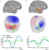
Simulation of forward projections of two different cortical sources, one in transverse temporal cortex (A) and the other one in superior temporal cortex (B). Forward projections to the scalp show clear topographical differences, as illustrated by the voltage maps. Different example bipolar channels as indicated in green and blue on the 3D head model show differential sensitivity to the two sources. Source amplitudes are shown in arbitrary units. The line plots are scaled to maximum activity for each source.
Figure 4
Figure 4
Continuous cEEGrid EEG data (5 s) with eyes open and eyes closed of one participant for a cEEGrid on the right side. Below is the mean amplitude of the alpha band (8–12 Hz). The increase in alpha band activity is readily visible in the raw signal and in the amplitude of the alpha band activity as soon as the eyes are closed. The effect is wide spread and can be seen on all cEEGrid channels.
Figure 5
Figure 5
Grand average single channel result for an auditory attention paradigm as described in Bleichner et al. (2016). (A) cEEGrid channel pair that was used to compute data show in (B,C). (B) The grand average ERPs for attended tones (green) and unattended tones (red) showed clear differences in amplitude. Shaded gray areas indicate the standard error of the mean. (C) The effect size over time is given as Hedges' g (absolute) values.
Figure 6
Figure 6
Single subject sleep EEG recording over the course of a night. Shown are the electrode impedance values recorded in parallel with EEG. Indicated in red is the median impedance score for all channels.
Figure 7
Figure 7
Single subject sleep EEG recording. Shown are three right ear cEEGrid EEG channels (blue), the horizontal electrooculogram (green), the electrocardioagram (red), and the head motion, as measured by the Smarting amplifier gyroscope (purple). Different characteristic sleep patterns are clearly visible, such as (A) theta activity, (B) k-complexes, and (C) slow wave sleep.
Figure 8
Figure 8
Fifteen-second resting EEG data from a healthy boy, 7-years of age, with no history of epileptic seizures, whose older brother is diagnosed with rolando epilepsy. Note spike-wave EEG activity, indicated in gray. See Figure 2 for illustration of electrode labels on cEEGrid.
Figure 9
Figure 9
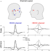
Lateral eye movements and eye blinks recorded simultaneously with cEEGrid and conventional electrooculogram (EOG) channels. (A) Channel layout for EOG and selected cEEGrid channels. The VEOG was the bipolar derivation between an above and a below eye electrode. The cVEOG was the bipolar derivation between the cEEGrid channels R2 and R7. The HEOG was the bipolar derivation between a channel next to the left and right eye. The cHEOG was the bipolar derivation between the cEEGrid channels R1 and R4. (B) Eye blinks are clearly reflected in a vertically oriented cEEGrid channel, whereas lateral eye movements are clearly reflected in a horizontally oriented cEEGrid channel. Note the similarity in morphology and latency between EOG and cEEGrid channels.
Figure 10
Figure 10
Single-subject Heart-rate variability analysis based on multi-channel cEEGrid recordings. (A) Linear decomposition of multi-channel cEEGrid signals with independent component analysis revealed one independent component representing heart-electrical activity, as indicated by prominent R-peaks (red circles). (B) Histogram of the RR interval, further supporting reliable R-peak detection. (C) Power spectrum analysis of inter-beat intervals, showing typical spectral signatures, such as 0.10 Hz activity.

References

    1. Alba N. A., Sclabassi R. J., Sun M., Cui X. T. (2010). Novel hydrogel-based preparation-free EEG electrode. IEEE Trans. Neural. Syst. Rehabil. Eng. 18, 415–423. 10.1109/TNSRE.2010.2048579 - DOI - PubMed
    1. Al-Yahya E., Dawes H., Smith L., Dennis A., Howells K., Cockburn J. (2011). Cognitive motor interference while walking: a systematic review and meta-analysis. Neurosci. Biobehav. Rev. 35, 715–728. 10.1016/j.neubiorev.2010.08.008 - DOI - PubMed
    1. Bertrand A., Mihajlovic V., Grundlehner B., Van Hoof C., Moonen M. (2013). Motion artifact reduction in EEG recordings using multi-channel contact impedance measurements, in 2013 IEEE Biomedical Circuits and Systems Conference (BioCAS), (IEEE; ) 258–261.
    1. Bleichner M. G., Lundbeck M., Selisky M., Minow F., Jager M., Emkes R., et al. . (2015). Exploring miniaturized EEG electrodes for brain-computer interfaces. An EEG you do not see? Physiol. Rep. 3:e12362. 10.14814/phy2.12362 - DOI - PMC - PubMed
    1. Bleichner M. G., Mirkovic B., Debener S. (2016). Identifying auditory attention with ear-EEG: cEEGrid versus high-density cap-EEG comparison. J. Neural Eng. 13:66004. 10.1088/1741-2560/13/6/066004 - DOI - PubMed