. 2017 May;112(3):33.
doi: 10.1007/s00395-017-0622-5. Epub 2017 Apr 24.

IL-10 improves cardiac remodeling after myocardial infarction by stimulating M2 macrophage polarization and fibroblast activation

Affiliations

IL-10 improves cardiac remodeling after myocardial infarction by stimulating M2 macrophage polarization and fibroblast activation

Mira Jung et al. Basic Res Cardiol. 2017 May.

Abstract

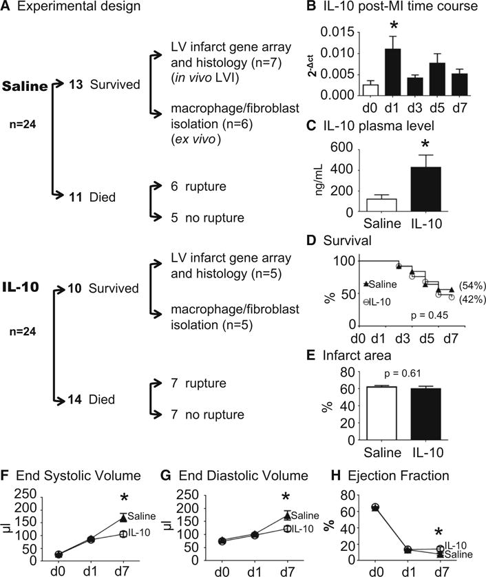
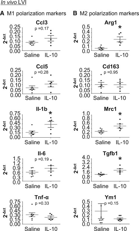
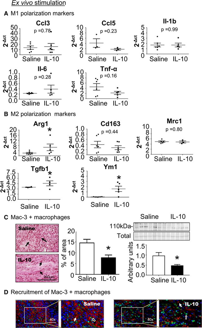
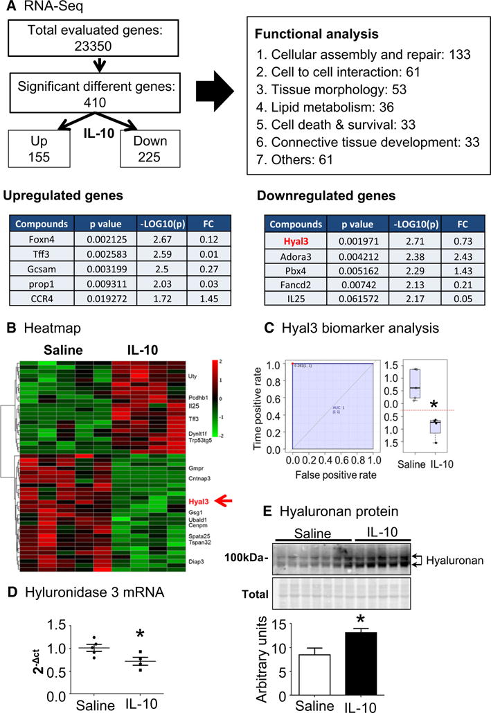
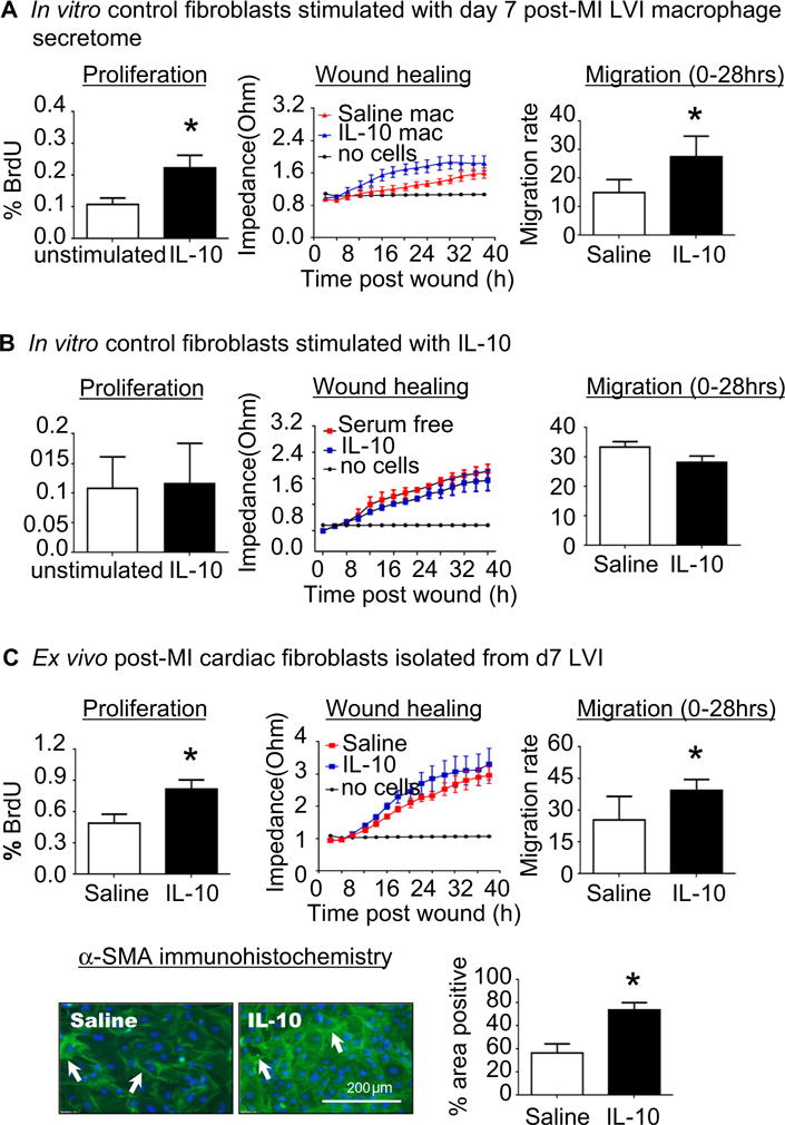
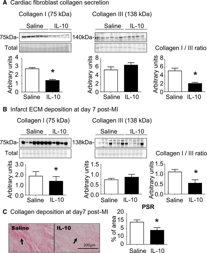
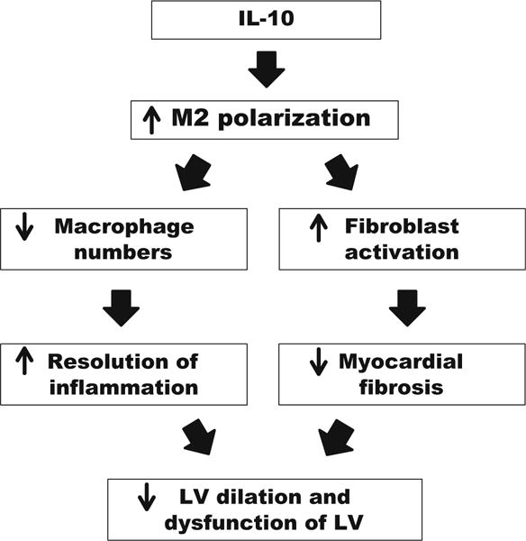
Inflammation resolution is important for scar formation following myocardial infarction (MI) and requires the coordinated actions of macrophages and fibroblasts. In this study, we hypothesized that exogenous interleukin-10 (IL-10), an anti-inflammatory cytokine, promotes post-MI repair through actions on these cardiac cell types. To test this hypothesis, C57BL/6J mice (male, 3- to 6-month old, n = 24/group) were treated with saline or IL-10 (50 μg/kg/day) by osmotic mini-pump infusion starting at day (d) 1 post-MI and sacrificed at d7 post-MI. IL-10 infusion doubled plasma IL-10 concentrations by d7 post-MI. Despite similar infarct areas and mortality rates, IL-10 treatment significantly decreased LV dilation (1.6-fold for end-systolic volume and 1.4-fold for end-diastolic volume) and improved ejection fraction 1.8-fold (both p < 0.05). IL-10 treatment attenuated inflammation at d7 post-MI, evidenced by decreased numbers of Mac-3-positive macrophages in the infarct (p < 0.05). LV macrophages isolated from d7 post-MI mice treated with IL-10 showed significantly elevated gene expression of M2 markers (Arg1, Ym1, and Tgfb1; all p < 0.05). We further performed RNA-seq analysis on post-MI cardiac macrophages and identified 410 significantly different genes (155 increased, 225 decreased by IL-10 treatment). By functional network analysis grouping, the majority of genes (133 out of 410) were part of the cellular assembly and repair functional group. Of these, hyaluronidase 3 (Hyal3) was the most important feature identified by p value. IL-10 treatment decreased Hyal3 by 28%, which reduced hyaluronan degradation and limited collagen deposition (all p < 0.05). In addition, in vivo IL-10 treatment increased fibroblast activation (proliferation, migration, and collagen production), an effect that was both directly and indirectly influenced by macrophage M2 polarization. Combined, our results indicate that in vivo infusion of IL-10 post-MI improves the LV microenvironment to dampen inflammation and facilitate cardiac wound healing.

Keywords: Collagen; Fibroblast; Hyaluronan; IL-10; Inflammation; Macrophage; Myocardial infarction.


Conflict of interest statement

Conflict of interest The authors declare that they have no conflict of interest.

Figures

Fig. 1
Fig. 1
IL-10 treatment improved post-MI LV physiology. LV dilation was decreased and ejection fraction increased in the IL-10-treated group at 7 days post-MI. a Experimental design. b IL-10 was elevated in the LV infarct (LVI) at d1 post-MI. c IL-10 infusion significantly elevated plasma IL-10 level at 7 days post-MI. IL-10 infusion did not affect d7 post-MI survival rate (c) and infarct area (d) compared to saline treatment. LV physiology was significantly improved in the IL-10 group compared to saline group. IL-10 treatment resulted in less LV dilation, noted by decreased end-systolic volume (e), end-diastolic volume (f) and increased ejection fraction (g). n = 24/group for survival analysis, n = 10–13/group for physiology measurements, * p < 0.05 vs. saline
Fig. 2
Fig. 2
IL-10 treatment enhanced the anti-inflammatory response in the LV infarct region (LVI) at d7 post-MI. a IL-10 did not affect M1 polarization marker expression. b IL-10 elevated anti-inflammatory (M2) marker expression (Arg1, Mrc1, and Tgfb1). n = 5–7/group, *p < 0.05 vs. saline
Fig. 3
Fig. 3
IL-10 treatment promoted M2 polarization and decreased cardiac macrophage numbers at 7 days post-MI. Macrophages were isolated from the infarct region at d7 post-MI and evaluated for M1 and M2 markers. a IL-10 infusion did not affect expression of M1 markers on ex vivo isolated macrophages. b IL-10 treatment stimulated macrophage polarization at d7 post-MI to an M2 phenotype, as isolated macrophages demonstrated elevated expression of Tgfb-1 and Ym1 compared to saline-treated mice. n = 5–7/group, *p < 0.05 vs. saline. c IL-10 treatment decreased macrophage numbers at d7 post-MI. Mac 3 in the LVI was quantified by immunohistochemistry; arrows indicate positive staining. Sample sizes are five images per section and n = 5 sections per group. *p < 0.05 vs. saline, scale bar is 200 μm. d IL-10 treatment had no effect on macrophage proliferation, as Mac-3 positive cells were not PCNA-positive. Open arrows indicate PCNA (green) positive staining, closed arrows indicate Mac-3 (red) positive staining and 4′,6-diamidine-2′-phenylindole dihydrochloride (DAPI; blue) was used to stain nuclei
Fig. 4
Fig. 4
IL-10 decreased hyaluronidase 3 (Hyal3) in cardiac macrophages, which resulted in less turnover of hyaluronic acid (HA). a RNA-Seq results and functional group analysis. b Heat mapping of the top 50 ranked (by p value and fold-change) genes that were differentially expressed in macrophages isolated from the infarcts of IL-10- and saline-treated mice. In the color legend, red represents up-regulation of genes compared to saline and green represents down-regulation of genes compared to saline. c Hyaluronidase 3 (Hyal3) was significantly reduced in LVI macrophages isolated from IL-10-treated mice. d RT-PCR results support decreased Hyal3 levels in LVI macrophages isolated from IL-10-treated mice. n = 4–5/group, *p < 0.05 vs. saline. e By immunoblotting, the IL-10 LVI showed increased full-length hyaluronan acid (HA) due to decreased HA turnover. n = 5–7/group, *p < 0.05 vs. saline
Fig. 5
Fig. 5
IL-10 treatment enhanced post-MI cardiac fibroblast activation and α-SMA expression through indirect effects on macrophage polarization. a d0 control cardiac fibroblasts were stimulated with the secretome of macrophages isolated from the d7 LV infarct (LVI) region. The post-MI macrophage secretome from IL-10-treated mice activated fibroblasts, resulting in increased proliferation and migration rates. n = 4–5/group, *p < 0.05 vs. saline. b Control cardiac fibroblasts stimulated with IL-10 in vitro (50 ng/mL) showed no effect on proliferation or migration rates. n = 3/group. c Cardiac fibroblasts isolated from d7 LVI showed increased proliferation and migration and α-SMA expression in the IL-10-treated group compared to the saline group. n = 4–7/group, *p < 0.05 vs. saline, scale bar is 200 μm
Fig. 6
Fig. 6
IL-10 treatment reduced the ratio of collagen I to collagen III, resulting in decreased fibrosis noted by reduced picrosirius red (PSR) staining. a Cardiac fibroblasts isolated from IL-10-treated LVI showed increased collagen I secretion, with no effect on collagen III secretion. The ratio of collagen I to collagen III was significantly reduced by IL-10 treatment. n = 5–6/group, *p < 0.05 vs. saline. b IL-10 treatment significantly decreased the ratio of collagen I to collagen III in the LVI in vivo, consistent with ex vivo findings. For immunoblotting, lanes were normalized by total protein for that lane. n = 5–7/group, *p < 0.05 vs. saline. c Picrosirius red staining showed reduced collagen accumulation in the IL-10-treated LVI at d7 post-MI. Sample sizes are five images for section and n = 5 sections per group. *p < 0.05 vs. saline, scale bar is 200 μm
Fig. 7
Fig. 7
Result’s summary. IL-10 treatment improved LV physiology by reducing LV dilation post-MI. At d7 post-MI, IL-10 infusion significantly increased M2 polarization of cardiac macrophages, leading to reduced macrophage numbers and cardiac fibroblast activation. This resulted in accelerated inflammation resolution and reduced collagen accumulation. Taken together, our results indicate that the improvement seen with IL-10 treatment was the result of direct actions on macrophage polarization and direct and indirect actions on fibroblast activation

Similar articles

Cited by

References

    1. Abe S, Steinmann BU, Wahl LM, Martin GR. High cell density alters the ratio of type III to I collagen synthesis by fibroblasts. Nature. 1979;279:442–444. doi: 10.1038/279442a0. - DOI - PubMed
    1. Balaji S, King A, Marsh E, LeSaint M, Bhattacharya SS, Han N, Dhamija Y, Ranjan R, Le LD, Bollyky PL, Crombleholme TM, Keswani SG. The role of interleukin-10 and hyaluronan in murine fetal fibroblast function in vitro: implications for recapitulating fetal regenerative wound healing. PLoS One. 2015;10:e0124302. doi: 10.1371/journal.pone.0124302. - DOI - PMC - PubMed
    1. Beam J, Botta A, Ye J, Soliman H, Matier BJ, Forrest M, MacLeod KM, Ghosh S. Excess linoleic acid increases collagen I/III ratio and “stiffens” the heart muscle following high fat diets. J Biol Chem. 2015;290:23371–23384. doi: 10.1074/jbc.M115.682195. - DOI - PMC - PubMed
    1. Berg DJ, Kuhn R, Rajewsky K, Muller W, Menon S, Davidson N, Grunig G, Rennick D. Interleukin-10 is a central regulator of the response to LPS in murine models of endotoxic shock and the Shwartzman reaction but not endotoxin tolerance. J Clin Investig. 1995;96:2339–2347. doi: 10.1172/JCI118290. - DOI - PMC - PubMed
    1. Christia P, Bujak M, Gonzalez-Quesada C, Chen W, Dobaczewski M, Reddy A, Frangogiannis NG. Systematic characterization of myocardial inflammation, repair, and remodeling in a mouse model of reperfused myocardial infarction. J Histochem Cytochem. 2013;61:555–570. doi: 10.1369/0022155413493912. - DOI - PMC - PubMed

Publication types