. 2017 May;7(1):41.
doi: 10.1007/s13205-017-0677-x. Epub 2017 Apr 24.

Imaging the accumulated intracellular microalgal lipids as a response to temperature stress

Affiliations

Imaging the accumulated intracellular microalgal lipids as a response to temperature stress

Khaled N M Elsayed et al. 3 Biotech. 2017 May.

Abstract

Over the last few decades, many scientists considered microalgae as promising actors for future biofuels because of the high lipid productivity inside their cells. Moreover, much attention has been paid to algal lipids as they can be used in biodiesel production. In this study, we optimized the different suitable conditions such as incubation time, incubation temperature, Dimethylesulfoxide and Nile red concentrations of the lipophilic fluorescence dye Nile red as an excellent and fast vital stain to detect and quantify intracellular lipids. This was achieved using the green alga Nannochloropsis salina. In addition, investigating the accumulation of lipid vesicles inside different isolated microalgal species as a response to temperature stress. Furthermore, the confocal laser scanning microscopy (LS510) for imaging and measuring the size and volume of the accumulated lipid vesicles was used.

Keywords: Fluorescence dye; Microalgae; Neutral lipids; Nile red (NR); Scanning microscopy.


Conflict of interest statement

The authors declare that they have no conflict of interest.

Figures

Fig. 1
Fig. 1
Effect of different parameters on Nile red assay a NR concentration µg ml−1(w/v), b DMSO concentration % (v/v), c Incubation time (min) and d Incubation temperature (°C) on the fluorescence intensity (a.u.) of the green alga N. salina as a model organism. The excitation and emission wavelengths used were 530 and 580 nm, respectively, the OD700 of the algal suspension was 1. All the tested samples expressed as mean values with standard deviations of three replicates
Fig. 2
Fig. 2
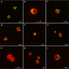
Visualization of lipid vesicles under temperature stress. Confocal fluorescence microscopy study of lipid content in different algal species: aMicractinium sp. YACCYB33, bSynechocystis sp. PAK12, cPedinomonas noctilucae C34, dChlorella sorokiniana KLL-G018, eSynechocystis sp. ElfSCS31, fChlorella variabilis NC64A, gSynechocystis sp. PCC 6803, hDesmodesmus pannonicus GM4n, iDesmodesmus intermedius CCAP 258/36. Lipophilic fluorescent dye Nile red is used as a vital stain for visualization of the oil-containing lipid bodies. The overlay shows auto-fluorescence of chlorophyll in chloroplasts (red) and Nile red stained lipid bodies (green) within algal cells. In some cases, lipid bodies appear yellow due to their spatial overlap with chloroplasts. Excitation of Nile Red was at 488 nm, emission was collected using a 530–600 nm band-pass filter. Excitation of chlorophyll fluorescence was at 633 nm, emission was imaged through a 650 nm long-pass filter

Similar articles

Cited by

References

    1. Abou-Shanab RAI, Matter IA, Kim SN, Oh YK, ChoiI Jeon BH. Characterization and identification of lipid-producing microalgae species isolated from a freshwater lake. Biomass Bioenerg. 2011;35(7):3079–3085. doi: 10.1016/j.biombioe.2011.04.021. - DOI
    1. Akimoto S, Mimuro M. Application of time-resolved polarization fluorescence spectroscopy in the femtosecond range to photosynthetic systems. Photochem Photobiol. 2007;83:163–170. - PubMed
    1. Bertozzini E, Galluzzi L, Penna A, Magnani M. Applications of the standard addition method for the absolute quantification of neutral lipids in microalgae using Nile red. J Microbiol Meth. 2011;87(1):17–23. doi: 10.1016/j.mimet.2011.06.018. - DOI - PubMed
    1. Brown WJ, Sullivan TR, Greenspan P. Nile red staining of lysosomal phospholipid inclusions. Histochemistry. 1992;97(4):349–354. doi: 10.1007/BF00270037. - DOI - PubMed
    1. Cabanelas IT, Van der Zwart M, Kleinegris DM, Barbosa MJ, Wijffels RH. Rapid method to screen and sort lipid accumulating microalgae. Bioresour Technol. 2015;18:47–52. doi: 10.1016/j.biortech.2014.10.057. - DOI - PubMed

LinkOut - more resources