

Elevated glypican-1 expression is associated with an unfavorable prognosis in pancreatic ductal adenocarcinoma
- PMID: 28440066
- PMCID: PMC5463070
- DOI: 10.1002/cam4.1064
Elevated glypican-1 expression is associated with an unfavorable prognosis in pancreatic ductal adenocarcinoma
Abstract
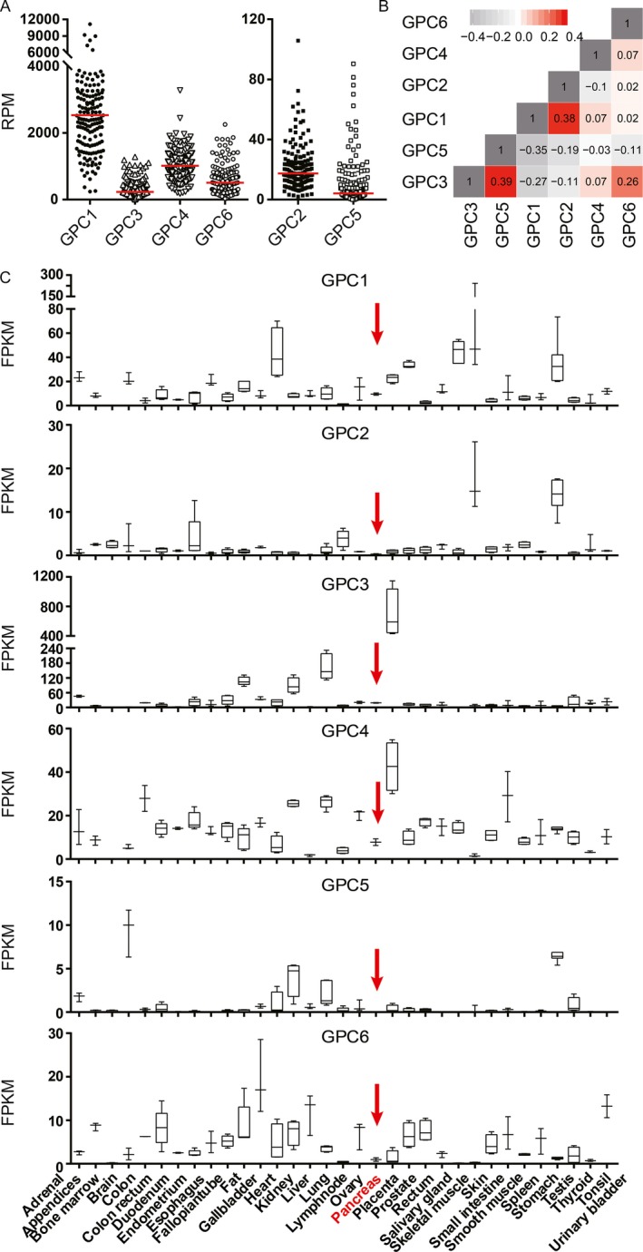
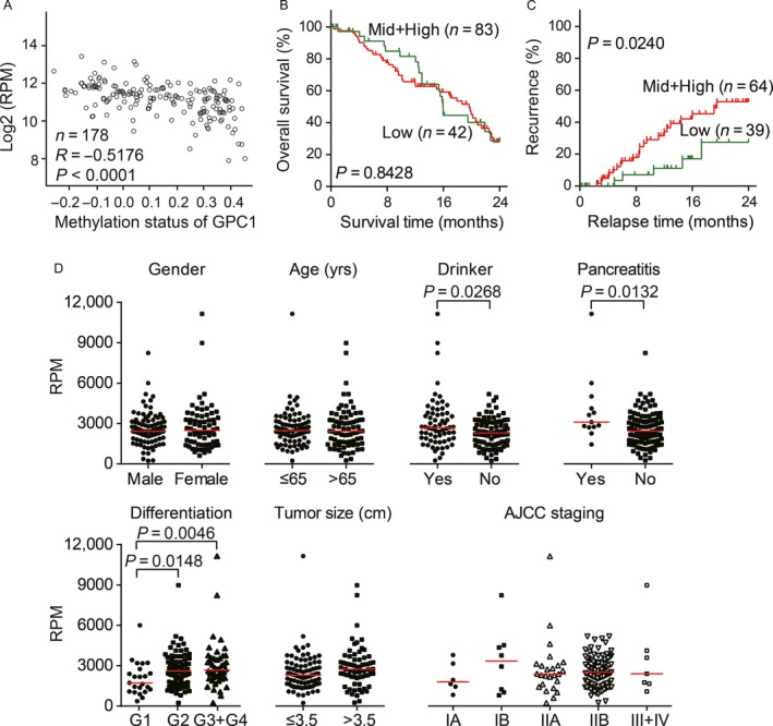
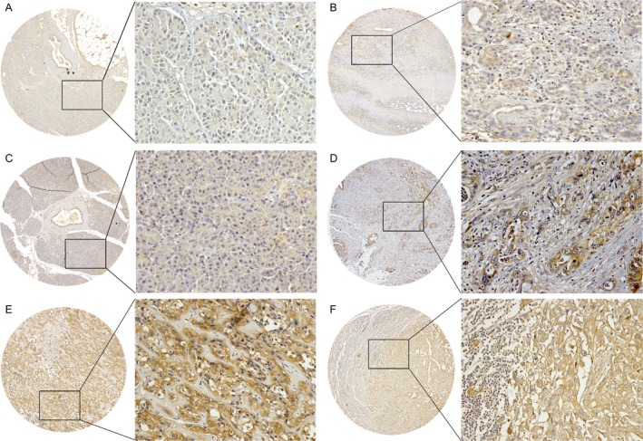
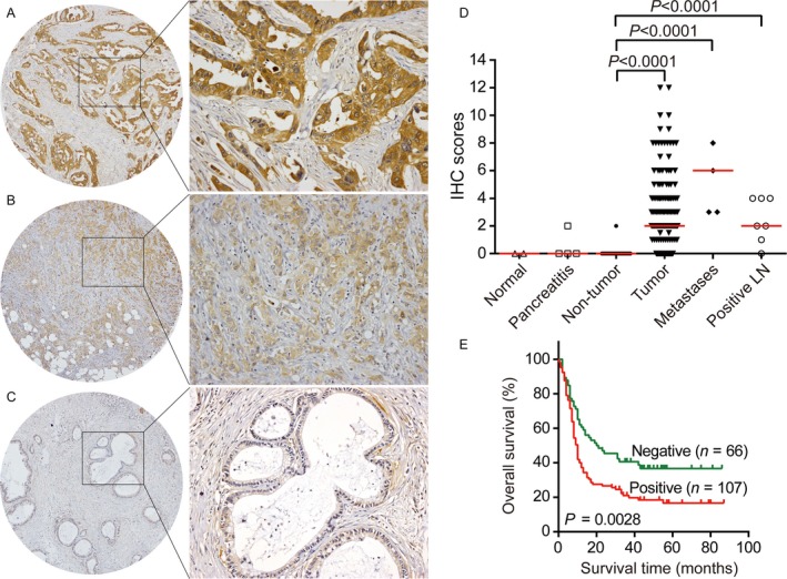
Pancreatic ductal adenocarcinoma (PDAC) is the most lethal cancer in humans, with a 5-year survival rate of <5%. Recently, glypican-1 (GPC1)-expressing circulating exosomes were found to be a promising diagnostic tool for PDAC. However, the aberrant expression of GPC1 has not been systematically evaluated in large-scale clinical samples of PDAC. Here, we performed a comprehensive analysis of GPC1 mRNA and protein expression features. Included in this study were 178 PDAC patients from the cancer genome atlas (TCGA) and 186 subjects whose tissues were used in immunohistochemical staining assays. We demonstrated that GPC1 mRNA was silenced in normal pancreata; however, it was re-expressed in PDAC tissues probably because of the promoter hypomethylation. The GPC1 protein was barely expressed in the normal and adjacent noncancerous pancreata. In tumor tissues, 59.7% (111/186) of the detected samples showed positive expression. Notably, GPC1 was elevated in 63.6% (34/55) of early stage cases. High levels of GPC1 were associated with poorer differentiation and larger tumor diameters. Kaplan-Meier analysis showed a significant difference in overall survival between the groups categorized by GPC1 expression (P = 0.0028). Multivariate analyses indicated that GPC1 was a significant risk factor for poor overall survival with a 1.82-fold increase in the hazard ratio (P = 0.0022). In conclusion, during pancreatic tumorigenesis, GPC1 was ectopically expressed and served as an independent poor prognostic factor. Our findings highlighted the alluring prospect of GPC1 as an early diagnostic and prognostic marker as well as a therapeutic target for PDAC.
Keywords: Biomarker; gene expression; glypican-1; pancreatic ductal adenocarcinoma; prognostic factor.
© 2017 The Authors. Cancer Medicine published by John Wiley & Sons Ltd.
Figures
Similar articles
-
High levels of serum glypican-1 indicate poor prognosis in pancreatic ductal adenocarcinoma.Cancer Med. 2018 Nov;7(11):5525-5533. doi: 10.1002/cam4.1833. Epub 2018 Oct 24. Cancer Med. 2018. PMID: 30358133 Free PMC article.
-
Correlation of glypican-1 expression with TGF-beta, BMP, and activin receptors in pancreatic ductal adenocarcinoma.Int J Oncol. 2006 Nov;29(5):1139-48. Int J Oncol. 2006. PMID: 17016645
-
Extracellular Vesicular Analysis of Glypican 1 mRNA and Protein for Pancreatic Cancer Diagnosis and Prognosis.Adv Sci (Weinh). 2024 Mar;11(11):e2306373. doi: 10.1002/advs.202306373. Epub 2024 Jan 10. Adv Sci (Weinh). 2024. PMID: 38204202 Free PMC article.
-
The next "sweet" spot for pancreatic ductal adenocarcinoma: Glycoprotein for early detection.Mass Spectrom Rev. 2023 Mar;42(2):822-843. doi: 10.1002/mas.21748. Epub 2021 Nov 12. Mass Spectrom Rev. 2023. PMID: 34766650 Free PMC article. Review.
-
The Oncoprotein Mucin 1 in Pancreatic Cancer Onset and Progression: Potential Clinical Implications.Biomolecules. 2025 Feb 13;15(2):275. doi: 10.3390/biom15020275. Biomolecules. 2025. PMID: 40001578 Free PMC article. Review.
Cited by
-
Inhibition of glypican-1 expression induces an activated fibroblast phenotype in a human bone marrow-derived stromal cell-line.Sci Rep. 2021 Apr 29;11(1):9262. doi: 10.1038/s41598-021-88519-7. Sci Rep. 2021. PMID: 33927256 Free PMC article.
-
Development of a reliable assay to measure glypican-1 in plasma and serum reveals circulating glypican-1 as a novel prostate cancer biomarker.Oncotarget. 2018 Apr 27;9(32):22359-22367. doi: 10.18632/oncotarget.25009. eCollection 2018 Apr 27. Oncotarget. 2018. PMID: 29854284 Free PMC article.
-
IFN‑γ induces apoptosis in gemcitabine‑resistant pancreatic cancer cells.Mol Med Rep. 2024 May;29(5):76. doi: 10.3892/mmr.2024.13200. Epub 2024 Mar 15. Mol Med Rep. 2024. PMID: 38488034 Free PMC article.
-
The Glycocalyx and Its Role in Vascular Physiology and Vascular Related Diseases.Cardiovasc Eng Technol. 2021 Feb;12(1):37-71. doi: 10.1007/s13239-020-00485-9. Epub 2020 Sep 21. Cardiovasc Eng Technol. 2021. PMID: 32959164 Free PMC article. Review.
-
Identification and Validation of Three PDAC Subtypes and Individualized GSVA Immune Pathway-Related Prognostic Risk Score Formula in Pancreatic Ductal Adenocarcinoma Patients.J Oncol. 2021 Dec 27;2021:4986227. doi: 10.1155/2021/4986227. eCollection 2021. J Oncol. 2021. PMID: 34987579 Free PMC article.
References
-
- Torre, L. A. , Bray F., Siegel R. L., Ferlay J., Lortet‐Tieulent J., and Jemal A.. 2015. Global cancer statistics, 2012. CA Cancer J. Clin. 65:87–108. - PubMed
-
- Scara, S. , Bottoni P., and Scatena R.. 2015. CA 19‐9: biochemical and clinical aspects. Adv. Exp. Med. Biol. 867:247–260. - PubMed
-
- Bergquist, J. R. , Puig C. A., Shubert C. R., Groeschl R. T., Habermann E. B., Kendrick M. L., et al. 2016. Carbohydrate antigen 19‐9 elevation in anatomically resectable, early stage pancreatic cancer is independently associated with decreased overall survival and an indication for neoadjuvant therapy: a national cancer database study. J. Am. Coll. Surg. 223:52–65. - PubMed
MeSH terms
Substances
LinkOut - more resources
Full Text Sources
Other Literature Sources
Medical
Research Materials
