Review
. 2017 Nov;43(6):779-798.
doi: 10.1080/1040841X.2017.1313814. Epub 2017 Apr 25.

Strategies in Ebola virus disease (EVD) diagnostics at the point of care

Affiliations
Review

Strategies in Ebola virus disease (EVD) diagnostics at the point of care

Chad T Coarsey et al. Crit Rev Microbiol. 2017 Nov.

Abstract

Ebola virus disease (EVD) is a devastating, highly infectious illness with a high mortality rate. The disease is endemic to regions of Central and West Africa, where there is limited laboratory infrastructure and trained staff. The recent 2014 West African EVD outbreak has been unprecedented in case numbers and fatalities, and has proven that such regional outbreaks can become a potential threat to global public health, as it became the source for the subsequent transmission events in Spain and the USA. The urgent need for rapid and affordable means of detecting Ebola is crucial to control the spread of EVD and prevent devastating fatalities. Current diagnostic techniques include molecular diagnostics and other serological and antigen detection assays; which can be time-consuming, laboratory-based, often require trained personnel and specialized equipment. In this review, we discuss the various Ebola detection techniques currently in use, and highlight the potential future directions pertinent to the development and adoption of novel point-of-care diagnostic tools. Finally, a case is made for the need to develop novel microfluidic technologies and versatile rapid detection platforms for early detection of EVD.

Keywords: Ebola virus; diagnostics; immunoassay; microfluidics; polymerase chain reaction.


Conflict of interest statement

Disclosure statement

Authors declare no financial conflict of interest.

Figures
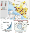
Figure 1
Figure 1
The current unprecedented and past EVD epidemiological outbreak maps and models; (a) image of the regions affected by the 2014 West African Ebola Outbreak (Aylward et al. 2014). Regional maps of confirmed and probable cases, clockwise from top right: (regions with declared epidemics) Guinea, Sierra Leone and Liberia; (regions with subsequent transmission events) Mali, Texas and New York in the USA and Spain. Reprinted with copyright permission from Aylward et al. (2014). (b) 2014 outbreak growth of confirmed, probable and suspected cases of EVD in each affected country under epidemic crisis (Sierra Leone, Liberia and Guinea). Reprinted with copyright permission from Gire et al. (2014). (c) Temporal sequence of Ebola infection within a 48-h duration in an infected cell, demonstrating the events prior to detectable threshold. Green and orange particles represent common serological targets, VP40 and GP, respectively. Viral RNA is represented by blue and red transcripts. Reprinted with copyright permission from Roca et al. (2015).
Figure 2
Figure 2
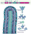
The Genomic and Protein maps of Ebolavirus (Alazard-Dany et al. 2006). (top) The overall ~19kbp genomic structure of Ebola with: a nucleoprotein (NP), a polymerase cofactor (VP35), a matrix protein (VP40), a transmembrane glycoprotein (GP), the GP gene also codes for an edited mRNA for a soluble glycoprotein (sGP), a transcriptional activator (VP30), a viral envelope-associated protein (VP24) and a RNA–dependent RNA polymerase (L). (b) A rendered Ebola virus protein schematic, displaying the viral proteins in their appropriate roles of the overall viral structure. Reprinted with copyright permission from Lee and Saphire (2009).
Figure 3
Figure 3
RT-LAMP final cyclic reaction and elongation steps; (a) following the FIP primer binding, there is another round of polymerization. A reverse transcriptase binds the FIP primer making a cDNA copy. This step is continued with the BIP primer, which makes a template DNA strand from cDNA polymerization. The outside reaction is repeated in multiple rounds, allowing for mass amplification of the Ebola RNA target. Reprinted with copyright permission from Notomi et al. (2000). (b) Results of RT-LAMP for Ebola (Kurosaki et al. 2007) showing sensitivity of the amplified in vitro Ebola RNA at only 20 copies. (c) The duration of the Ebola genome amplification by RT-LAMP have shown to detect Ebola within 20 min. Reprinted with copyright permission from Kurosaki et al. (2007).
Figure 4
Figure 4
Magnetic bead actuation on a chip and whole-blood sampling isotachophoresis; (a) Functionalized magnetic beads with anti-CD4-antibodies in an HIV ELISA detection assay. Magnetic beads bind and capture CD-4 cells, and are actuated to each reaction chamber when a magnetic field is applied a colour change indicates a seropositive reaction in the final well. Reprinted with copyright permission from Wang et al. (2014). (b) Complex isotachophoresis on-a-chip designed to capture and isolate parasite DNA from infected leukocytes, using electric fields to generate electrophoretic separation of ionic samples. Reprinted with copyright permission from Marshall et al. (2011).
Figure 5
Figure 5
Immuno-PCR (iPCR) assay: iPCR technique with both variations of Enzyme-Linked Immunosorbent Assay (ELISA), the standard for Ebola serological diagnosis; iPCR differs from ELISA by using DNA amplification of an indicator a DNA for ultrasensitive. Reprinted with copyright permission from Akter et al. (2014). (b) The sensitivity of iPCR technique is enhanced using functionalized gold nanoparticle conjugated with multiple single stranded DNA oligonucleotides. Reprinted with copyright permission from Chen et al. (2009).
Figure 6
Figure 6
Alternative biosensing technologies for rapid and early diagnosis of Ebola. (a) Quartz crystal microbalance immunosensors are composed of a gold electrode layer with surface antibody capture agents, placed on top of a piezoelectric quartz crystal. Resonance frequency changes are measured as target binds to the antibody-coated surface, allowing for label-free detection. Reprinted with copyright permission from Kierny et al. (2012). (b) Surface plasmon resonance (SPR) biosensor schematic, composed of a channel with specific antibodies fixed on a gold layer. A measurement in changes of light reflected from an optic source allows for potentially highly sensitive diagnostic capability. Reprinted with copyright permission from Cooper (2002). (c) A nanostructured photonic crystal-based sensor functionalized with gp120 to selectively capture and detect HIV copies from whole blood. Reprinted with copyright permission from Shafiee et al. (2014). (d) Break junction functionalized with aptamer to capture cancer biomarkers through electrical impedance. Reprinted with copyright permission from Ilyas et al. (2012).

Similar articles

  • An Outbreak of Ebola Virus Disease in the Lassa Fever Zone.
    Goba A, Khan SH, Fonnie M, Fullah M, Moigboi A, Kovoma A, Sinnah V, Yoko N, Rogers H, Safai S, Momoh M, Koroma V, Kamara FK, Konowu E, Yillah M, French I, Mustapha I, Kanneh F, Foday M, McCarthy H, Kallon T, Kallon M, Naiebu J, Sellu J, Jalloh AA, Gbakie M, Kanneh L, Massaly JL, Kargbo D, Kargbo B, Vandi M, Gbetuwa M, Gevao SM, Sandi JD, Jalloh SC, Grant DS, Blyden SO, Crozier I, Schieffelin JS, McLellan SL, Jacob ST, Boisen ML, Hartnett JN, Cross RW, Branco LM, Andersen KG, Yozwiak NL, Gire SK, Tariyal R, Park DJ, Haislip AM, Bishop CM, Melnik LI, Gallaher WR, Wimley WC, He J, Shaffer JG, Sullivan BM, Grillo S, Oman S, Garry CE, Edwards DR, McCormick SJ, Elliott DH, Rouelle JA, Kannadka CB, Reyna AA, Bradley BT, Yu H, Yenni RE, Hastie KM, Geisbert JB, Kulakosky PC, Wilson RB, Oldstone MB, Pitts KR, Henderson LA, Robinson JE, Geisbert TW, Saphire EO, Happi CT, Asogun DA, Sabeti PC, Garry RF; Viral Hemorrhagic Fever Consortium. Goba A, et al. J Infect Dis. 2016 Oct 15;214(suppl 3):S110-S121. doi: 10.1093/infdis/jiw239. Epub 2016 Jul 11. J Infect Dis. 2016. PMID: 27402779 Free PMC article.
  • Comparative Evaluation of the Diagnostic Performance of the Prototype Cepheid GeneXpert Ebola Assay.
    Jansen van Vuren P, Grobbelaar A, Storm N, Conteh O, Konneh K, Kamara A, Sanne I, Paweska JT. Jansen van Vuren P, et al. J Clin Microbiol. 2016 Feb;54(2):359-67. doi: 10.1128/JCM.02724-15. Epub 2015 Dec 4. J Clin Microbiol. 2016. PMID: 26637383 Free PMC article.
  • Diagnostics in Ebola Virus Disease in Resource-Rich and Resource-Limited Settings.
    Shorten RJ, Brown CS, Jacobs M, Rattenbury S, Simpson AJ, Mepham S. Shorten RJ, et al. PLoS Negl Trop Dis. 2016 Oct 27;10(10):e0004948. doi: 10.1371/journal.pntd.0004948. eCollection 2016 Oct. PLoS Negl Trop Dis. 2016. PMID: 27788135 Free PMC article. Review.
  • Lateral Flow Immunoassays for Ebola Virus Disease Detection in Liberia.
    Phan JC, Pettitt J, George JS, Fakoli LS 3rd, Taweh FM, Bateman SL, Bennett RS, Norris SL, Spinnler DA, Pimentel G, Sahr PK, Bolay FK, Schoepp RJ. Phan JC, et al. J Infect Dis. 2016 Oct 15;214(suppl 3):S222-S228. doi: 10.1093/infdis/jiw251. Epub 2016 Jul 20. J Infect Dis. 2016. PMID: 27443616 Free PMC article.
  • Towards detection and diagnosis of Ebola virus disease at point-of-care.
    Kaushik A, Tiwari S, Dev Jayant R, Marty A, Nair M. Kaushik A, et al. Biosens Bioelectron. 2016 Jan 15;75:254-72. doi: 10.1016/j.bios.2015.08.040. Epub 2015 Aug 20. Biosens Bioelectron. 2016. PMID: 26319169 Free PMC article. Review.

Cited by

References

    1. Akter F, Mie M, Kobatake E. DNA-based immunoassays for sensitive detection of protein. Sens Actuators B Chem. 2014;202:1248–1256.
    1. Alazard-Dany N, Terrangle MO, Volchkov V. Ebola et Marburg: les hommes contre-attaquent. M/S: Méd Sci. 2006;22:405–410. - PubMed
    1. Asghar W, El Assal R, Shafiee H, Pitteri S, Paulmurugan R, Demirci U. Engineering cancer microenvironments for in vitro 3-D tumor models. Mater Today. 2015;18:539–553. - PMC - PubMed
    1. Asghar W, Ilyas A, Billo JA, Iqbal SM. Shrinking of solid-state nanopores by direct thermal heating. Nanoscale Res Lett. 2011;6:1–6. - PMC - PubMed
    1. Asghar W, Ilyas A, Deshmukh RR, Sumitsawan S, Timmons RB, Iqbal SM. Pulsed plasma polymerization for controlling shrinkage and surface composition of nanopores. Nanotechnology. 2011;22:285304. - PubMed

MeSH terms

LinkOut - more resources