

"Clinical" cytology for endoscopists: A practical guide
- PMID: 28440233
- PMCID: PMC5418972
- DOI: 10.4103/eus.eus_21_17
"Clinical" cytology for endoscopists: A practical guide
Abstract
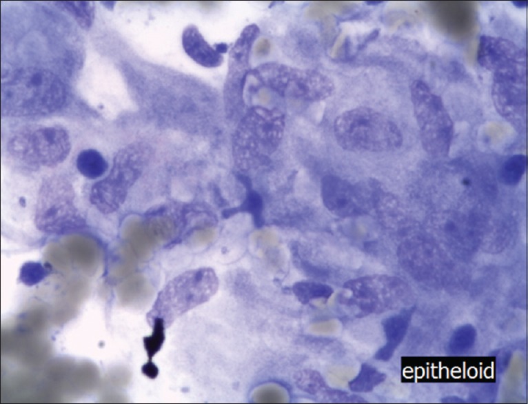
Clinical cytology was originally used by clinicians to provide rapid diagnosis. However, with advancing medical subspecialization, few clinicians interpret cytology themselves these days, for example, gynecologists, hematologists, urologists, and occasional gastroenterologist (mainly in Asian countries). Cytological assessment enjoyed a renaissance with the development of endoscopic ultrasound (EUS)-guided fine-needle aspiration (FNA). Subsequently, pathologists, most of them more experienced in histology, had to take over. Recently, it has been shown that in-room cytology can be easily performed by the endoscopist themselves for initial evaluation of the quality of the EUS-FNA specimen and an initial diagnosis distinguishing benign or malignant cells. Bringing cytology back to the clinician has some advantages but does not substitute the professional cytopathologist. This report has written to lower the threshold for the clinician to find his way back to the microscope, which may improve both their diagnostic yield and assessment of EUS-FNA sample quality.
Conflict of interest statement
There are no conflicts of interest.
Figures
Similar articles
-
Touch imprint cytology on endoscopic ultrasound fine-needle biopsy provides comparable sample quality and diagnostic yield to standard endoscopic ultrasound fine-needle aspiration specimens in the evaluation of solid pancreatic lesions.Cytopathology. 2019 Mar;30(2):179-186. doi: 10.1111/cyt.12662. Epub 2018 Dec 21. Cytopathology. 2019. PMID: 30484917
-
Improved diagnostic yield of endoscopic ultrasound-fine needle biopsy with histology specimen processing.World J Gastrointest Endosc. 2020 Aug 16;12(8):212-219. doi: 10.4253/wjge.v12.i8.212. World J Gastrointest Endosc. 2020. PMID: 32879656 Free PMC article.
-
Fluorescence cytology with 5-aminolevulinic acid in EUS-guided FNA as a method for differentiating between malignant and benign lesions (with video).Gastrointest Endosc. 2015;81(6):1457-62. doi: 10.1016/j.gie.2015.01.031. Epub 2015 Apr 9. Gastrointest Endosc. 2015. PMID: 25865388
-
Endoscopic ultrasound-guided fine-needle aspiration biopsy and trucut biopsy in gastroenterology - An overview.Best Pract Res Clin Gastroenterol. 2009;23(5):743-59. doi: 10.1016/j.bpg.2009.05.006. Best Pract Res Clin Gastroenterol. 2009. PMID: 19744637 Review.
-
[EUS-FNA: how to improve biopsy results? An evidence based review].Z Gastroenterol. 2014 Sep;52(9):1081-92. doi: 10.1055/s-0034-1385133. Epub 2014 Sep 8. Z Gastroenterol. 2014. PMID: 25198088 Review. German.
Cited by
-
Elastography of the Pancreas, Current View.Clin Endosc. 2019 Nov;52(6):533-540. doi: 10.5946/ce.2018.156. Epub 2019 Jul 17. Clin Endosc. 2019. PMID: 31311914 Free PMC article.
-
Rapid on-site evaluation (ROSE) with EUS-FNA: The ROSE looks beautiful.Endosc Ultrasound. 2019 Sep-Oct;8(5):283-287. doi: 10.4103/eus.eus_65_19. Endosc Ultrasound. 2019. PMID: 31603143 Free PMC article. No abstract available.
-
Comparison of EUS-guided conventional smear and liquid-based cytology in pancreatic lesions: A systematic review and meta-analysis.Endosc Int Open. 2020 Nov;8(11):E1611-E1622. doi: 10.1055/a-1240-0027. Epub 2020 Oct 22. Endosc Int Open. 2020. PMID: 33140017 Free PMC article.
-
Discriminating chronic pancreatitis from pancreatic cancer: Contrast-enhanced EUS and multidetector computed tomography in direct comparison.Endosc Ultrasound. 2018 Nov-Dec;7(6):395-403. doi: 10.4103/eus.eus_24_18. Endosc Ultrasound. 2018. PMID: 30246709 Free PMC article.
-
Stereomicroscopic on-site evaluation in endoscopic ultrasound-guided tissue acquisition of upper gastrointestinal subepithelial lesions.Clin Endosc. 2024 Jan;57(1):89-95. doi: 10.5946/ce.2022.288. Epub 2023 Apr 18. Clin Endosc. 2024. PMID: 37070203 Free PMC article.
References
-
- Chang KJ, Nguyen P, Erickson RA, et al. The clinical utility of endoscopic ultrasound-guided fine-needle aspiration in the diagnosis and staging of pancreatic carcinoma. Gastrointest Endosc. 1997;45:387–93. - PubMed
-
- Jenssen C, Faiss S, Nürnberg D. Complications of endoscopic ultrasound and endoscopic ultrasound-guided interventions – Results of a survey among German centers. Z Gastroenterol. 2008;46:1177–84. - PubMed
-
- Eloubeidi MA, Tamhane A. Prospective assessment of diagnostic utility and complications of endoscopic ultrasound-guided fine needle aspiration. Results from a newly developed academic endoscopic ultrasound program. Dig Dis. 2008;26:356–63. - PubMed
-
- Pang NK, Chin SY, Nga ME, et al. Comparative validation of c-kit exon 11 mutation analysis on cytology samples and corresponding surgical resections of gastrointestinal stromal tumours. Cytopathology. 2009;20:297–303. - PubMed
-
- Spieler P, Ammann M, Schönegg R. Fine-needle aspiration cytology. Aspects of a minimally invasive diagnostic procedure. Pathologe. 2007;28:325–33. - PubMed
Publication types
LinkOut - more resources
Full Text Sources
Other Literature Sources
