

Histochemical and Immunohistochemical Study of α-SMA, Collagen, and PCNA in Epithelial Ovarian Neoplasm
- PMID: 28440973
- PMCID: PMC5464482
- DOI: 10.22034/APJCP.2017.18.3.667
Histochemical and Immunohistochemical Study of α-SMA, Collagen, and PCNA in Epithelial Ovarian Neoplasm
Abstract
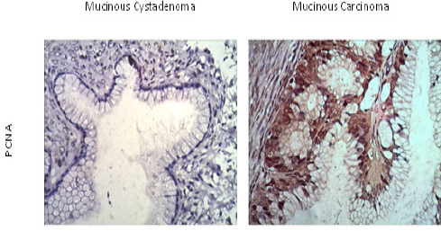
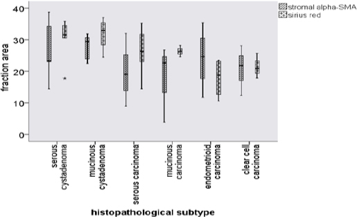
Background: Alpha-smooth muscle actin (α-SMA) is an isoform of actin, positive in myofibroblasts and is an epithelial to mesenchymal transition (EMT) marker. EMT is a process by which tumor cells develop to be more hostile and able to metastasize. Progression of tumor cells is always followed by cell composition and extracellular matrix component alteration. Increased α-SMA expression and collagen alteration may predict the progressivity of ovarian neoplasms. Objective: The aim of this research was to analyse the characteristic of α-SMA and collagen in tumor cells and stroma of ovarian neoplasms. In this study, PCNA (proliferating cell nuclear antigen) expression was also investigated. Methods: Thirty samples were collected including serous, mucinous, endometrioid, and clear cell subtypes. The expression of α-SMA and PCNA were calculated in cells and stroma of ovarian tumors. Collagen was detected using Sirius Red staining and presented as area fraction. Results: The overexpressions of α-SMA in tumor cells were only detected in serous and clear cell ovarian carcinoma. The histoscore of α-SMA was higher in malignant than in benign or borderline ovarian epithelial neoplasms (105.3±129.9 vs. 17.3±17.1, P=0.011; mean±SD). Oppositely, stromal α-SMA and collagen area fractions were higher in benign than in malignant tumors (27.2±6.6 vs 20.5±8.4, P=0.028; 31.0±5.6 vs. 23.7±6.4, P=0.04). The percentages of epithelial and stromal PCNA expressions were not significantly different between benign and malignant tumors. Conclusion: Tumor cells of serous and clear cell ovarian carcinoma exhibit mesenchymal characteristic as shown by α-SMA positive expression. This expression might indicate that these subtypes were more aggressive. This research showed that collagen and α-SMA area fractions in stroma were higher in benign than in malignant neoplasms.
Keywords: Ovarian neoplasm; α-SMA; collagen; PCNA; epithelial to mesenchymal transition.
10.22034/APJCP.2017.18.3.667
Figures


References
-
- Barboza CA, Pereira Pinto L, Freitas Rde A, et al. Proliferating cell nuclear antigen (PCNA) and p53 protein expression in ameloblastoma and adenomatoid odontogenic tumor. Braz Dent J. 2005;16:56–61. - PubMed
-
- Berny W, Weiser A, Markowska-Woyciechowska A, et al. Analysis of PCNA, Ki67, AgNOR and p53 expression in brain glial tumors. Neurol Neurochir Pol. 2004;38:457–63. - PubMed
-
- Cherng S, Young J, Ma H. Alpha-smooth muscle actin (α-SMA) J Am Sci. 2008;4:10.
-
- Ding L, Zhang Z, Shang D, et al. alpha-Smooth muscle actin-positive myofibroblasts, in association with epithelial-mesenchymal transition and lymphogenesis, is a critical prognostic parameter in patients with oral tongue squamous cell carcinoma. J Oral Pathol Med. 2014;43:335–43. - PubMed
LinkOut - more resources
Full Text Sources
Other Literature Sources
Miscellaneous