. 2017 Apr 25;12(4):e0176132.
doi: 10.1371/journal.pone.0176132. eCollection 2017.

Creating and parameterizing patient-specific deep brain stimulation pathway-activation models using the hyperdirect pathway as an example

Affiliations

Creating and parameterizing patient-specific deep brain stimulation pathway-activation models using the hyperdirect pathway as an example

Kabilar Gunalan et al. PLoS One. .

Abstract

Background: Deep brain stimulation (DBS) is an established clinical therapy and computational models have played an important role in advancing the technology. Patient-specific DBS models are now common tools in both academic and industrial research, as well as clinical software systems. However, the exact methodology for creating patient-specific DBS models can vary substantially and important technical details are often missing from published reports.

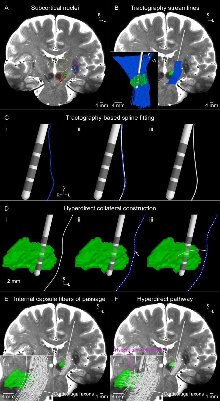
Objective: Provide a detailed description of the assembly workflow and parameterization of a patient-specific DBS pathway-activation model (PAM) and predict the response of the hyperdirect pathway to clinical stimulation.

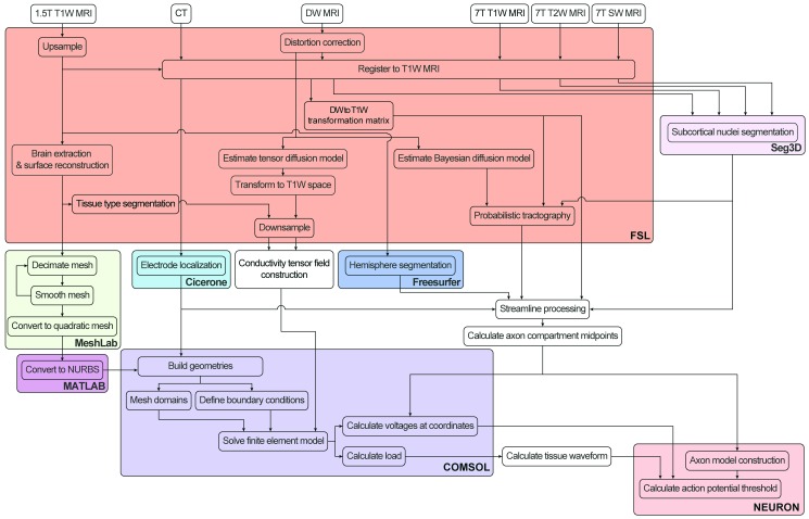
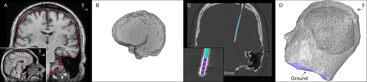
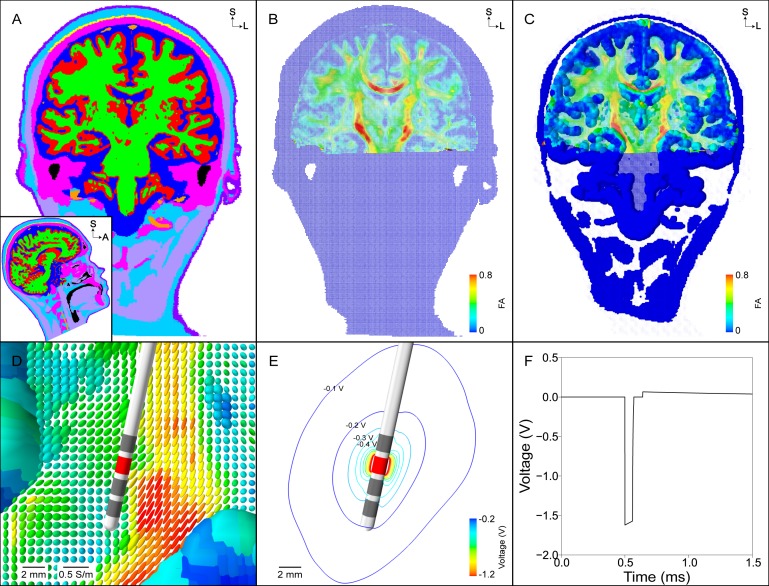
Methods: Integration of multiple software tools (e.g. COMSOL, MATLAB, FSL, NEURON, Python) enables the creation and visualization of a DBS PAM. An example DBS PAM was developed using 7T magnetic resonance imaging data from a single unilaterally implanted patient with Parkinson's disease (PD). This detailed description implements our best computational practices and most elaborate parameterization steps, as defined from over a decade of technical evolution.

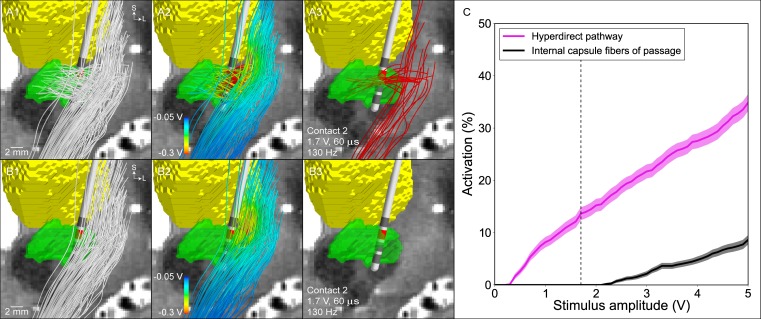
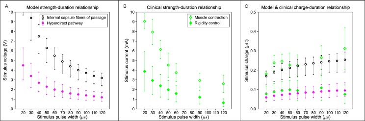
Results: Pathway recruitment curves and strength-duration relationships highlight the non-linear response of axons to changes in the DBS parameter settings.

Conclusion: Parameterization of patient-specific DBS models can be highly detailed and constrained, thereby providing confidence in the simulation predictions, but at the expense of time demanding technical implementation steps. DBS PAMs represent new tools for investigating possible correlations between brain pathway activation patterns and clinical symptom modulation.


Conflict of interest statement

Competing Interests: We have read the journal's policy and the authors of this manuscript have the following competing interests: CCM, NH, GS, and YD are shareholders in Surgical Information Sciences, Inc. CCM is a paid consultant to Boston Scientific Neuromodulation. AC is currently an employee of Medtronic Neuromodulation. This does not alter our adherence to PLOS ONE policies on sharing data and materials.

Figures

Fig 1
Fig 1. Scientific workflow for development of pathway-activation models.
Color shading corresponds to the software program used for each step. Patient images are processed and tractography is performed in FSL (red). The finite element model is constructed and solved in COMSOL (purple). The axon model is constructed and the threshold stimulus amplitude for action potential generation is solved for in NEURON (pink). We automated many of the steps using custom MATLAB, Python, NEURON, and Bash scripts.
Fig 2
Fig 2. Finite element model boundaries.
(A) The non-skull stripped 1.5T T1-weighted (T1W) image is used to extract the inner skull surface (red). (B) Inner skull surface mesh from (A) prior to any processing. (C) An oblique coronal view of the post-operative CT image, co-registered to the pre-operative T1W image, that is used to localize the four collinear electrode contacts. The inset shows the artifact of the 4 electrode contacts and a 3-dimensional rendering of the model Medtronic 3389 DBS electrode fit to the electrode artifact. (D) Domains of the finite element model, including the electrode, brain, and head. The neck region of the head surface mesh is set to 0 V under the monopolar configuration (blue).
Fig 3
Fig 3. Finite element model and DBS voltage distribution.
(A) Segmentation of the head into different tissue types (grey matter–red, white matter–green, cerebrospinal fluid–dark blue, muscle–light purple, tendon–yellow, bone–pink, fat–light blue, skin–dark purple, intervertebral disks–not visible, blood–orange, air–black). (B) Conductivity tensors within the head normalized by their volume. Anisotropic conductivity tensors are constructed within the brain using the eigenvectors of the diffusion tensors and a scalar mapping of the diffusion eigenvalues. Each tensor is colored according to its fractional anisotropy. (C) Same tensors from (B) but scaled so that the relative differences in conductivities can be visualized. (D) Zoomed view of tensors from (C) near the DBS electrode. (E) Isolines of the voltage distribution generated by a -1.7 V stimulus at contact 2. (F) The stimulus waveform at the electrode-tissue interface generated by the implantable pulse generator.
Fig 4
Fig 4. Tractography-based axon model of the hyperdirect pathway and internal capsule fibers of passage.
(A) Subcortical nuclei outlined on the T2-weighted coronal image (subthalamic nucleus [STN]–green, substantia nigra–orange, red nucleus–red, thalamus–yellow, putamen–purple, globus pallidus externus–light blue, globus pallidus internus–dark blue). (B) Tractography-generated corticofugal streamlines. Inset is a sagittal view of the resulting streamlines. (C) A smoothing spline (white) is fit to an example tractography-generated streamline (blue). (D) The hyperdirect pathway axon is comprised of a collateral that branches off of a (i) corticofugal axon at a (ii) node of Ranvier (blue spheres) and (iii) terminates in a random voxel (red) within the STN. An example population of (E) 100 internal capsule fibers of passage and (F) 100 hyperdirect pathway axons. The inset in (F) shows that each hyperdirect pathway axon is comprised of a corticofugal axon with a branching collateral that terminates within the STN, whereas the inset in (E) shows that the internal capsule fibers of passage do not have a collateral.
Fig 5
Fig 5. Model predictions for the activation of the hyperdirect pathway and internal capsule fibers of passage.
Representative population of (A1) 100 hyperdirect pathway axons and (B1) 100 internal capsule fibers of passage (subthalamic nucleus–green, thalamus–yellow). (A2), (B2) The voltage distribution generated by -1.7 V applied at contact 2 is interpolated along the streamlines. (A3), (B3) The voltage distribution is used to stimulate the axon models, and those axons that are activated by the clinically effective stimulation setting (-1.7 V, 60 μs, 130 Hz) are shown in red. (C) Percent activation of each pathway as a function of the stimulation amplitude (contact 2 [cathode], IPG case [anode], 60 μs, 130 Hz). The dashed vertical line is the clinically effective stimulation amplitude.
Fig 6
Fig 6. Model and clinical strength-duration and charge-duration curves.
(A) Model threshold amplitudes for activation of the hyperdirect pathway (pink filled circle) and internal capsule fibers of passage (black open circle) at 15 ± 5% and 10 ± 5%, respectively. (B) Clinically-measured threshold amplitudes for DBS-induced rigidity control (green filled diamond) and muscle contractions (green open diamond) [42]. (C) Total charge injected during the cathodic phase of the stimulus for the threshold amplitudes shown in A and B.

References

    1. Lozano AM, Lipsman N. Probing and regulating dysfunctional circuits using deep brain stimulation. Neuron. 2013. February 6;77(3):406–24. doi: 10.1016/j.neuron.2013.01.020 - DOI - PubMed
    1. Welter ML, Schüpbach M, Czernecki V, Karachi C, Fernandez-Vidal S, Golmard JL, et al. Optimal target localization for subthalamic stimulation in patients with Parkinson disease. Neurology. 2014. April 15;82(15):1352–61. doi: 10.1212/WNL.0000000000000315 - DOI - PMC - PubMed
    1. Eisenstein SA, Koller JM, Black KD, Campbell MC, Lugar HM, Ushe M, et al. Functional anatomy of subthalamic nucleus stimulation in Parkinson disease. Ann Neurol. 2014. August;76(2):279–95. doi: 10.1002/ana.24204 - DOI - PMC - PubMed
    1. Riva-Posse P, Choi KS, Holtzheimer PE, McIntyre CC, Gross RE, Chaturvedi A, et al. Defining critical white matter pathways mediating successful subcallosal cingulate deep brain stimulation for treatment-resistant depression. Biol Psychiatry. 2014. December 15;76(12):963–9. doi: 10.1016/j.biopsych.2014.03.029 - DOI - PMC - PubMed
    1. Ranck JB Jr. Which elements are excited in electrical stimulation of mammalian central nervous system: a review. Brain Res. 1975. November 21;98(3):417–40. - PubMed