

The effect of parental factors in children with large cup-to-disc ratios
- PMID: 28441443
- PMCID: PMC5404865
- DOI: 10.1371/journal.pone.0175900
The effect of parental factors in children with large cup-to-disc ratios
Abstract
Background: To investigate large cup-to-disc ratios (CDR) in children and to determine the relationship between parental CDR and clinical characteristics associated with glaucoma.
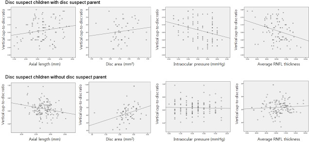
Methods: Two hundred thirty six children aged 6 to 12 years with CDR ≥ 0.6 were enrolled in this study. Subjects were classified into two groups based on parental CDR: disc suspect children with disc suspect (CDR ≥0.6) parents and disc suspect children without disc suspect parents. Ocular variables were compared between the two groups.
Results: Of the 236 disc suspect children, 100 (42.4%) had at least one disc suspect parent. Intraocular pressure (IOP) was higher in disc suspect children with disc suspect parents (16.52±2.66 mmHg) than in disc suspect children without disc suspect parents (14.38±2.30 mmHg, p = 0.023). In the group with disc suspect parents, vertical CDR significantly correlated with IOP (R = -0.325, p = 0.001), average retinal nerve fiber layer (RNFL) thickness (R = -0.319, p = 0.001), rim area (R = -0.740, p = 0.001), and cup volume (R = 0.499, p = 0.001). However, spherical equivalent (R = 0.333, p = 0.001), AL (R = -0.223, p = 0.009), and disc area (R = 0.325, p = 0.001) significantly correlated with vertical CDR in disc suspect children without disc suspect parents, in contrast to those with disc suspect parents. Larger vertical CDR was associated with the presence of disc suspect parents (p = 0.001), larger disc area (p = 0.001), thinner rim area (p = 0.001), larger average CDR (p = 0.001), and larger cup volume (p = 0.021).
Conclusions and relevance: Family history of large CDR was a significant factor associated with large vertical CDR in children. In children with disc suspect parents, there were significant correlations between IOP and average RNFL thickness and vertical CDR.
Conflict of interest statement
Figures
Similar articles
-
Lamina Cribrosa Depth is Associated With the Cup-to-Disc Ratio in Eyes With Large Optic Disc Cupping and Cup-to-Disc Ratio Asymmetry.J Glaucoma. 2016 May;25(5):e536-45. doi: 10.1097/IJG.0000000000000387. J Glaucoma. 2016. PMID: 26859358
-
Change of ocular parameters in children with large cup-to-disc ratio and interocular cup-to-disc ratio asymmetry.Graefes Arch Clin Exp Ophthalmol. 2021 Nov;259(11):3453-3459. doi: 10.1007/s00417-021-05274-1. Epub 2021 Jun 18. Graefes Arch Clin Exp Ophthalmol. 2021. PMID: 34142187
-
Cup-to-disc ratio, intraocular pressure, and primary open-angle glaucoma in retinal venous occlusion.Ophthalmology. 2002 Feb;109(2):282-6. doi: 10.1016/s0161-6420(01)00922-8. Ophthalmology. 2002. PMID: 11825809
-
The Relationship Between Optic Nerve Cup-to-Disc Ratio and Retinal Nerve Fiber Layer Thickness in Suspected Pediatric Glaucoma.J Pediatr Ophthalmol Strabismus. 2020 Mar 1;57(2):90-96. doi: 10.3928/01913913-20200117-02. J Pediatr Ophthalmol Strabismus. 2020. PMID: 32203592
-
The Heidelberg retina tomograph ancillary study to the European glaucoma prevention study: study design and baseline factors.Acta Ophthalmol. 2013 Dec;91(8):e612-9. doi: 10.1111/aos.12180. Epub 2013 May 25. Acta Ophthalmol. 2013. PMID: 23710686 Clinical Trial.
Cited by
-
A Case Report on Premature Twins: Primary Congenital Glaucoma or Large Cupping Disks Mimicking Primary Congenital Glaucoma?Cureus. 2021 Aug 11;13(8):e17108. doi: 10.7759/cureus.17108. eCollection 2021 Aug. Cureus. 2021. PMID: 34527493 Free PMC article.
-
Prevalence of fundus changes in healthy school-aged children and adolescents aged 5-19 years in Beijing.Eye (Lond). 2025 Jun;39(8):1624-1630. doi: 10.1038/s41433-025-03711-7. Epub 2025 Feb 26. Eye (Lond). 2025. PMID: 40011737
-
Optic disc parameters and associations with early life exposures in over 3000 12-year-old children: findings from the ALSPAC cohort.Eye (Lond). 2025 Jun;39(8):1592-1598. doi: 10.1038/s41433-025-03716-2. Epub 2025 Feb 22. Eye (Lond). 2025. PMID: 39987338 Free PMC article.
References
-
- Siatkowski RM, Good WV, Summers CG, Quinn GE, Tung B. Clinical characteristics of children with severe visual impairment but favorable retinal structural outcomes from the Early Treatment for Retinopathy of Prematurity (ETROP) study. Journal of AAPOS: the official publication of the American Association for Pediatric Ophthalmology and Strabismus / American Association for Pediatric Ophthalmology and Strabismus. 2013;17(2):129–34. Epub 2013/03/26. PubMed Central PMCID: PMCPMC4381920. - PMC - PubMed
-
- Flores-Rodriguez P, Gili P, Martin-Rios MD. Ophthalmic features of optic disc drusen. Ophthalmologica Journal international d'ophtalmologie International journal of ophthalmology Zeitschrift fur Augenheilkunde. 2012;228(1):59–66. Epub 2012/05/16. doi: 10.1159/000337842 - DOI - PubMed
-
- O'Gallagher MK, McLoone EM. Optic disc morphology in porencephaly. European journal of ophthalmology. 2012;22(5):840–2. Epub 2012/03/20. doi: 10.5301/ejo.5000142 - DOI - PubMed
-
- Jacobson L, Hellstrom A, Flodmark O. Large cups in normal-sized optic discs: a variant of optic nerve hypoplasia in children with periventricular leukomalacia. Archives of ophthalmology (Chicago, Ill: 1960). 1997;115(10):1263–9. Epub 1997/10/24. - PubMed
MeSH terms
LinkOut - more resources
Full Text Sources
Other Literature Sources
Medical
