. 2017 Apr 25;12(4):e0176002.
doi: 10.1371/journal.pone.0176002. eCollection 2017.

Crucial roles of Pox neuro in the developing ellipsoid body and antennal lobes of the Drosophila brain

Affiliations

Crucial roles of Pox neuro in the developing ellipsoid body and antennal lobes of the Drosophila brain

Shilpi Minocha et al. PLoS One. .

Abstract

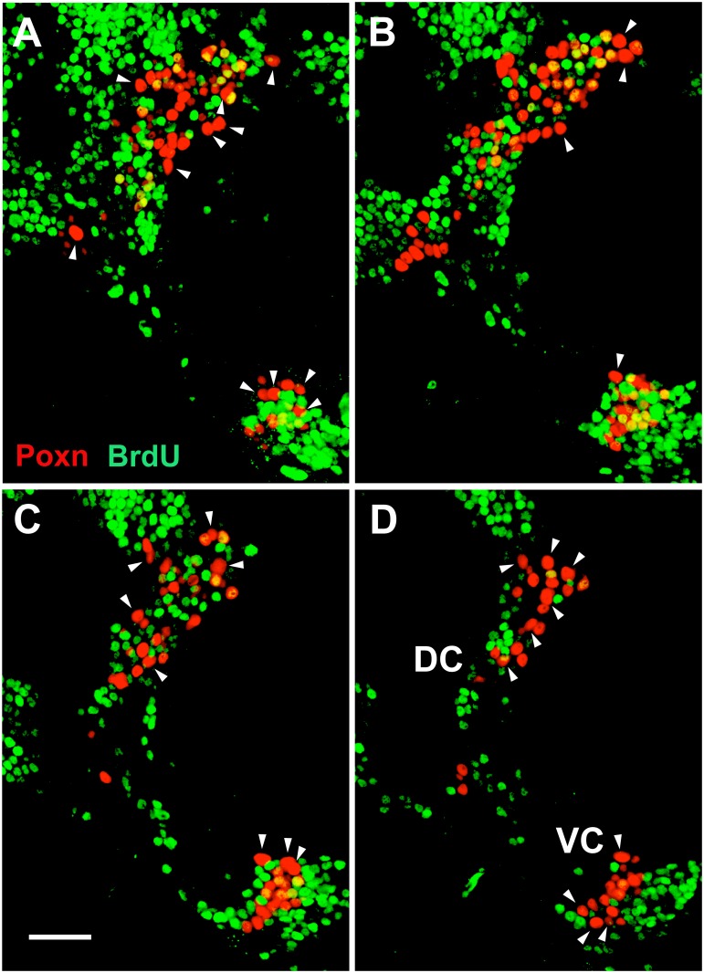
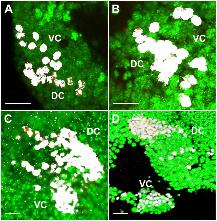
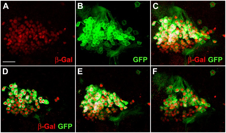
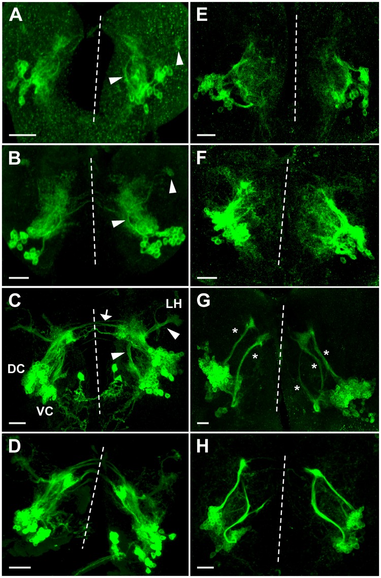
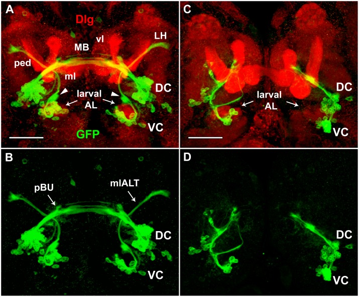
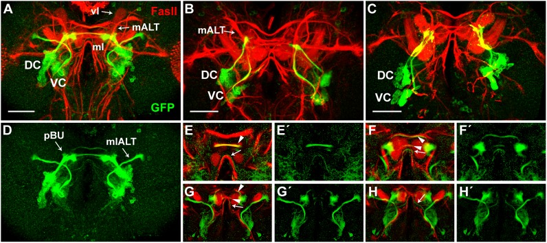
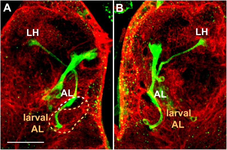
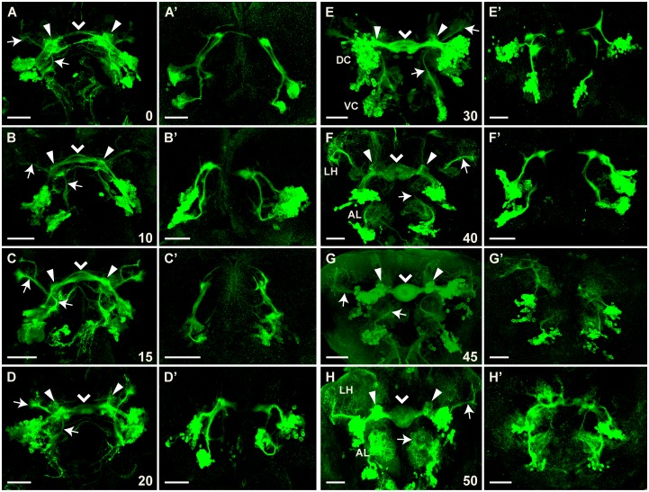
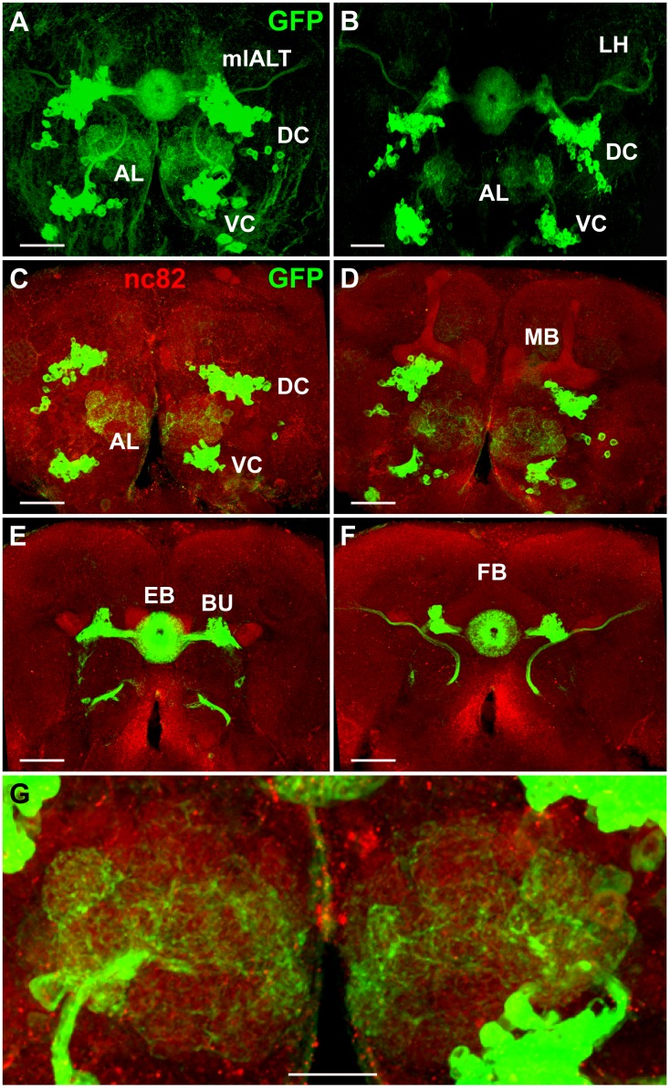
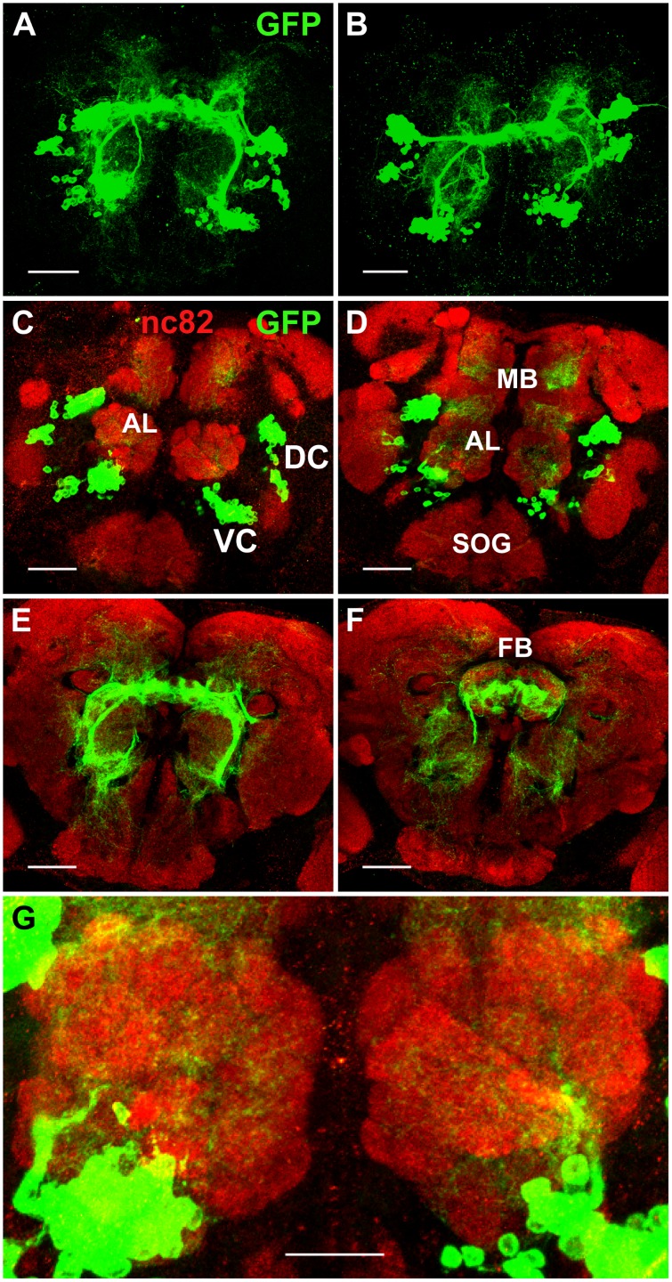
The paired box gene Pox neuro (Poxn) is expressed in two bilaterally symmetric neuronal clusters of the developing adult Drosophila brain, a protocerebral dorsal cluster (DC) and a deutocerebral ventral cluster (VC). We show that all cells that express Poxn in the developing brain are postmitotic neurons. During embryogenesis, the DC and VC consist of only 20 and 12 neurons that express Poxn, designated embryonic Poxn-neurons. The number of Poxn-neurons increases only during the third larval instar, when the DC and VC increase dramatically to about 242 and 109 Poxn-neurons, respectively, virtually all of which survive to the adult stage, while no new Poxn-neurons are added during metamorphosis. Although the vast majority of Poxn-neurons express Poxn only during third instar, about half of them are born by the end of embryogenesis, as demonstrated by the absence of BrdU incorporation during larval stages. At late third instar, embryonic Poxn-neurons, which begin to express Poxn during embryogenesis, can be easily distinguished from embryonic-born and larval-born Poxn-neurons, which begin to express Poxn only during third instar, (i) by the absence of Pros, (ii) their overt differentiation of axons and neurites, and (iii) the strikingly larger diameter of their cell bodies still apparent in the adult brain. The embryonic Poxn-neurons are primary neurons that lay out the pioneering tracts for the secondary Poxn-neurons, which differentiate projections and axons that follow those of the primary neurons during metamorphosis. The DC and the VC participate only in two neuropils of the adult brain. The DC forms most, if not all, of the neurons that connect the bulb (lateral triangle) with the ellipsoid body, a prominent neuropil of the central complex, while the VC forms most of the ventral projection neurons of the antennal lobe, which connect it ipsilaterally to the lateral horn, bypassing the mushroom bodies. In addition, Poxn-neurons of the VC are ventral local interneurons of the antennal lobe. In the absence of Poxn protein in the developing brain, embryonic Poxn-neurons stall their projections and cannot find their proper target neuropils, the bulb and ellipsoid body in the case of the DC, or the antennal lobe and lateral horn in the case of the VC, whereby the absence of the ellipsoid body neuropil is particularly striking. Poxn is thus crucial for pathfinding both in the DC and VC. Additional implications of our results are discussed.


Conflict of interest statement

Competing Interests: The authors have declared that no competing interests exist.

Figures

Fig 1
Fig 1. At least half of the Poxn-expressing cells in the adult brain completed their last S-phase during embryogenesis.
BrdU fed throughout larval development and incorporated into DNA was analyzed in an adult Ore-R brain in CLSM sections at 63x magnification. Poxn-neurons of one brain hemisphere, stained for Poxn (red) and incorporated BrdU (green), are shown in 1 μm sections at 4–5 μm (A), 7–8 μm (B), 10–11 μm (C), and 13–14 μm (D) of a Z-stack extending from 0 (anterior) to 30 μm (posterior). Colocalization of BrdU with Poxn was analyzed in each Poxn-labeled nucleus by careful inspection of single confocal layers. Arrowheads point at the 20 and 12 largest Poxn-nuclei of the DC and VC, respectively, all of which are free of BrdU, and thus are thought to correspond to the 20 (DC) and 12 (VC) Poxn-neurons that express Poxn during embryogenesis and lack Pros in late third instar larval brains (S5, S7A and S7B Figs, Table 1). Note that some of these Poxn-nuclei appear smaller than other BrdU-negative Poxn-nuclei in the same section but are actually larger, as evident from adjacent sections (not shown). Scale bar: 20 μm.
Fig 2
Fig 2. Cells expressing Poxn in larval and adult brains are post-mitotic neurons.
(A–D) Colocalization (white) in nuclei of Poxn (red) and Elav (green) proteins, visualized by immunofluorescent staining, is shown in wild-type (Ore-R) brain hemispheres of first (A), second (B), and late third instar larvae (C), and of adults (D). Staining of all Poxn-nuclei with Elav was corroborated by visual inspection in single layers of the Z-stacks. Panels show maximum intensity projections of CLSM sections of Z-stacks extending over 43 μm (A), 45 μm (B), and 50 μm (C) at 63x magnification, and of a Z-stack extending over 26 μm at 40x magnification (D). Scale bars: 10 μm (A–C) and 20 μm (D).
Fig 3
Fig 3. Most Poxn-neurons survive metamorphosis and continue to express Poxn in the adult brain.
(A–C) The DC of Poxn-neurons, visualized by immunofluorescent staining for β-Gal (red) and GFP (green), is shown in the brain of a w1118 UAS-Flp/+; tub-Gal80ts/+; Act5C>polyA>lacZ.nls1 Poxn-Gal4-13-1/Poxn-CD8::GFP female. A maximum intensity projection of the same CLSM sections of an entire Z-stack is shown in the red channel (A), green channel (B), and in both channels (C) at 63x magnification. β-Gal-positive nuclei (red) belong to cells that expressed Poxn at the time of heat shock during the third larval instar (feeding stage). The membrane-associated CD8::GFP fusion protein labels Poxn-expressing cells at the time of fixation. (DF) Maximum intensity projections of substacks at 0–5 μm (D), 5–10 μm (E), and 10–18 μm (F) of the Z-stack extending from 0 (anterior) to 31 μm (posterior) shown in (C), which includes all Poxn-nuclei. Virtually all β- Gal-labeled neurons also express GFP. Similar results were obtained for the VC of Poxn-neurons (data not shown). Scale bar: 20 μm.
Fig 4
Fig 4. Projection patterns of Poxn-neurons in wild type and Poxn mutants during larval development.
(A–H) Poxn-neurons are visualized by the expression of Poxn-CD8::GFP and immunofluorescent staining for GFP in first (A,E), late second (B,F), and late third instar (C,D,G,H) brains of w1118; Poxn-CD8::GFP (A-C), w1118; PoxnΔM22-B5; Poxn-CD8::GFP (E-G), w1118; PoxnΔM22-B5 Poxn-SuperA-158; Poxn-CD8::GFP (D), and w1118; PoxnΔM22-B5 Poxn-Sbl-107; Poxn-CD8::GFP (H) larvae. Note that the projection patterns of the latter two resemble those in wild-type (C) and Poxn mutant brains (G), respectively. Arrowheads in (A-C) point to arc-like projections of the VC and their targets in the lateral protocerebrum, and arrow in (C) points to tracts of the SEC, emanating from the DC of wild-type brains. Asterisks in (G) mark aberrant projections from both Poxn clusters in a Poxn mutant brain. Dashed lines indicate midlines of the flattened brains viewed along the anteroposterior axis. Panels show maximum intensity projections of CLSM sections of Z-stacks extending over 41 μm (A), 50 μm (B), 53 μm (C), 49 μm (D), 75 μm (E), 36 μm (F), 48 μm (G), and 77 μm (H) at 20x magnification. Panel H shows the same brain as Fig 6B. DC, dorsal cluster of Poxn-neurons; LH, lateral horn; VC, ventral cluster of Poxn-neurons. Scale bars: 20 μm.
Fig 5
Fig 5. Projections of Poxn-neurons do not express Dlg in wild-type and Poxn mutant brains of late third instar larvae.
Poxn-neurons, immunostained for the expression of Poxn-CD8::GFP (green), and neuropils, immunostained for the expression of Dlg (red), are shown in brains of w1118; Poxn-CD8::GFP (A,B) and w1118; PoxnΔM22-B5 Poxn-Sbl-107; Poxn-CD8::GFP (C,D) late third instar larvae, in the red and green channel (A,C) and only in the green channel (B,D) at 40x magnification. To improve the visibility of the axon tracts and neuropils, especially the larval ALs, substacks extending from 26 μm (anterior) to 72 μm (posterior) (A,B) and from 18 μm (anterior) to 67 μm (posterior) (C,D) of Z-stacks, extending over 87 μm, are shown as maximum intensity projections of CLSM sections. Although these substacks exclude many cell bodies of the Poxn-neurons visible in the excluded substacks, they include all neurite projections. The mlALT originating from the VC crosses behind the commissural tracts emanating from the DC. Arrowheads in (A) point at ‘swellings’ of the mlALT preceding dendrite formation in the region of the future AL. Screening through single stacks shows no colocalization of GFP and Dlg in (A) and (C). DC and VC, dorsal and ventral cluster of Poxn-neurons; larval AL, larval antennal lobe; LH, lateral horn; mlALT, mediolateral antennal lobe tract; MB, larval mushroom bodies; vl, vertical lobe of MB; ml, medial lobe of MB; ped, pedunculus of MB; pBU, primordial bulb. Scale bars: 50 μm.
Fig 6
Fig 6. Projections of Poxn-neurons and axonal tracts labeled by FasII in wild-type and Poxn mutant brains of late third instar larvae.
(A-D) Poxn-neurons, immunostained for the expression of Poxn-CD8::GFP (green), and neuropils, immunostained for the expression of FasII (red), are shown in brains of w1118; Poxn-CD8::GFP (A,D) and w1118; PoxnΔM22-B5 Poxn-Sbl-107; Poxn-CD8::GFP (B,C) late third instar larvae in the red and green channel (A-C) and only in the green channel (D) at 40x magnification. The two Poxn mutant brains display the two types of mutant projection patterns commonly observed: projections from the VC seem to follow the mALT instead of the mlALT (B), or adopt an entirely different path, eventually running parallel to the projections from the DC before they stall (C). Scale bars: 50 μm. (E-H') Central parts of substacks of (A) are shown from posterior to anterior at 10–15 μm (E,E'), 23–28 μm (F,F'), 28–30 μm (G,G'), and 30–35 μm (H,H') as maximum intensity projections of CLSM sections of a Z-stack extending over 85 μm in both channels (E-H) and only in the green channel (E'-H') at 40x magnification. Poxn-neuron tracts that pass through the SEC and do or do not co-express FasII are indicated by arrowheads and arrows, respectively. DC, dorsal cluster of Poxn-neurons; vl, vertical lobe of mushroom bodies; mALT, middle antennal lobe tract; mlALT, mediolateral antennal lobe tract; ml, medial lobe of mushroom bodies; pBU, primordial bulb; VC, ventral cluster of Poxn-neurons.
Fig 7
Fig 7. Projection pattern of Poxn-neurons with respect to glia in late third instar larval brain.
(A,B) Left (A) and right (B) hemispheres of w1118; UAS-CD2; repo-Gal4/Poxn-CD8::GFP late third instar larval brain, immunostained for GFP (green), labeling Poxn-neurons, and CD2 (red) in glial membranes [60]. The axons of the Poxn-neurons project along the lateral region of the larval AL (surrounded by dashed orange line in A), exhibit a striking ‘swelling’ at the developing adult AL (surrounded by dashed white line) but no arborization, and follow the mlALT to target the LH. No arborization is detected at the larval AL. To optimize the visibility of the axon tracts of the Poxn-neurons from the VC along the larval AL and developing adult AL to the LH, substacks extending from 30 μm (anterior) to 56 μm (posterior) (A) and from 48 μm (anterior) to 76 μm (posterior) (B) of a Z-stack, extending over 85 μm, are shown as maximum intensity projections of CLSM sections at 40x magnification. Although these substacks exclude the cell bodies of the Poxn-neurons, they include all neurite projections. Scale bar: 50 μm.
Fig 8
Fig 8. Formation of ellipsoid body depends strongly on Poxn function.
Brains of w1118; Poxn-CD8::GFP (A-H) or w1118; PoxnΔM22-B5 Poxn-Sbl-107; Poxn-CD8::GFP (A’-H’) pupae, immunostained for GFP at 0 h APF (A,A’), 10 h APF (B,B’), 15 h APF (C,C’), 20 h APF (D,D’), 30 h APF (E, E’), 40 h APF (F,F’), 45 h APF (G,G’), and 50 h APF (H,H’), are shown as maximum intensity projections of confocal Z-stacks at 20x magnification (panels except E, G, and G’ show substacks that remove some of the cell bodies but improve the visibility of projections). Z-stacks extended over 89 μm (A), 67 μm (B), 82 μm (C), 52 μm (D), 72 μm (E), 54 μm (F), 79 μm (G), 51 μm (H), 44 μm (A’), 65 μm (B’), 90 μm (C’), 88 μm (D’), 56 μm (E’), 46 μm (F’), 102 μm (G’), and 66 μm (H’). Arrows point to the bilateral arc-like mlALTs visible at all pupal stages. Filled arrowheads point to the arborizations at the developing bulbs, while arrowheads point to the midline of the forming ellipsoid body neuropil. Note that arborizations at the lateral horn (LH) and antennal lobe (AL) become prominent around 40 h APF in the wild-type brain (F-H), while they are slightly delayed in mutant brains where they occur at the end of the stalled projections (G’,H’). Pupal stages, measured as hours APF, imply a developmental temporal variation corresponding to about 5%. Scale bars: 50 μm.
Fig 9
Fig 9. Projection pattern of Poxn-neurons in adult brains.
(A,B) Projections of Poxn-neurons in w1118; Poxn-CD8::GFP (A) and w1118; PoxnΔM22-B5 Poxn-SuperA; Poxn-CD8::GFP (B) adult brains (dorsal side up), immunostained for GFP, are shown as maximum intensity projections of Z-stacks, extending over 76 μm at 40x magnification (A) and extending over 75 μm at 20x magnification (B). Projection patterns in the brain of Poxn mutants rescued by the complete Poxn transgene Poxn-SuperA (B) are similar to those in the wild-type brain (A). (CF) Substacks of (A), immunostained for GFP (green) and nc82 (red), are shown from anterior to posterior at 6–14 μm (C), 14–25 μm (D), 34–43 μm (E), and 43–66 μm (F). Note that what may look like cell bodies at the BU in (A,B,E,F) are the bush-like postsynaptic arborizations of the Poxn-R neurons at the glomeruli of the ipsilateral BU [10,14,61]. (G) Enlarged region of substacks of (A) at 6–35 μm, illustrating the dendritic arborizations of Poxn-neurons invading many glomeruli of the ALs. AL, antennal lobe; DC, dorsal cluster; EB, ellipsoid body; FB, fan-shaped body; LH, lateral horn; BU, bulb; MB, mushroom bodies; mlALT, mediolateral antennal lobe tract; VC, ventral cluster. Scale bars: 50 μm (A–F) and 25 μm (G).
Fig 10
Fig 10. Projection patterns of Poxn-neurons in Poxn mutant adult brains.
(A,B) Projections of Poxn-neurons in w1118; PoxnΔM22-B5 Poxn-Sbl-107; Poxn-CD8::GFP (A) and w1118; PoxnΔM22-B5; Poxn-CD8::GFP (B) Poxn mutant brains (dorsal side up), immunostained for GFP, show similar patterns at maximum intensity projection of Z-stacks extending over 85 μm (A) and 109 μm (B) at 40x magnification. (CF) Substacks of (A), immunostained for GFP (green) and nc82 (red), are shown from anterior to posterior at 7–18 μm (C), 18–28 μm (D), 36–43 μm (E), and 43–49 μm (F). (G) Enlarged region of substacks of (A) at 0–31 μm, illustrating the reduced arborizations of Poxn-neurons that fail to invade the glomeruli of the ALs. AL, antennal lobe; DC, dorsal cluster; FB, fan-shaped body; MB, mushroom bodies; SOG, subesophageal ganglion; VC, ventral cluster. Scale bars: 50 μm (A–F) and 25 μm (G).

Similar articles

Cited by

References

    1. Truman JW (1990) Metamorphosis of the central nervous system of Drosophila. J Neurobiol 21: 1072–1084. 10.1002/neu.480210711 - DOI - PubMed
    1. Ito K, Hotta Y (1992) Proliferation pattern of postembryonic neuroblasts in the brain of Drosophila melanogaster. Dev Biol 149: 134–148. - PubMed
    1. Levine RB, Morton DB, Restifo LL (1995) Remodeling of the insect nervous system. Curr Opin Neurobiol 5: 28–35. - PubMed
    1. Consoulas C, Duch C, Bayline RJ, Levine RB (2000) Behavioral transformations during metamorphosis: Remodeling of neural and motor systems. Brain Res Bull 53: 571–583. - PubMed
    1. Consoulas C, Restifo LL, Levine RB (2002) Dendritic remodeling and growth of motoneurons during metamorphosis of Drosophila melanogaster. J Neurosci 22: 4906–4917. - PMC - PubMed

MeSH terms

Substances

LinkOut - more resources