. 2017 May 23;56(20):2612-2626.
doi: 10.1021/acs.biochem.7b00283. Epub 2017 May 9.

Caenorhabditis elegans PRMT-7 and PRMT-9 Are Evolutionarily Conserved Protein Arginine Methyltransferases with Distinct Substrate Specificities

Affiliations

Caenorhabditis elegans PRMT-7 and PRMT-9 Are Evolutionarily Conserved Protein Arginine Methyltransferases with Distinct Substrate Specificities

Andrea Hadjikyriacou et al. Biochemistry. .

Abstract

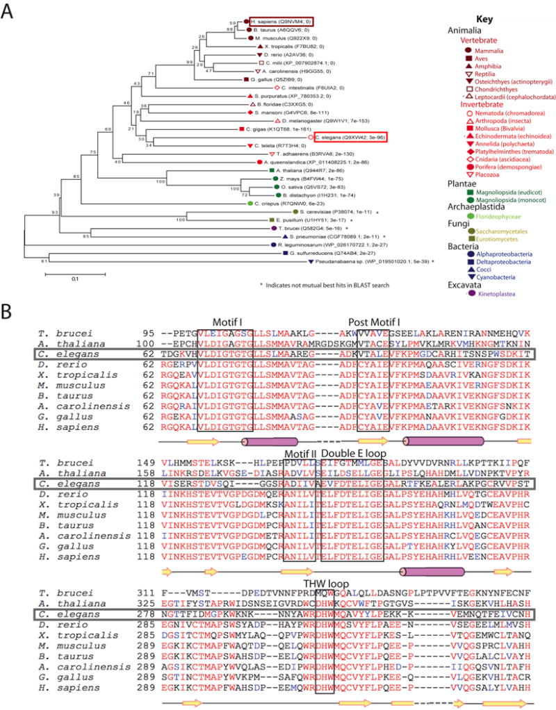
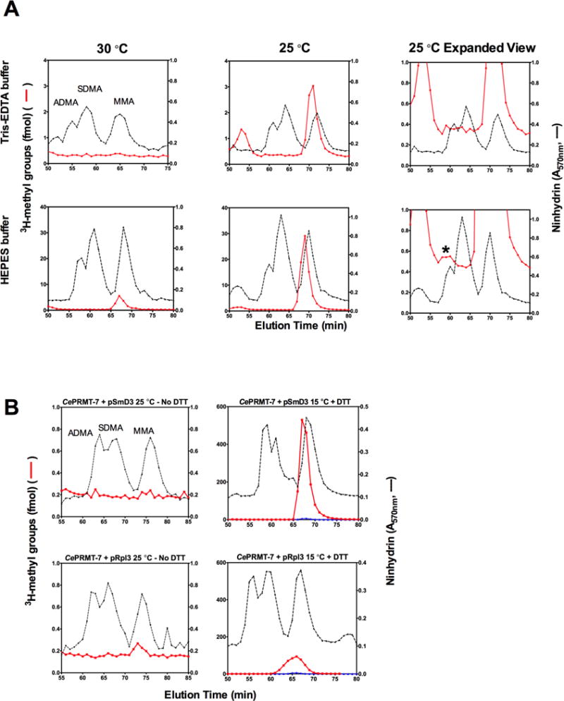
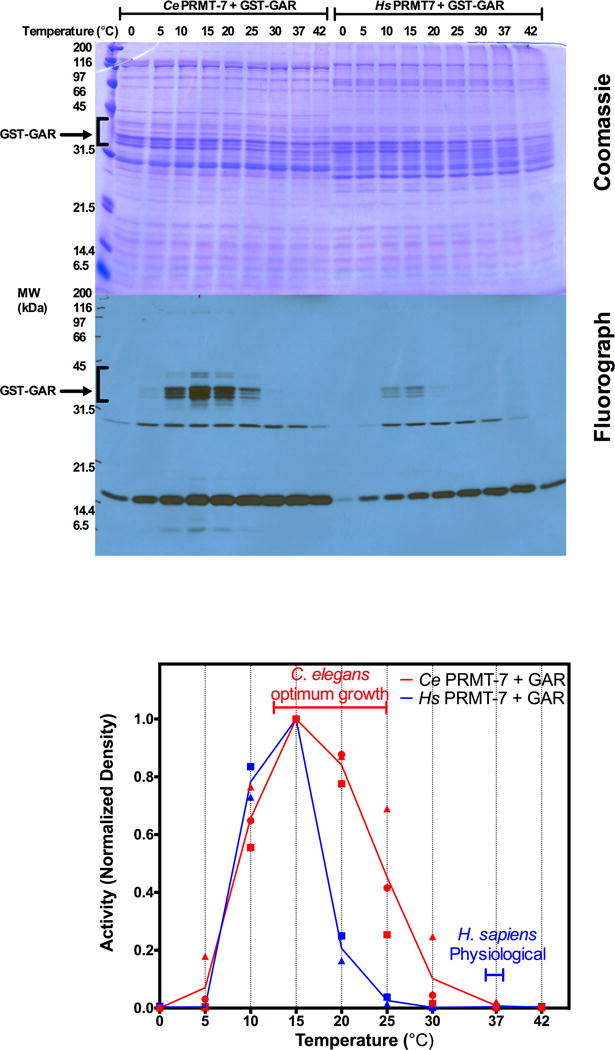
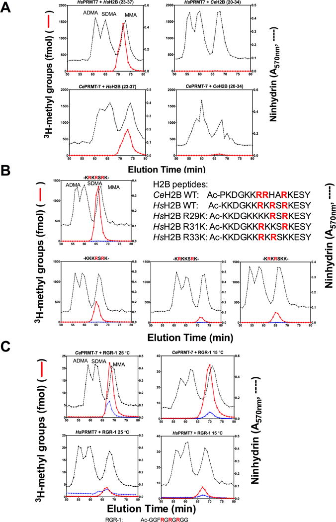
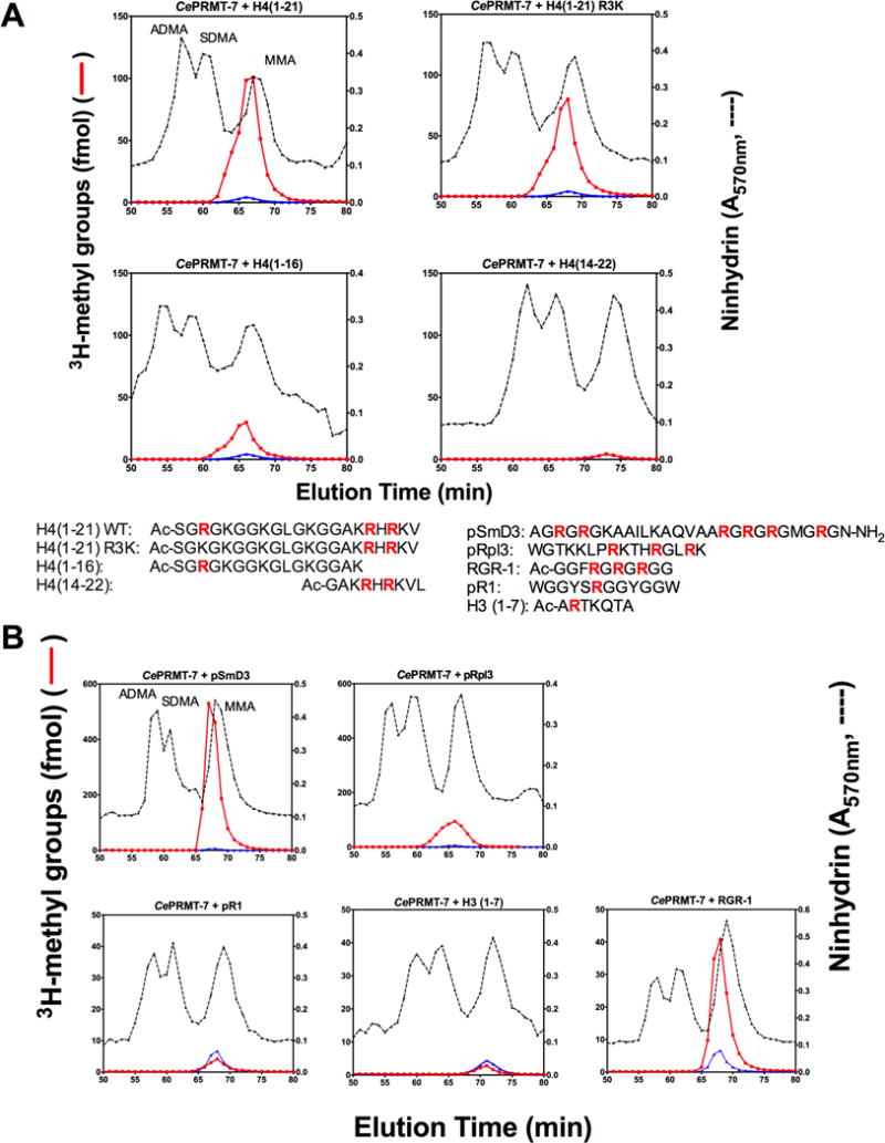
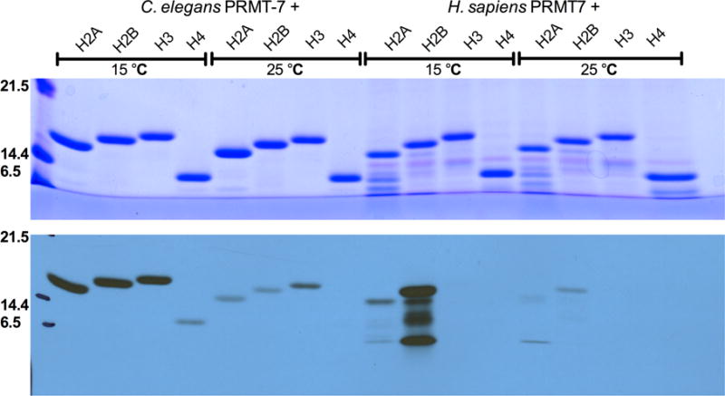
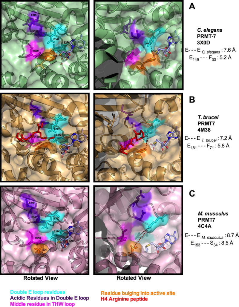
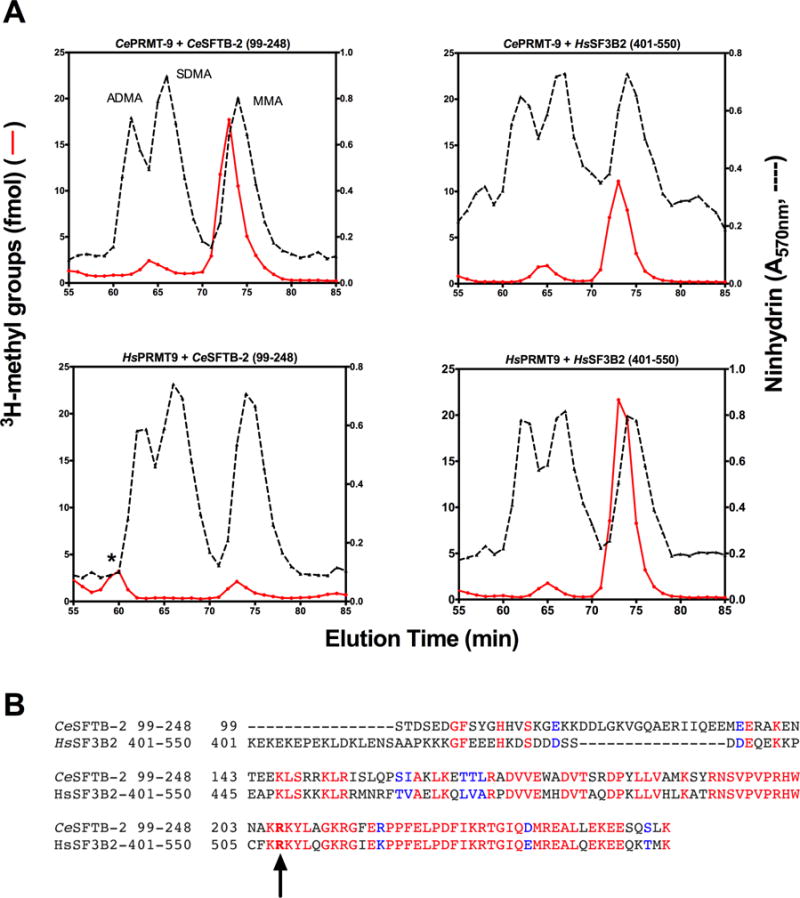
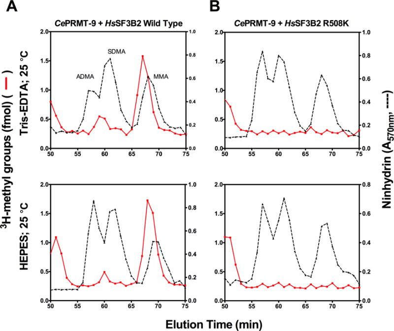
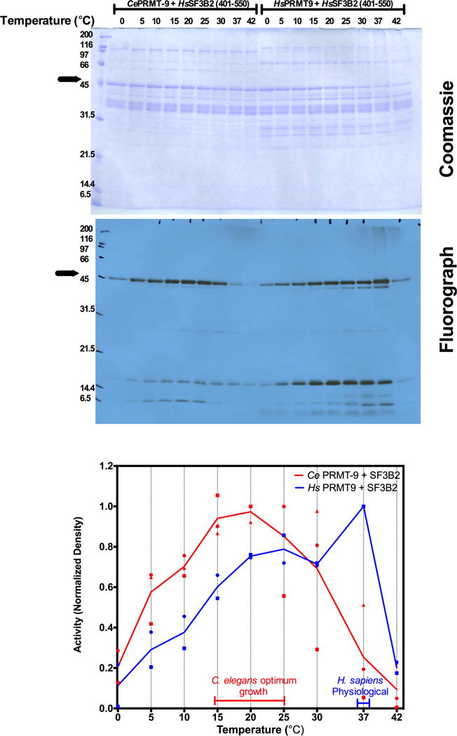
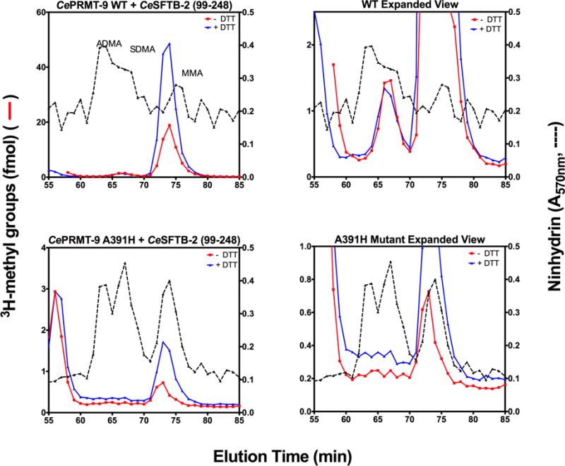
Caenorhabditis elegans protein arginine methyltransferases PRMT-7 and PRMT-9 are two evolutionarily conserved enzymes, with distinct orthologs in plants, invertebrates, and vertebrates. Biochemical characterization of these two enzymes reveals that they share much in common with their mammalian orthologs. C. elegans PRMT-7 produces only monomethylarginine (MMA) and preferentially methylates R-X-R motifs in a broad collection of substrates, including human histone peptides and RG-rich peptides. In addition, the activity of the PRMT-7 enzyme is dependent on temperature, the presence of metal ions, and the reducing agent dithiothreitol. C. elegans PRMT-7 has a substrate specificity and a substrate preference different from those of mammalian PRMT7, and the available X-ray crystal structures of the PRMT7 orthologs show differences in active site architecture. C. elegans PRMT-9, on the other hand, produces symmetric dimethylarginine and MMA on SFTB-2, the conserved C. elegans ortholog of human RNA splicing factor SF3B2, indicating a possible role in the regulation of nematode splicing. In contrast to PRMT-7, C. elegans PRMT-9 appears to be biochemically indistinguishable from its human ortholog.


Conflict of interest statement

Notes

The authors declare that they have no conflicts of interest with the contents of this article.

Figures

Figure 1
Figure 1. Evolutionary conservation of PRMT7 across the various kingdoms of life
A. Phylogenetic tree based on human PRMT7. UniProt IDs of representative orthologs from Animalia, Plantae, Archaeplastida, Fungi, Bacteria, and Excavata are shown with their E values based on a protein BLAST search against the human species. The phylogenetic tree was constructed after amino acid sequences were aligned using MUSCLE in MEGA6 software as described in. Each ortholog sequence was then subjected to protein BLAST against the human protein database. All proteins were mutual best hits with the exception of those species marked with an asterisk. The number shown next to each branch is the percent of replicate trees with the same clustering in 500 bootstrap test replicates. The scale bar indicates the fraction of amino acid differences for each entry. H. sapiens PRMT7 is boxed in dark red, and C. elegans PRMT-7 is boxed in red. B. Partial sequence alignment of the major motifs in PRMT7 orthologs across vertebrates, invertebrates, plants, and excavata (T. brucei) using the EMBL-EBI Clustal Omega Multiple Sequence Alignment software. The major distinguishing motifs of protein arginine methyltransferases are boxed in black, including Motif I, Post Motif I, Motif II, the Double E loop, and the THW loop. Red letters indicate identity and blue letters indicate similar amino acid properties. Secondary structure is indicated on the bottom with beta strands in yellow and alpha-helices in magenta based on the structure of the M. musculus PRMT7 (PDB: 4C4A,). The C. elegans PRMT-7 sequence is boxed in dark gray.
Figure 2
Figure 2. C. elegans PRMT-7 produces MMA and its activity is temperature and metal ion dependent
A. Amino acid analysis of3H-methylation reactions of C. elegans GST-PRMT-7 (2 μg) with GST-GAR (5 μg) after a 20 h reaction at 30 °C (left column) or 25 °C (middle and right columns) in either Tris-EDTA buffer (100 mM Tris-HCl, 100 mM NaCl, 2 mM EDTA, pH 8.0) (top row), or 50 mM potassium HEPES, 10 mM NaCl, pH 8.2 (bottom row) as indicated in the “Experimental Procedures” section. The final concentration of DTT in these reactions (from the GST-GAR preparation) was 0.16 mM. After trichloroacetic acid precipitation and acid hydrolysis, 1 μmol of each non-radiolabeled methylated arginine standard (ADMA, SDMA and MMA) was added to the hydrolyzed pellet and amino acid analysis was performed as in the “Experimental Procedures” section. Dashed black lines indicate ninhydrin absorbance of the methylarginine standards with the peaks of ADMA, SDMA, and MMA identified using 50 μl of each fraction. Red lines indicate the elution of the radiolabeled methylated amino acids from the hydrolysates of the reactions. Radioactivity from 950 μl aliquots of each fraction is given as the average of three 5-min counting cycles using liquid scintillation counting. Radioactive methylated amino acids elute approximately 1 min before the non-radiolabeled standard due to the tritium isotope effect. The right panels show an expanded view of the data from the middle columns at 25 °C to demonstrate that the enzyme only produces MMA under both buffer conditions. These reactions were replicated three independent times. The asterisk indicates the peak eluting prior to the position expected for3H-ADMA; this peak is not consistently observed in the replicates. B. PRMT-7 enzyme activity is dependent on presence of DTT. Enzyme was reacted with 12.5 μM pSmD3 (top) or pRpl3 (bottom) peptides at 25 °C in 50 mM potassium HEPES, 10 mM NaCl, pH 8.2 (left column), or at 15 °C in the same buffer with the addition of 1 mM DTT. Results are presented as shown in panel A; the blue line in the right hand panels indicates radioactivity for incubations where no peptide substrate was added. These reactions were replicated twice with no DTT and once with DTT.
Figure 3
Figure 3. PRMT-7 has a temperature dependence consistent with C. elegans physiology
Methylation reactions consisting of 5 μg GST-GAR substrate and 2 μg of enzyme (C. elegans GST-PRMT-7 or H. sapiens GST-PRMT7) were incubated for 20 h at the indicated temperatures in reaction buffer containing 50 mM potassium HEPES, 10 mM NaCl, pH 8.2, with 0.7 μM [methyl-3H]AdoMet in a final reaction volume of 60 μl. The final concentration of DTT in these reactions (from the GST-GAR preparation) was 0.16 mM. Reactions were quenched by the addition of SDS sample loading buffer and polypeptides separated by 12.6% Tris-glycine SDS-PAGE followed by autoradiography as described in “Experimental Procedures.” The dried gel was then exposed to Denville E3012 autoradiography film for 21 d at −80 °C. Molecular weight positions are shown from approximately 2 μg of unstained SDS-PAGE broad range marker (BioRad, catalog no, 161-0317). This full experiment was independently replicated and a further third replicate using only the C. elegans enzyme was also done. Densitometry was done using ImageJ software on scanned images of the films and quantified as relative density and normalized to the highest peak of GST-GAR radioactivity in each individual film (C. elegans, red symbols; human, blue symbols – different symbols used to indicate each replicate experiment). Lines are drawn for the averaged normalized values. The optimal growth temperatures for C. elegans and humans are indicated. Lower bands shown on the fluorography are nonspecific and not dependent on the enzyme activity.
Figure 4
Figure 4. C. elegans PRMT-7 has a similar R-X-R substrate specificity as the human PRMT7 enzyme
A. Amino acid analysis of reactions consisting of 2 μg of human GST-PRMT7 or C. elegans PRMT-7 enzymes, reacted with 12.5 μM of synthetic human histone H2B (23–37) peptide, or a synthetic peptide containing the equivalent sequence from C. elegans histone H2B (20–34) at 15 °C as described in “Experimental Procedures,” in 50 mM potassium HEPES buffer, 10 mM NaCl, pH 8.0, containing 1 mM DTT for 20 h. Reactions were repeated three times for human histone H2B (23–37) and twice for C. elegans H2B (20–34) at this temperature and also replicated at 25 °C once. B. Amino acid analysis of reactions containing 2 μg of C. elegans GST-PRMT-7 enzyme reacted with 12.5 μM synthetic human histone H2B peptides (Wild type, top; R29K, R31K and R33K bottom row) for 20 h, using the same conditions described above in panel A. These reactions were single replicates. C. Amino acid analysis of reactions containing 2 μg of C. elegans or human PRMT-7/7 enzymes, reacted with 12.5 μM of synthetic peptide RGR-1 (a sequence containing an R-X-R sequence where X = G) at 25 °C (left) or 15 °C (right), under the same reaction conditions as described in A. Reactions were replicated once and the reaction of C. elegans with RGR-1 was replicated an additional two times.
Figure 5
Figure 5. C. elegans PRMT-7 has a preference for R-X-R motifs in various peptide substrates
A. C. elegans PRMT-7 (2 μg) enzyme was reacted with 12.5 μM various synthetic human histone H4 peptides (1–21 wild type; 1–21 R3K; 1–16 wild type; and 14–22) at 15 °C in the reaction conditions of 50 mM potassium HEPES, 10 mM NaCl, pH 8.0, with 1 mM DTT for 20 h. Amino acid analysis was done as described above and in “Experimental Procedures.” Reactions were replicated twice. B. C. elegans PRMT-7 enzyme (2 μg) was reacted with 12.5 μM of various synthetic peptides (pSmD3, pRpl3, pR1, Histone H3 (1–7) and RGR-1), at the conditions described in A. Reactions were single replicates. The reactions in the top panels for pSmD3 and pRpl3 are the same panels used in Fig. 2B right column.
Figure 6
Figure 6. C. elegans PRMT-7 has a distinct substrate specificity than the mammalian ortholog for mammalian histones
Reactions consisting of 5 μg of substrate (recombinant human histones from New England BioLabs (H2A: M2502S; H2B: M2505S; H3.3: M2507S; H4: M2504S) were reacted with 2 μg of C. elegans GST-PRMT-7 or human GST-PRMT7 at 15 °C or 25 °C as described in “Experimental Procedures”. Reactions were stopped by the addition of sample loading buffer and polypeptides separated as described in “Experimental Procedures”. The dried gel was then exposed to autoradiography film for 3 d at −80 °C. Molecular weight positions are shown from approximately 2 μg of unstained SDS-PAGE broad range marker as in Fig. 3. The results shown here were replicated three times for C. elegans PRMT-7 and two times for human PRMT7. Radiolabeled bands migrating more rapidly than histone H2B in the lane with human PRMT7 appear to be proteolytic fragments.
Figure 7
Figure 7. Active site architecture of C. elegans, trypanosome, and mammalian PRMT7s
A. Pymol surface and cartoon representation of the active site of C. elegans PRMT-7 (3X0D,). Flanking glutamate double E loop residues are highlighted in cyan (E140, E149), internal loop acidic residues in purple (D143, E145), and a THW loop residue in magenta (H300). The residue highlighted in orange (F33) protrudes from a helix to form a cavity with E149. The distance from the side chain carboxyl carbon of E140 to the side chain carboxyl carbon of E149 is 7.6 Å, and distance from the side chain carboxyl carbon of E149 to the closest side chain carbon atom in F33 is 5.2 Å. S-Adenosylhomocysteine (SAH) is shown in CPK coloring. B. Pymol surface and cartoon representation of the active site of T. brucei PRMT7 (4M38,), highlighting residues corresponding to those shown in panel A. Flanking double E loop residues E172 and E181 are highlighted in cyan, G175 and M177 (residues corresponding to D143 and E145 residues in C. elegans) are highlighted in purple, and the Q329 THW loop residue is highlighted in magenta. The residue highlighted in orange (F71) corresponds to F33 in the C. elegans structure and also protrudes from a helix to form a cavity with E181. The distances between the flanking double E residues and E181-F71 are given as in panel A. The arginine-3 residue of the co-crystallized histone H4 peptide is modeled in red. C. Pymol surface and cartoon representation of active site of M. musculus PRMT7 (4C4A,), highlighting residues corresponding to those shown in panels A and B. The distances between the flanking double E residues and E153-S34 are given as in panels A and B. We note that the S34 side chain oxygen is shown in two conformations.
Figure 8
Figure 8. C. elegans PRMT-9 symmetrically dimethylates SFTB-2, the C. elegans ortholog of the mammalian SF3B2
A. Amino acid analysis was done as in Fig. 2 on methylation reactions consisting of 5 μg of substrate (C. elegans GST-tagged SFTB-2 fragment 99–248 or H. sapiens GST-tagged SF3B2 fragment 401–550) and approximately 2 μg of enzyme (C. elegans GST-tagged PRMT-9 enzyme or H. sapiens GST-tagged PRMT9 enzyme) that were incubated for 20 h at 25 °C (nematode enzyme) or 37 °C (human enzyme) in a reaction buffer consisting of 50 mM potassium HEPES, 10 mM NaCl, pH 8.2, with a final concentration of 0.7 μM [methyl-3H]AdoMet in a volume of 60 μl. The reactions were quenched by the addition of final concentration 12.5% trichloroacetic acid and acid hydrolyzed as described in the materials and methods. The asterisk indicates a radioactive peak eluting prior to the expected position of3H-ADMA. The experiment was replicated twice. B. Sequence alignment of C. elegans SFTB-2 (UniProt ID: O16997) residues 99–248 and human SF3B2 (UniProt ID: Q13435, bottom). Red letters indicate identity and blue letters indicate similar amino acid properties. Bolded R and black arrow indicates methylation site for the enzyme.
Figure 9
Figure 9. C. elegans PRMT-9 methylates SF3B2 at the same R508 site that is methylated by the mammalian PRMT9 enzyme
A. Amino acid analysis of methylation reaction consisting of 2 μg of C. elegans GST-PRMT-9 enzyme with 5 μg of human GST-SF3B2 (401–550) wild type at 25°C for 20 h in either 100 mM Tris-EDTA buffer pH 8.0 (top panel) and 50 mM potassium HEPES, 10 mM NaCl, pH 8.2 (bottom panel). Reactions were quenched by the addition of final concentration of 12.5% trichloroacetic acid and acid hydrolyzed as described above. Reactions were duplicated twice. B. Methylation reactions of C. elegans PRMT-9 enzyme was reacted with human GST-SF3B2 (401–550) R508K mutant fragment, in the same reaction conditions described in part A at 25 °C for 20 h. Reactions were duplicated twice.
Figure 10
Figure 10. Substrate specificity of C. elegans PRMT-9
A. Methylation reactions for amino acid analysis were set up using 2 μg of C. elegans PRMT-9 with 5 μg of various substrates (GST-GAR, top left; GST-SF3B2 (401–550), top right; recombinant human histone H2A, bottom left; recombinant human histone H2B bottom right), in 100 mM Tris-EDTA buffer, pH 8.0, for 6 h at 30 °C, conditions defined in Takahashi et al.. These reactions using these conditions were single replicates. B. In vitro methylation reactions consisting of 2 μg of C. elegans PRMT-9 or human PRMT9 enzyme were reacted with 5 μg of substrate (GST-GAR, His-Rbp16, or GST-SFTB-2 (C. elegans fragment 99–248) or GST-SF3B2 (human fragment 401–550) for 20 h at 25 °C (C. elegans) or 37 °C (human) in 50 mM potassium HEPES, 10 mM NaCl, 1 mM DTT, pH 8.2 with 0.7 μM [methyl-3H]AdoMet. Sample loading buffer was added to stop the reactions and polypeptides were separated using SDS-PAGE, using the same methods described in Fig. 3. The gel was then dried and exposed to film for 7 days at −80 °C. Coomassie stained gel is shown on top, and the fluorographic exposure is shown below. The molecular weight standards are shown as described in Fig. 3. This experiment was replicated independently using amino acid analysis.
Figure 11
Figure 11. Temperature dependence of C. elegans PRMT-9
Methylation reactions consisting of 5 μg of substrate (H. sapiens GST-tagged SF3B2 fragment 401–550) were reacted with 2 μg of enzyme (both C. elegans and H. sapiens PRMT9 enzymes) at the various temperatures indicated for 20 h at the reaction conditions of 50 mM potassium HEPES, 10 mM NaCl, pH 8.2 with 0.7 μM [methyl-3H]AdoMet in a final reaction volume of 60 μl. Reactions were quenched by the addition of sample loading buffer and proteins were separated using SDS-PAGE using the same methods described in Fig. 3. The gel was dried and exposed to autoradiography film for 21 d at −80 °C. Coomassie gel (top panel) and fluorograph (lower panel) are shown. Molecular weight positions are shown from approximately 2 μg of unstained SDS-PAGE broad range marker as described in Figure 3. This experiment was replicated independently and a third replicate using the C. elegans PRMT-9 enzyme was done. The bottom panel indicates the densitometry of the bands comparing methylation activity of the C. elegans enzyme to the human enzyme. Densitometry was done using ImageJ software on the scanned images of the films and quantified similarly to Fig. 3 (C. elegans, red symbols; human, blue symbols). A solid line is drawn for the average of the normalized values of the replicates. The optimal growth temperatures for C. elegans and humans are indicated.
Figure 12
Figure 12. C. elegans PRMT-9 THW loop residues are important for conferring SDMA specificity and a small DTT effect
C. elegans PRMT-9 enzyme (2 μg) wild type (top row) or THW loop mutant A391H (bottom row) was reacted with approximately 5 μg of C. elegans SFTB-2 for 20 h at 25 °C in the reaction buffer containing 50 mM potassium HEPES, 10 mM NaCl, pH 8.0, with (blue line) or without 1 mM DTT (red line), as indicated. Mutation of the THW loop residue A391 to the conserved histidine residue abolishes the SDMA activity, producing only MMA, consistent with previous work with the human enzyme PRMT9 C431H mutant. These reactions were single replicates.

Similar articles

Cited by

References

    1. Wang Y, Wang J, Chen C, Chen Y, Li C. A novel BLAST-Based Relative Distance (BBRD) method can effectively group members of protein arginine methyltransferases and suggest their evolutionary relationship. Mol Phylogenet Evol. 2015;84:101–111. - PubMed
    1. Wang YC, Li C. Evolutionarily conserved protein arginine methyltransferases in non-mammalian animal systems. FEBS J. 2012;279:932–945. - PubMed
    1. Fuhrmann J, Clancy KW, Thompson PR. Chemical biology of protein arginine modifications in epigenetic regulation. Chem Rev. 2015;115:5413–5461. - PMC - PubMed
    1. Bedford MT, Clarke SG. Protein arginine methylation in mammals : Who, what, and why. Mol Cell. 2009;33:1–13. - PMC - PubMed
    1. Yang Y, Hadjikyriacou A, Xia Z, Gayatri S, Kim D, Zurita-Lopez C, Kelly R, Guo A, Li W, Clarke SG, Bedford MT. PRMT9 is a Type II methyltransferase that methylates the splicing factor SAP145. Nat Commun. 2015;6:6428. - PMC - PubMed

Publication types

MeSH terms

Substances