Review
. 2017 Apr 24;27(8):R318-R326.
doi: 10.1016/j.cub.2017.02.061.

The Cytoskeleton-Autophagy Connection

Affiliations
Review

The Cytoskeleton-Autophagy Connection

David J Kast et al. Curr Biol. .

Abstract

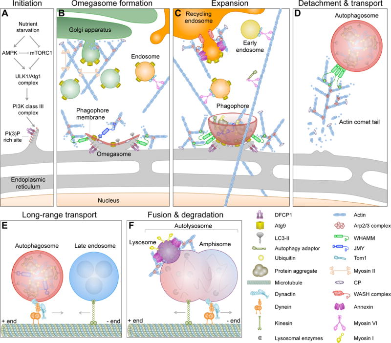
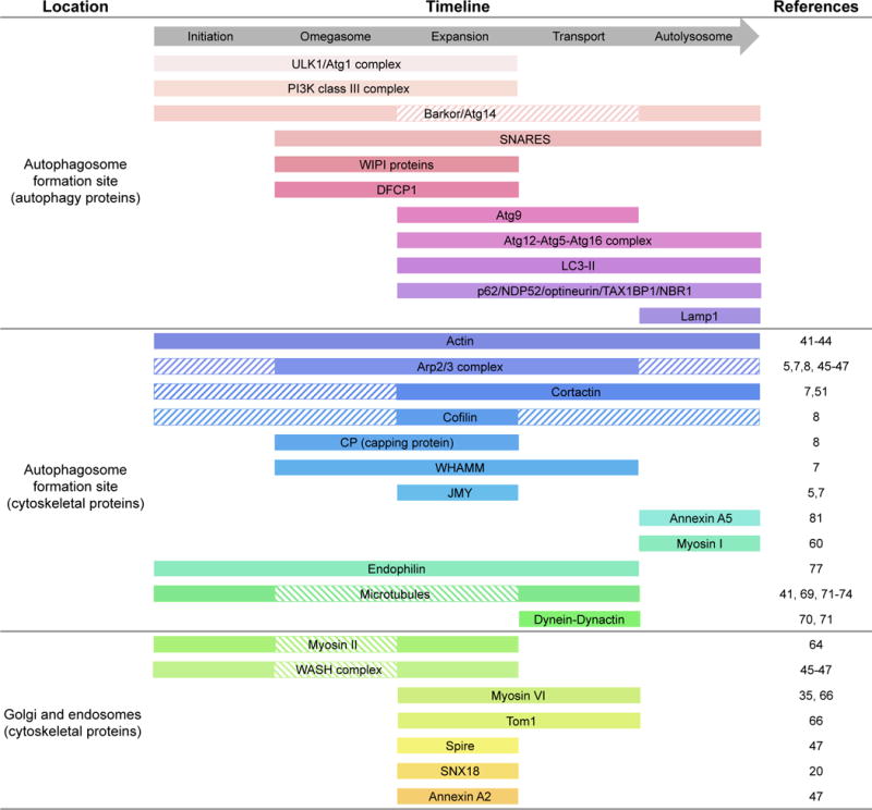
Actin cytoskeleton dynamics play vital roles in most forms of intracellular trafficking by promoting the biogenesis and transport of vesicular cargoes. Mounting evidence indicates that actin dynamics and membrane-cytoskeleton scaffolds also have essential roles in macroautophagy, the process by which cellular waste is isolated inside specialized vesicles called autophagosomes for recycling and degradation. Branched actin polymerization is necessary for the biogenesis of autophagosomes from the endoplasmic reticulum (ER) membrane. Actomyosin-based transport is then used to feed the growing phagophore with pre-selected cargoes and debris derived from different membranous organelles inside the cell. Finally, mature autophagosomes detach from the ER membrane by an as yet unknown mechanism, undergo intracellular transport and then fuse with lysosomes, endosomes and multivesicular bodies through mechanisms that involve actin- and microtubule-mediated motility, cytoskeleton-membrane scaffolds and signaling proteins. In this review, we highlight the considerable progress made recently towards understanding the diverse roles of the cytoskeleton in autophagy.


Figures

Figure 1
Figure 1
Stages of autophagy and the link to the cytoskeleton. (A) During nutrient starvation, the class III PI3K complex is recruited to the endoplasmic reticulum (ER), where it creates a PI(3)P-rich site that seeds the formation of the omegasome. Key autophagy proteins, such as WIPI1, WIPI2 and DFCP1, and the Arp2/3 complex NPF WHAMM are recruited to these sites. WHAMM activates the Arp2/3 complex to form an actin-branched network that provides mechanical forces for the formation of the omegasome. (B) A membrane cisterna called the phagophore (or isolation membrane) begins to form and expand on the omegasome. The expansion of the phagophore requires Atg9-rich membranes derived from different sources, including endosomes and the Golgi apparatus. LC3-II inserts into the phagophore membrane, where it serves as an anchor for autophagy adaptor proteins, and may also recruit JMY, another NPF of the Arp2/3 complex. (C) Together, JMY and WHAMM induce the formation of a branched actin network needed for the expansion of the phagophore membrane. Ubiquitinated cargo from different sources, including mitochondria and protein aggregates, is delivered to the growing phagophore using actomyosin-based transport. BAR domain proteins and annexins contribute to membrane remodeling and fusion events during expansion. (D) The mature autophagosome detaches from the omegasome and is initially transported using an actin comet tail mechanism. (E) The autophagosome is then transported over longer distances by dynein-dynactin along microtubules. (F) The autophagosome fuses with a late endosome to form an amphisome, which then fuses with an acidic lysosome to form an autolysosome. These fusion events depend on membrane-cytoskeleton adaptors, such as annexins, SNARE proteins, actin and myosin I. The acidic pH of the autolysosome activates hydrolytic enzymes that degrade the contents of the autophagosome.
Figure 2
Figure 2
Autophagy timeline showing the arrival and departure of mammalian cytoskeleton assembly factors. Solid bars indicate the timing of appearance of autophagy and cytoskeletal proteins according to published evidence (references listed on the right), whereas striped bars indicate the time of arrival based on the known functions of proteins for which direct evidence is still lacking.

References

    1. Anitei M, Hoflack B. Bridging membrane and cytoskeleton dynamics in the secretory and endocytic pathways. Nat Cell Biol. 2011;14:11–19. - PubMed
    1. Suetsugu S, Gautreau A. Synergistic BAR-NPF interactions in actin-driven membrane remodeling. Trends Cell Biol. 2012;22:141–150. - PubMed
    1. Kaksonen M, Toret CP, Drubin DG. Harnessing actin dynamics for clathrin-mediated endocytosis. Nat Rev Mol Cell Biol. 2006;7:404–414. - PubMed
    1. Egea G, Lazaro-Dieguez F, Vilella M. Actin dynamics at the Golgi complex in mammalian cells. Curr Opin Cell Biol. 2006;18:168–178. - PubMed
    1. Coutts AS, La Thangue NB. Actin nucleation by WH2 domains at the autophagosome. Nat Commun. 2015;6:7888. - PMC - PubMed

LinkOut - more resources