

In Vitro and In Vivo Inhibitory Effects of α-Mangostin on Cholangiocarcinoma Cells and Allografts
- PMID: 28441703
- PMCID: PMC5464488
- DOI: 10.22034/APJCP.2017.18.3.707
In Vitro and In Vivo Inhibitory Effects of α-Mangostin on Cholangiocarcinoma Cells and Allografts
Abstract
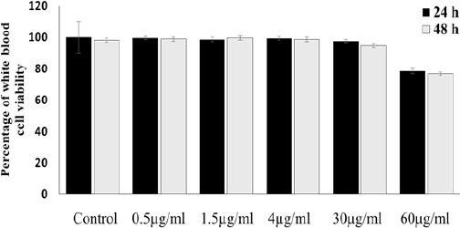
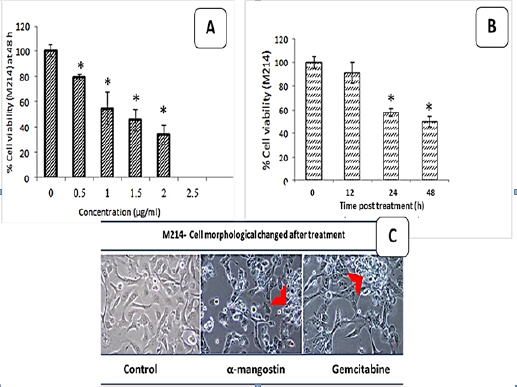
We investigated the anti-cholangiocarcinoma effect of α-mangostin from Garcinia mangostana pericarp extract (GM) in a human cholangiocarcinoma (CCA) cell line and a hamster CCA allograft model. In vitro, human CCA cells were treated with GM at various concentrations and for different time periods; then cell-cycle arrest and apoptosis were evaluated using flow cytometry, and metastatic potential with wound healing assays. In vivo, hamster allografts were treated with GM, gemcitabine (positive control) and a placebo (negative control) for 1 month; tumor weight and volume were then determined. Histopathological features and immunostaining (CK19 and PCNA) characteristics were examined by microscopy. The present study found that α-mangostin could: inhibit CCA cell proliferation by inducing apoptosis through the mitochondrial pathway; induce G1 cell-cycle arrest; and inhibit metastasis. Moreover, α-mangostin could inhibit CCA growth, i.e. reduce tumor mass (weight and size) and alter CCA pathology, as evidenced by reduced positive staining for CK19 and PCNA. The present study thus suggested that α-mangostin is a promising anti-CCA compound whose ready availability in tropical countries might indicate use for prevention and treatment of CCA.
Keywords: Inhibitory effect; cholangiocarcinoma; α-mangostin.
Creative Commons Attribution License
Figures


Similar articles
-
α-Mangostin extracted from the pericarp of the mangosteen (Garcinia mangostana Linn) reduces tumor growth and lymph node metastasis in an immunocompetent xenograft model of metastatic mammary cancer carrying a p53 mutation.BMC Med. 2011 Jun 3;9:69. doi: 10.1186/1741-7015-9-69. BMC Med. 2011. PMID: 21639868 Free PMC article.
-
Antitumor and apoptosis-inducing effects of α-mangostin extracted from the pericarp of the mangosteen fruit (Garcinia mangostana L.)in YD-15 tongue mucoepidermoid carcinoma cells.Int J Mol Med. 2016 Apr;37(4):939-48. doi: 10.3892/ijmm.2016.2517. Epub 2016 Mar 4. Int J Mol Med. 2016. PMID: 26951885 Free PMC article.
-
Potential of Alpha-Mangostin-Loaded PLGA Nanoparticles for Cholangiocarcinoma Treatment.Polymers (Basel). 2022 Oct 20;14(20):4444. doi: 10.3390/polym14204444. Polymers (Basel). 2022. PMID: 36298022 Free PMC article.
-
The Potential of α-Mangostin from Garcinia mangostana as an Effective Antimicrobial Agent-A Systematic Review and Meta-Analysis.Antibiotics (Basel). 2022 May 26;11(6):717. doi: 10.3390/antibiotics11060717. Antibiotics (Basel). 2022. PMID: 35740124 Free PMC article. Review.
-
Nanoformulations of α-Mangostin for Cancer Drug Delivery System.Pharmaceutics. 2021 Nov 24;13(12):1993. doi: 10.3390/pharmaceutics13121993. Pharmaceutics. 2021. PMID: 34959275 Free PMC article. Review.
Cited by
-
α-Mangostin: A Xanthone Derivative in Mangosteen with Potent Anti-Cancer Properties.Biomolecules. 2024 Oct 30;14(11):1382. doi: 10.3390/biom14111382. Biomolecules. 2024. PMID: 39595559 Free PMC article. Review.
-
Exploring the antineoplastic potential of α-mangostin in breast cancer.Nat Prod Bioprospect. 2025 Jul 3;15(1):43. doi: 10.1007/s13659-025-00528-5. Nat Prod Bioprospect. 2025. PMID: 40608162 Free PMC article. Review.
-
Synthesis and Properties of α-Mangostin and Vadimezan Conjugates with Glucoheptoamidated and Biotinylated 3rd Generation Poly(amidoamine) Dendrimer, and Conjugation Effect on Their Anticancer and Anti-Nematode Activities.Pharmaceutics. 2022 Mar 10;14(3):606. doi: 10.3390/pharmaceutics14030606. Pharmaceutics. 2022. PMID: 35335982 Free PMC article.
-
Recent updates on metabolite composition and medicinal benefits of mangosteen plant.PeerJ. 2019 Jan 31;7:e6324. doi: 10.7717/peerj.6324. eCollection 2019. PeerJ. 2019. PMID: 30755827 Free PMC article.
-
α-Mangostin Nanoparticles Cytotoxicity and Cell Death Modalities in Breast Cancer Cell Lines.Molecules. 2021 Aug 24;26(17):5119. doi: 10.3390/molecules26175119. Molecules. 2021. PMID: 34500560 Free PMC article. Review.
References
-
- Aukkanimart R, Boonmars T, Sriraj P, et al. Anthelmintic, anti-inflammatory and antioxidant effects of Garcinia mangostana extract in hamster opisthorchiasis. Exp Parasitol. 2015;30:5–13. - PubMed
-
- Chamadol N, Pairojkul C, Khuntikeo N, et al. Histological confirmation of periductal fibrosis from ultrasound diagnosis in cholangiocarcinoma patients. J Hepatobiliary Pancreat Sci. 2014;21:316–22. - PubMed
-
- Charernsriwilaiwat N, Rojanarata T, Ngawhirunpat T, Sukma M, Opanasopit P. Electrospun chitosan-based nanofiber mats loaded with Garcinia mangostana extracts. Int J Pharm. 2013;16:333–43. - PubMed
-
- Gomez-Lazaro M, Galindo MF, Melero-Fernandez de Mera RM, et al. Reactive oxygen species and p38 mitogen-activated protein kinase activate Bax to induce mitochondrial cytochrome c release and apoptosis in response to malonate. Mol Pharmaco. 2007;71:736–43. - PubMed
LinkOut - more resources
Full Text Sources
Miscellaneous