. 2017 Apr 24;9(4):90.
doi: 10.3390/v9040090.

Replication and Oncolytic Activity of an Avian Orthoreovirus in Human Hepatocellular Carcinoma Cells

Affiliations

Replication and Oncolytic Activity of an Avian Orthoreovirus in Human Hepatocellular Carcinoma Cells

Robert A Kozak et al. Viruses. .

Abstract

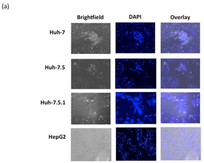
Oncolytic viruses are cancer therapeutics with promising outcomes in pre-clinical and clinical settings. Animal viruses have the possibility to avoid pre-existing immunity in humans, while being safe and immunostimulatory. We isolated an avian orthoreovirus (ARV-PB1), and tested it against a panel of hepatocellular carcinoma cells. We found that ARV-PB1 replicated well and induced strong cytopathic effects. It was determined that one mechanism of cell death was through syncytia formation, resulting in apoptosis and induction of interferon stimulated genes (ISGs). As hepatitis C virus (HCV) is a major cause of hepatocellular carcinoma worldwide, we investigated the effect of ARV-PB1 against cells already infected with this virus. Both HCV replicon-containing and infected cells supported ARV-PB1 replication and underwent cytolysis. Finally, we generated in silico models to compare the structures of human reovirus- and ARV-PB1-derived S1 proteins, which are the primary targets of neutralizing antibodies. Tertiary alignments confirmed that ARV-PB1 differs from its human homolog, suggesting that immunity to human reoviruses would not be a barrier to its use. Therefore, ARV-PB1 can potentially expand the repertoire of oncolytic viruses for treatment of human hepatocellular carcinoma and other malignancies.

Keywords: avian orthoreovirus; hepatitis C virus; hepatocellular carcinoma; oncolytic virus; syncytia.


Conflict of interest statement

The authors declare no conflict of interest.

Figures

Figure 1
Figure 1
Evaluation of avian orthoreovirus (ARV-PB1) replication in cell lines. (a) Cell viability in human liver cell lines was measured at 96 hours post-infection (h.p.i.) with PrestoBlueTM Cell Viability Reagent (Life Technologies) and data was normalized to uninfected controls. Experiments for each cell line were performed a minimum of five times, and error bars represent the standard deviation. (b) ARV-PB1 titers at various time points, in cell lines following infection at a multiplicity of infection (MOI) of 5. The experiment was performed in duplicate and data are from one representative experiment. (c) Cytopathic effect following crystal violet staining in Huh-7.5 cells at 72 h.p.i. Similar results were observed in the other liver cell lines tested, which included Huh-7, Huh-7.5.1 and HepG2. Experiments were performed in triplicate, and one representative picture is shown. (d) Cell viability in other cancer cell lines infected at an MOI of 10 was measured at 96 h.p.i. with PrestoBlueTM Cell Viability Reagent (Life Technologies) and data was normalized to uninfected controls. The origin of the analyzed cell lines is described in Table S2. Experiments for the ID8 cell line was performed twice, all other cell lines were repeated a minimum of three times. Data represent the mean, and error bars represent the standard deviation.
Figure 2
Figure 2
Cytotoxicity assessment of ARV-PB1. (a) Primary hepatocytes were infected at multiplicities of infection (MOI) of 10 and 100. After 1 h virus adsorption, the inoculum was removed and cells were washed with phosphate buffered saline (PBS) and fresh medium was added. Cell viability was determined at 96 h.p.i. by PrestoBlueTM Cell Viability Reagent (Life Technologies). Data were normalized to uninfected controls. Experiments were performed four times, and data represent the mean while error bars represent the standard deviation. (b) Primary hepatocytes were infected with ARV-PB1 at an MOI of 10 as described above and virus titers were examined at 0 and 96 h.p.i.
Figure 3
Figure 3
Syncytia formation in liver cell lines. (a) Cells were infected at an MOI of 5 for 72 h followed by fixation with formalin and 4',6-diamidino-2-phenylindole (DAPI) staining. (b) No syncytial formation was observed in uninfected cells. Cells were analyzed by fluorescence and bright-field microscopy (100× magnification).
Figure 3
Figure 3
Syncytia formation in liver cell lines. (a) Cells were infected at an MOI of 5 for 72 h followed by fixation with formalin and 4',6-diamidino-2-phenylindole (DAPI) staining. (b) No syncytial formation was observed in uninfected cells. Cells were analyzed by fluorescence and bright-field microscopy (100× magnification).
Figure 4
Figure 4
Analysis of apoptosis in hepatocellular carcinoma cells. Huh-7.5 cells were either mock infected or infected with ARV-PB1 at a MOI of 5 for 72 h. Subsequently, mechanisms of cell death were determined by annexin V and 7-AAD staining according to the manufacturer’s instructions. The percentages of viable (annexin V− 7-AAD−), early apoptotic (annexin V+ 7-AAD−), late apoptotic (annexin V+ 7-AAD+) and necrotic (annexin V− 7-AAD+) cells were determined. Data represent the mean from experiments performed in triplicate. Error bars represent standard deviations. Samples were compared using Student’s t-test with significant differences indicated by * p ≤ 0.05.
Figure 5
Figure 5
Expression of interferon-stimulated genes (ISGs). Huh-7 cells (5 × 10⁵) were infected at a MOI of 5 and messenger RNA (mRNA) was collected for analysis at 6 hours post-infection. Transcription of the analyzed ISGs was analyzed by quantitative real-time polymerase chain reaction (PCR) with glyceraldehyde 3-phosphate dehydrogenase (GAPDH) as a housekeeping gene and expressed as a fold-change compared to uninfected cells (35). Data represent the mean from six replicate experiments, with error bars showing the standard error of the mean. Samples were compared using Student’s t-test with significant differences indicated by * p ≤ 0.05.
Figure 6
Figure 6
ARV-PB1 activity in HCV-replicon-containing cells. (a) Cell viability in BB7 cells infected at differing MOIs was measured at 96 hours post-infection (h.p.i.) using PrestoBlueTM Cell Viability Reagent (Life Technologies). Data were normalized to uninfected controls. The experiment was performed at three times, and error bars represent the standard error of the mean. (b) ARV-PB1 viral titers in BB7 cells at various time points following infection at a MOI of 5. (c) Fluorescent and bright-field microscopy (100× magnification) to view DAPI staining and evaluate syncytia in BB7 cells infected with ARV-PB1 (MOI of 5) at 96 h.p.i. The experiment was performed in triplicate and data are from one representative experiment. (d) BB7 cells were either uninfected or infected with ARV-PB1 (MOI of 5) for three days. Subsequently, the cells were fixed and treated with annexin V and 7-AAD. The percentages of viable (annexin V− 7-AAD−), early apoptotic (annexin V+ 7-AAD−), late apoptotic (annexin V+ 7-AAD+) and necrotic (annexin V− 7-AAD+) cells were determined. Data represent the means from experiments performed in triplicate. Error bars show standard deviations. Samples were compared using Student’s t-test with significant differences indicated by * p ≤ 0.05.
Figure 7
Figure 7
ARV-PB1 activity in JFH-1 -infected cells. (a) Huh-7.5.1 cells were infected with JFH-1 and 24 h later treated with ARV-PB1 at differing MOIs. At 48 hours post-treatment, cells were fixed and stained with crystal violet to evaluate cytopathic effect. The experiment was performed in triplicate. (b) Induction of syncytia in JFH-1-infected cells by ARV-PB1. Cells were infected with ARV-PB1 (MOI of 5) for 48 h. Subsequently, cells were fixed with formalin, stained with DAPI and analyzed by fluorescent and bright-field microscopy (100× magnification). (c) Measurement of remaining viable cells following treatment with various MOIs at 48h post-infection. The experiment was performed at four times, and error bars represent the standard deviation. Samples were compared using Student’s t-test with significant differences indicated by * p ≤ 0.05.
Figure 8
Figure 8
In silico structure generation of the S1 attachment protein of ARV-PB1. (a) I-TASSER-generated ARV-PB1 (blue), manually superimposed with avian reovirus σ C117-326, Protein Data Bank (PDB) ID 2JJL (pink), identified as the most closely related crystal structure. (b) Tertiary structural alignment of the S1 protein of ARV-PB1 and 2JJL. Conserved sequence regions are highlighted in black. (c) I-TASSER-generated ARV-PB1 (blue), manually superimposed with mammalian orthoreovirus 3 (Dearing strain), PDB ID 1KKE (green), using 1KKE as a generation template. (d) Tertiary structural alignment of the S1 protein of ARV-PB1 and 1KKE. Conserved sequence regions are highlighted in black, and the epitope targets of neutralizing antibodies are highlighted in red.
Figure 8
Figure 8
In silico structure generation of the S1 attachment protein of ARV-PB1. (a) I-TASSER-generated ARV-PB1 (blue), manually superimposed with avian reovirus σ C117-326, Protein Data Bank (PDB) ID 2JJL (pink), identified as the most closely related crystal structure. (b) Tertiary structural alignment of the S1 protein of ARV-PB1 and 2JJL. Conserved sequence regions are highlighted in black. (c) I-TASSER-generated ARV-PB1 (blue), manually superimposed with mammalian orthoreovirus 3 (Dearing strain), PDB ID 1KKE (green), using 1KKE as a generation template. (d) Tertiary structural alignment of the S1 protein of ARV-PB1 and 1KKE. Conserved sequence regions are highlighted in black, and the epitope targets of neutralizing antibodies are highlighted in red.

Similar articles

Cited by

References

    1. Bakiri L., Wagner E.F. Mouse models for liver cancer. Mol. Oncol. 2013;7:206–223. doi: 10.1016/j.molonc.2013.01.005. - DOI - PMC - PubMed
    1. Shah H.A., Heathcote J., Feld J.J. A Canadian screening program for hepatitis C: Is now the time? CMAJ. 2013;185:1325–1328. doi: 10.1503/cmaj.121872. - DOI - PMC - PubMed
    1. Russell S.J., Peng K.W., Bell J.C. Oncolytic virotherapy. Nat. Biotechnol. 2012;30:658–670. doi: 10.1038/nbt.2287. - DOI - PMC - PubMed
    1. Altomonte J., Marozin S., Schmid R.M., Ebert O. Engineered newcastle disease virus as an improved oncolytic agent against hepatocellular carcinoma. Mol. Ther. 2010;18:275–284. doi: 10.1038/mt.2009.231. - DOI - PMC - PubMed
    1. Unno Y., Shino Y., Kondo F., Igarashi N., Wang G., Shimura R., Yamaguchi T., Asano T., Saisho H., Sekiya S., et al. Oncolytic viral therapy for cervical and ovarian cancer cells by Sindbis virus AR339 strain. Clin. Cancer Res. 2005;11:4553–4560. doi: 10.1158/1078-0432.CCR-04-2610. - DOI - PubMed

Publication types

Substances