. 2017 Apr 26;9(1):30.
doi: 10.1186/s13195-017-0257-7.

Evaluation of a DNA Aβ42 vaccine in adult rhesus monkeys (Macaca mulatta): antibody kinetics and immune profile after intradermal immunization with full-length DNA Aβ42 trimer

Affiliations

Evaluation of a DNA Aβ42 vaccine in adult rhesus monkeys (Macaca mulatta): antibody kinetics and immune profile after intradermal immunization with full-length DNA Aβ42 trimer

Doris Lambracht-Washington et al. Alzheimers Res Ther. .

Abstract

Background: Aggregated amyloid-β peptide 1-42 (Aβ42), derived from the cellular amyloid precursor protein, is one of the pathological hallmarks of Alzheimer's disease (AD). Although active immunization against Aβ42 peptide was successful in AD mouse models and led to removal of plaques and improved memory, a similar clinical trial in humans (Aβ42 peptide immunization with QS-21 adjuvant) was stopped in phase II, when 6% of the treated patients developed encephalitis. Currently ongoing passive immunizations with the injection of preformed monoclonal antibodies against different epitopes within the Aβ1-42 peptide, which do not lead to activation of the immune system, have shown some effects in slowing AD pathology. Active DNA Aβ42 immunizations administered with the gene gun into the skin are noninflammatory because they activate a different T-cell population (Th2) with different cytokine responses eliciting a different humoral immune response. We present our findings in rhesus macaques that underwent the DNA Aβ42 immunization via gene gun delivery into the skin.

Methods: Six rhesus monkeys received two different doses of a DNA Aβ42 trimer vaccine. The humoral immune response was analyzed from blood throughout the study, and cellular immune responses were determined in peripheral blood mononuclear cells (PBMCs) after three and six immunizations.

Results: DNA Aβ42 trimer immunization led to high titer antibody responses in the nonhuman primate (NHP) model. Antibodies generated in the rhesus monkeys following DNA Aβ42 immunization detected amyloid plaques consisting of human Aβ42 peptide in the brain of the triple-transgenic AD mouse model. T-cell responses showed no interferon (IFN)-γ- and interleukin (IL)-17-producing cells from PBMCs in Enzyme-Linked ImmunoSpot assays after three immunization time points. At six immunization time points, IFN-γ- and IL-17-producing cells were found in immunized animals as well as in control animals and were thus considered nonspecific and not due to the immunization regimen. IFN-γ and IL-17 secretion in response to Aβ42 peptide restimulation became undetectable after a 3-month rest period.

Conclusions: Intradermal DNA Aβ42 immunization delivered with the gene gun produces a high antibody response in NHPs and is highly likely to be effective and safe in a clinical AD prevention trial in patients.

Keywords: Alzheimer’s disease; Amyloid-β; Antibody response; DNA vaccination; Immunotherapy; Nonhuman primates; Th2 immune response.


Figures
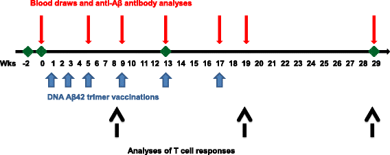
Fig. 1
Fig. 1
Illustration of the time line of the performed experiments and analyses of the immune responses in rhesus macaques. Aβ42 Amyloid-β peptide 1–42
Fig. 2
Fig. 2
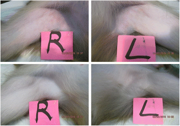
Photographs showing the axillary area of two animals that received DNA amyloid-β peptide 1–42 immunizations delivered with the gene gun 2 weeks past the second immunization. L Left side, R Right side
Fig. 3
Fig. 3
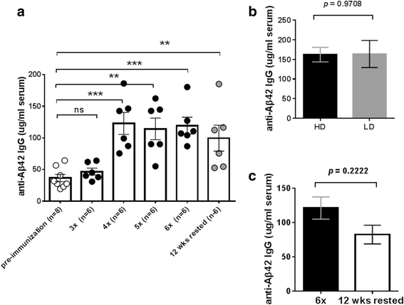
Antibody production in rhesus macaques after the different DNA amyloid-β peptide 1–42 (Aβ42) immunization time points. a Anti-Aβ42 antibody levels in DNA Aβ42-immunized rhesus monkeys (n = 6) are shown for blood drawn prior to the immunizations, from blood of animals immunized three, four, five, and six times, and from blood drawn following a 3-month rest period. b Antibody levels in animals that had received the high dose (16 μg of DNA per immunization time point, black bar) or the low dose (8 μg of DNA per immunization time point, gray bar) did not differ significantly following the sixth immunization time point. c Antibody levels declined slightly with the 3-month rest period
Fig. 4
Fig. 4
Antibody titers in the rhesus monkeys immunized six times. Serial dilutions of plasma samples from the six immunizations are shown. Immunization time points were tested for binding to amyloid-β peptide 1–42 (Aβ42) peptide in two-step enzyme-linked immunosorbent assays. a Antibody titers for anti-Aβ42 antibodies of the immunoglobulin A (IgA) isotype. b Antibody titers for antibodies of the IgG isotype. For comparison with antibody titers prior to the immunizations and to antibody titers following the second immunizations, these titers were included in the graph of the IgG titer analysis (red and green lines). Individual animals are shown with specific symbols as indicated on the figure. Animals receiving the high-dose (16 μg of DNA per immunization) and low-dose (8 μg of DNA/immunization) immunizations are indicated by HD and LD, respectively. c Comparison of IgG and IgA antibody titers in the immunized monkeys. Mean, SD, and SEM values are given in the table below the graph
Fig. 5
Fig. 5
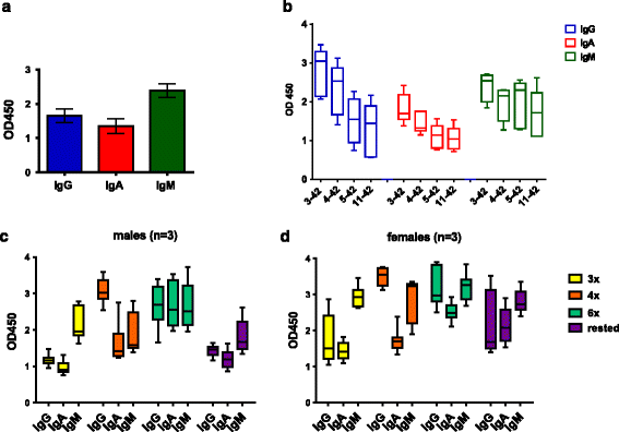
Antibody isotypes of anti-Aβ antibodies produced in the Rhesus monkey. a The humoral immune response generated anti-Aβ IgG, - IgM, and - IgA antibodies in the immunized monkeys. Plasma samples had been used in a 1:1000 dilution. b Binding of the different antibody isotypes to truncated Aβx–42 peptides was tested from plasma after six immunizations (dilution 1:500). c, d Anti-Aβ42 IgG, -IgA, and -IgM antibody isotype responses were compared for different time points (3x yellow bars, 4x orange bars, 6x light green bars, and three months rest purple bars) in male C and female D Rhesus macaques. Results are shown in box & whiskers graphs from minimum to maximum values. Median levels were indicated with the horizontal line, the Standard Error Mean (SEM) is shown for all columns above and below the boxes
Fig. 6
Fig. 6
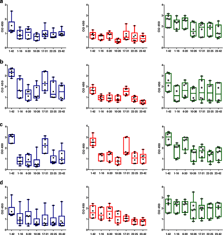
Amyloid-β peptide 1–42 (Aβ42) epitope binding of immunoglobulin G (IgG), IgA, and IgM isotypes at different time points of the immunization schedule. A panel of shorter Aβ peptides (Aβ1–16, Aβ6–20, Aβ10–26, Aβ17–31, Aβ22–35, and Aβ23–42) was used to detect linear epitope binding of antibodies generated in the animals after DNA Aβ42 immunization. a Aβ epitope binding pattern of IgG, IgA, and IgM antibodies observed after three immunizations. b Epitope binding pattern after four immunizations. c Epitope binding pattern found with six immunization time points. d Aβ epitope binding of antibodies of the IgG, IgA, and IgM isotypes after the 3-month rest period. Results are presented in box-and-whisker graphs with minimum to maximum values. Median levels are indicated by the horizontal lines, and the SEM is shown for all columns above and below the boxes. Each circle within the bars represents the value for one individual monkey that had received active DNA Aβ42 immunizations. IgG antibodies are indicated in blue, IgA antibodies are indicated in red, and IgM antibodies are indicated in green. OD 450 450-nm Optical density
Fig. 7
Fig. 7
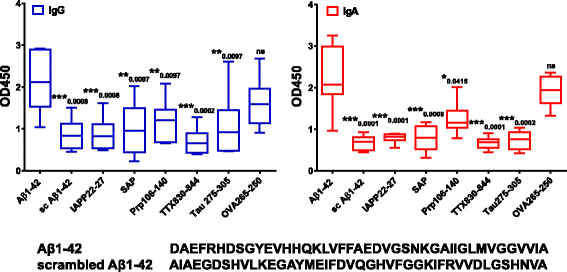
Specificity of antibodies in plasma of DNA amyloid-β peptide 1–42 (Aβ42) immunized rabbits to Aβ1–42. Plasma samples from the sixth immunization time point of the six immunized rhesus monkeys were tested for binding to a variety of different peptides (Aβ1–42, scrambled Aβ1–42, islet amyloid protein [IAPP], serum amyloid P component [SAP], prion protein fragments [Prp], tau peptide repeat 2 domain, tetanus toxin peptide [TTX], ovalbumin peptides [OVA]). Plasma was diluted 1:500 for these assays. In the comparison of binding to Aβ1–42, significant differences were found for binding of immunoglobulin G (IgG; blue bars) and IgA (red bars) antibodies in plasma to the distinct peptides tested (indicated by *, **, *** p values above the bars). Differences in IgG and IgA antibody binding in plasma samples from the immunized monkeys to nonrelated OVA peptides compared with binding to Aβ1–42 were nonsignificant (Mann-Whitney p values of 0.1246 and 0.5532, respectively). OD 450 450-nm Optical density
Fig. 8
Fig. 8
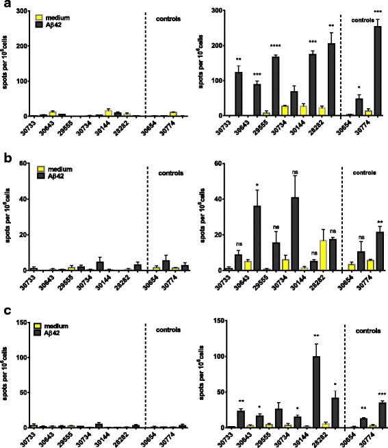
Interferon (IFN)-γ, interleukin (IL)-17, and IL-4 Enzyme-Linked ImmunoSpot assays for amyloid-β peptide 1–42 (Aβ42) peptide restimulated peripheral blood mononuclear cell cultures of DNA Aβ42-immunized rhesus monkeys. The first column shows the number of IFN-γ-, IL-17-, and IL-4-secreting cells after three immunization time points (48 h in cell culture, medium controls, and Aβ42 peptide restimulation). The second column provides results from the same analyses after the sixth immunization time point. Individual rhesus monkeys are indicated with numbers on the x-axis of all graphs. The y-axis of all graphs shows the number of cytokine-secreting cells (spots) per 106 cells. Increased numbers for all three cytokines were found in the immunized animals as well as in the nontreated control animals. Therefore, the cytokine secretion was considered as nonspecific and not due to the DNA Aβ42 immunization. a IL-17-secreting cells. b IFN-γ-secreting cells. c The number of IL-4-secreting cells per 106 splenocytes. * p < 0.05, ** p < 0.005, *** p  < 0.001, and **** p < 0.0001
Fig. 9
Fig. 9
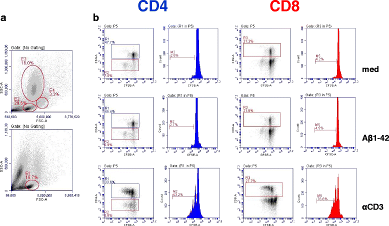
CD4 and CD8 T-cell proliferation in amyloid-β peptide 1–42 (Aβ42) peptide restimulated peripheral blood mononuclear cell cultures. a Mononuclear blood cells were separated on the basis of forward scatter (FSC) and side scatter (SSC). The upper histogram shows the pattern in fresh blood, and the lower histogram shows the cell populations after 6 days in culture. Gates for the carboxyfluorescein succinimidyl ester (CFSE) assay were set on lymphocytes (E1). b T-cell proliferation was analyzed using a CFSE dilution assay. Events were gated on CD4- and CD8-positive cells, and cytometric histograms are shown for medium controls, Aβ42 peptide restimulated cultures, and proliferation after anti-CD3 antibody stimulation. All samples were run in triplicates. No T-cell proliferation in response to Aβ42 peptide restimulation was found
Fig. 10
Fig. 10
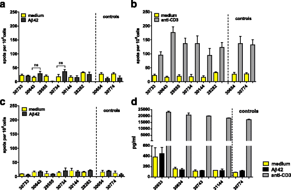
Interleukin (IL)-17 and IL-4 Enzyme-Linked ImmunoSpot (ELISPOT) assays, as well as interferon (IFN)-γ enzyme-linked immunosorbent assay (ELISA) from amyloid-β peptide 1–42 (Aβ42) peptide-restimulated peripheral blood mononuclear cell cultures of DNA Aβ42-immunized rhesus monkeys after a 3-month rest period. IL-17- and IL-4-secreting cells were analyzed in an ELISPOT assay (a–c). IFN-γ secretion was tested with a cytokine ELISA (d). The individual rhesus monkeys are indicated with numbers on the x-axis of all graphs. a–c The y-axis shows the number of cytokine-secreting cells (spots) per 106 cells. d The y-axis shows the amount of IFN-γ found (expressed in picograms per milliliter of culture supernatant)
Fig. 11
Fig. 11
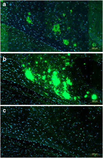
Antibodies from DNA amyloid-β peptide 1–42 (Aβ42)-immunized monkeys stain amyloid plaques containing human Aβ42 peptides in brain sections of triple-transgenic Alzheimer’s disease (3xTg-AD) mice. a Plaque staining in hippocampal area of 3xTg-AD mouse with plasma from animal 28282. b Control staining in parallel section with commercial anti-Aβ42 antibody (6E10). c No staining in parallel section that was incubated with the secondary antibody and detection reagents only. Scale bar indicates 100 μm

Similar articles

Cited by

References

    1. Schenk D, Barbour R, Dunn W, Gordon G, Grajeda H, Guido T, et al. Immunization with amyloid-β attenuates Alzheimer-disease-like pathology in the PDAPP mouse. Nature. 1999;400:173–7. doi: 10.1038/22124. - DOI - PubMed
    1. Games D, Bard F, Grajeda H, Guido T, Khan K, Soriano F, et al. Prevention and reduction of AD-type pathology in PDAPP mice immunized with Aβ1–42. Ann N Y Acad Sci. 2000;920:274–84. doi: 10.1111/j.1749-6632.2000.tb06936.x. - DOI - PubMed
    1. Morgan D, Diamond DM, Gottschall PE, Ugen KE, Dickey C, Hardy J, et al. Aβ peptide vaccination prevents memory loss in an animal model of Alzheimer’s disease. Nat. 2000;408:982–5. doi: 10.1038/35050116. - DOI - PubMed
    1. Schenk D. Amyloid-β immunotherapy for Alzheimer’s disease: the end of the beginning. Nat Rev Neurosci. 2002;3:824–8. doi: 10.1038/nrn938. - DOI - PubMed
    1. Orgogozo JM, Gilman S, Dartigues JF, Laurent B, Puel M, Kirby LC, et al. Subacute meningoencephalitis in a subset of patients with AD after Aβ42 immunization. Neurology. 2003;61:46–54. doi: 10.1212/01.WNL.0000073623.84147.A8. - DOI - PubMed