. 2018 Apr;67(4):606-615.
doi: 10.1136/gutjnl-2016-313584. Epub 2017 Apr 25.

Aspirin prevents NF-κB activation and CDX2 expression stimulated by acid and bile salts in oesophageal squamous cells of patients with Barrett's oesophagus

Affiliations

Aspirin prevents NF-κB activation and CDX2 expression stimulated by acid and bile salts in oesophageal squamous cells of patients with Barrett's oesophagus

Xiaofang Huo et al. Gut. 2018 Apr.

Abstract

Objective: In previous studies using oesophageal squamous cells from patients with Barrett's oesophagus (normal oesophageal squamous (NES)-B cells) and from patients without Barrett's oesophagus (NES-G cells), we showed that acid and bile salts induced caudal-related homeobox transcription factor 2 (CDX2) expression only in NES-B cells. CDX2, a transcription factor required to form intestinal epithelium, is a target of nuclear factor kappa-light-chain-enhancer of activated B cells (NF-κB) signalling, which can be inhibited by aspirin. We explored mechanisms underlying differences between NES-B and NES-G cells in CDX2 expression and effects of aspirin on that CDX2 expression.

Design: We exposed NES-B and NES-G cells to acid and bile salts, with and without aspirin, and evaluated effects on IκB-NF-κB-PKAc complex activation, p65 NF-κB subunit function, and CDX2 expression.

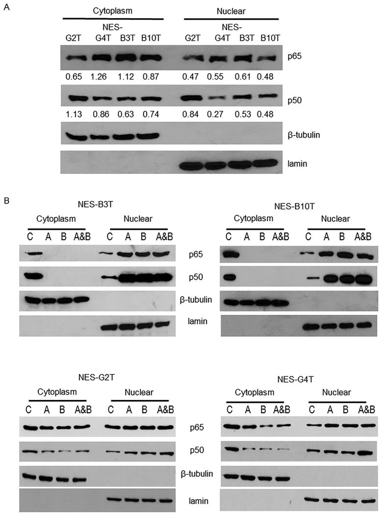
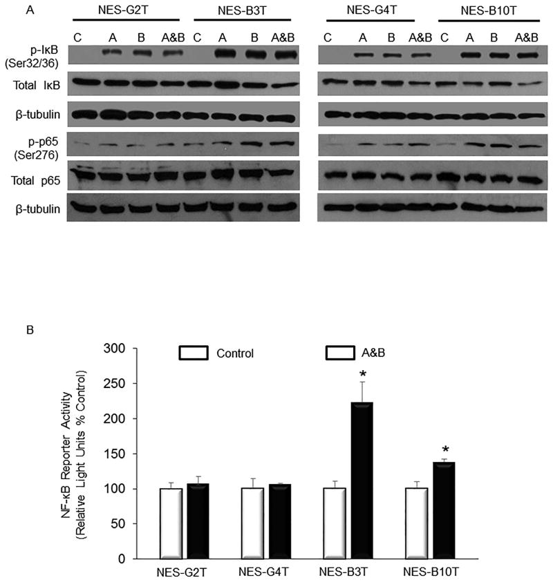
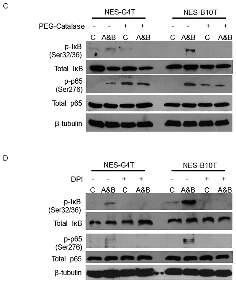
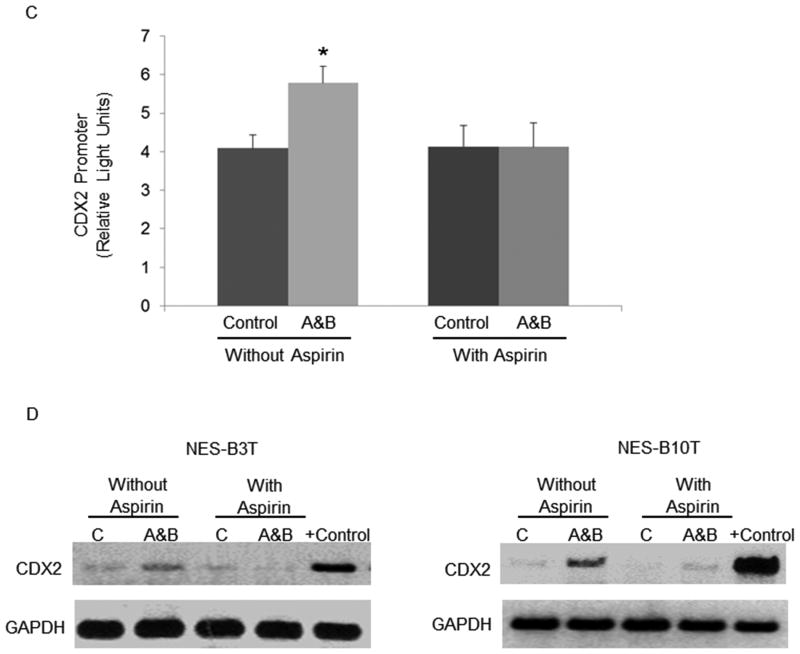
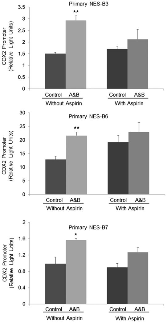
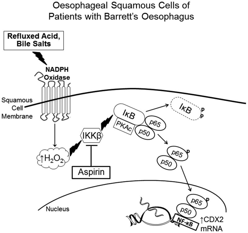
Results: In both NES-B and NES-G cells, acid and bile salts activated nicotinamide adenine dinucleotide phosphate oxidase to generate H2O2, which activated the IκB-NF-κB-PKAc complex. NES-B cells exhibited higher levels of phosphorylated IκB and p65 and greater NF-κB transcriptional activity than NES-G cells, indicating greater IκB-NF-κB-PKAc complex activation by acid and bile salts in NES-B cells, and p65 siRNA prevented their increased expression of CDX2. Aspirin blocked IκB phosphorylation, p65 nuclear translocation, CDX2 promoter activation and CDX2 expression induced by acid and bile salts in NES-B cells.

Conclusions: Differences between NES-B and NES-G cells in NF-κB activation by acid and bile salts can account for their differences in CDX2 expression, and their CDX2 expression can be blocked by aspirin. These findings might explain why some patients with GORD develop Barrett's oesophagus while others do not, and why aspirin might protect against development of Barrett's oesophagus.

Keywords: INFLAMMATION; OESOPHAGEAL DISEASE; OESOPHAGEAL REFLUX.


Conflict of interest statement

Competing interests: None declared.

Figures

Figure 1
Figure 1
Acid, bile salts and acidic bile salts markedly increase nuclear levels of p50 and p65 in NES-B cells. Representative experiments of Western blotting for cytoplasmic and nuclear p65 and p50 protein in NES-G and NES-B cell lines (A) at baseline (numbers depict semi-quantitative analyses with respect to loading controls shown beneath individual gels), and B) after exposure to acid, bile salts, and acidic bile salts. C) Representative experiments of immunofluorescence for cytoplasmic and nuclear p65 and p50 proteins in NES-G2T cells (scale bar = 50 μm). D) Acid, bile salts, and acidic bile salts do not increase DNA binding of either p50 or p65 to the CDX2 promoter in NES-G cell lines. Representative experiments of chromatin immune-precipitation (ChIP) assays for p50 and p65; IgG served as a control. Input demonstrates sheared chromatin without immune-precipitation added to the assays. Immunofluorescence images and Western blots shown are representative of the results from 2 independent experiments. C, untreated control; A, acid; B, bile salts; A&B, acidic bile salts; M, marker.
Figure 1
Figure 1
Acid, bile salts and acidic bile salts markedly increase nuclear levels of p50 and p65 in NES-B cells. Representative experiments of Western blotting for cytoplasmic and nuclear p65 and p50 protein in NES-G and NES-B cell lines (A) at baseline (numbers depict semi-quantitative analyses with respect to loading controls shown beneath individual gels), and B) after exposure to acid, bile salts, and acidic bile salts. C) Representative experiments of immunofluorescence for cytoplasmic and nuclear p65 and p50 proteins in NES-G2T cells (scale bar = 50 μm). D) Acid, bile salts, and acidic bile salts do not increase DNA binding of either p50 or p65 to the CDX2 promoter in NES-G cell lines. Representative experiments of chromatin immune-precipitation (ChIP) assays for p50 and p65; IgG served as a control. Input demonstrates sheared chromatin without immune-precipitation added to the assays. Immunofluorescence images and Western blots shown are representative of the results from 2 independent experiments. C, untreated control; A, acid; B, bile salts; A&B, acidic bile salts; M, marker.
Figure 2
Figure 2
Increases in nuclear levels of p65 are required for acid and bile salts to induce CDX2 promoter activation in oesophageal squamous cells. A) Western blot demonstrating p65 protein levels in whole cell lysates of NES-B10T cells transfected with control siRNA or p65 siRNA, untreated and following exposure to acid, bile salts and acidic bile salts. B) Representative Western blot demonstrating p50 nuclear protein levels in NES-B10T cells transfected with control siRNA or p65 siRNA, untreated and following exposure to acid, bile salts, and acidic bile salts. U, untreated; A, acid; B, bile salts; A&B, acidic bile salts. Western blots shown are representative of the results from 2 independent experiments. C) CDX2 promoter activation induced by acid and bile salts in NES-B10T containing control siRNA or p65 siRNA. Bar graphs represent the mean ±SEM. ***, p ≤ 0.001 compared with non-acidic bile salt treated controls.
Figure 3
Figure 3
NES-B cells exposed to acid, bile salts and acidic bile salts show a greater increase in p50/p65 heterodimer formation and a greater decrease in IκB- and PKAc-bound p65 in the cytoplasm than NES-G cells. Representative Western blots demonstrating A) immunoprecipitation (IP) for p50 and immunoblotting (IB) for p65 proteins and B) IP for p65 and IB for IκB and PKAc. C, untreated control; A, acid; B, bile salts; A&B, acidic bile salts. Western blots shown are representative of the results from 2 independent experiments.
Figure 4
Figure 4
Exposure to acid, bile salts and acidic bile salts causes a greater increase in IκB-NF-κB-PKAc complex activation in NES-B cells than in NES-G cells. A) Representative Western blots demonstrating acid and bile salt-induced phospho-IkB (Ser32/36) and phospho-p65 (Ser276) in NES cells. B) Luciferase reporter assay for NF-κB/p65 activity in response to acidic bile salts in NES cells. *, p < 0.05 compared with non-acidic bile salt treated controls. C&D) Acidic bile salts cause oesophageal squamous cells to generate H2O2 through NADPH oxidase, thereby activating the IκB-NF-κB-PKAc complex. Representative Western blots demonstrating inhibition of acidic bile salt-induced increases in phospho-IκB and phospho-p65 by C) PEG-catalase, a H2O2 scavenger, and D) DPI, an NADPH oxidase inhibitor. C, untreated controls; A, acid; B, bile salts; A&B, acidic bile salts. Western blots shown are representative of the results from 2 independent experiments.
Figure 4
Figure 4
Exposure to acid, bile salts and acidic bile salts causes a greater increase in IκB-NF-κB-PKAc complex activation in NES-B cells than in NES-G cells. A) Representative Western blots demonstrating acid and bile salt-induced phospho-IkB (Ser32/36) and phospho-p65 (Ser276) in NES cells. B) Luciferase reporter assay for NF-κB/p65 activity in response to acidic bile salts in NES cells. *, p < 0.05 compared with non-acidic bile salt treated controls. C&D) Acidic bile salts cause oesophageal squamous cells to generate H2O2 through NADPH oxidase, thereby activating the IκB-NF-κB-PKAc complex. Representative Western blots demonstrating inhibition of acidic bile salt-induced increases in phospho-IκB and phospho-p65 by C) PEG-catalase, a H2O2 scavenger, and D) DPI, an NADPH oxidase inhibitor. C, untreated controls; A, acid; B, bile salts; A&B, acidic bile salts. Western blots shown are representative of the results from 2 independent experiments.
Figure 5
Figure 5
Aspirin prevents the increase in phosphorylation of IκB, nuclear levels of p65 nuclear, CDX2 promoter activation, and CDX2 mRNA expression induced by acid and bile salt in NES-B cells. A) Representative Western blots demonstrating phospho- and total IkB serine 32/36 and phospho- and total p65 (Ser276) in NES-B10T cells. B) Immunofluorescence of nuclear p65 protein induced by acidic bile salts with and without treatment with aspirin in NES-B10T cells; DAPI demonstrates the number of cell nuclei in the same field (scale bar = 50 μm). C) CDX2 promoter activation induced by acidic bile salts with and without treatment with aspirin in NES-B10T cells. Bar graphs represent the mean ±SEM. *, p ≤ 0.05 compared with non-acidic bile salt treated controls D) PCR analysis of CDX2 mRNA expression induced by acidic bile salts in NES-B3T and NES-B10T cells with and without treatment with aspirin. C, untreated controls; A&B, acidic bile salts; the Barrett's epithelial cell line BAR-T served as a positive control for CDX2 mRNA expression. Western blots shown are representative of the results from 2 independent experiments.
Figure 5
Figure 5
Aspirin prevents the increase in phosphorylation of IκB, nuclear levels of p65 nuclear, CDX2 promoter activation, and CDX2 mRNA expression induced by acid and bile salt in NES-B cells. A) Representative Western blots demonstrating phospho- and total IkB serine 32/36 and phospho- and total p65 (Ser276) in NES-B10T cells. B) Immunofluorescence of nuclear p65 protein induced by acidic bile salts with and without treatment with aspirin in NES-B10T cells; DAPI demonstrates the number of cell nuclei in the same field (scale bar = 50 μm). C) CDX2 promoter activation induced by acidic bile salts with and without treatment with aspirin in NES-B10T cells. Bar graphs represent the mean ±SEM. *, p ≤ 0.05 compared with non-acidic bile salt treated controls D) PCR analysis of CDX2 mRNA expression induced by acidic bile salts in NES-B3T and NES-B10T cells with and without treatment with aspirin. C, untreated controls; A&B, acidic bile salts; the Barrett's epithelial cell line BAR-T served as a positive control for CDX2 mRNA expression. Western blots shown are representative of the results from 2 independent experiments.
Figure 6
Figure 6
Aspirin blocks CDX2 promoter activation by acidic bile salts in primary cultures of oesophageal squamous epithelial cells from patients with Barrett's oesophagus (NES-B3, NES-B6, NES-B7). Bar graphs represent the mean ±SEM. A&B, acidic bile salts; *, p<0.05 compared to non-acidic bile salt treated controls; **, p<0.01 compared to non-acidic bile salt treated controls.
Figure 7
Figure 7
Schematic demonstrating mechanism whereby refluxed acid and bile salts cause NF-κB pathway activation resulting in CDX2 expression in oesophageal squamous cells of patients with Barrett's oesophagus. Refluxed acid and bile salts stimulate NADPH oxidase to generate H2O2, which activates IKKβ, which in turn activates the IκB-NF-κB-PKAc complex through phosphorylation of IκB. This leads to the degradation of IκB, which releases the p50/p65 heterodimer. PKAc in the activated IκB-NF-κB-PKAc complex phosphorylates p65, inducing further formation of p50/p65 heterodimers, which translocate to the nucleus to stimulate transcription of NF-κB p50 target genes (e.g.CDX2) that might play a role in columnar metaplasia. By blocking the activity of IKKβ, aspirin can stop NF-κB signaling and CDX2 expression. Thus, aspirin might protect against the development of Barrett's oesophagus in GORD patients whose squamous epithelium is predisposed to heal reflux esophagitis through metaplasia rather than squamous cell regeneration.

Similar articles

Cited by

References

    1. Ronkainen J, Aro P, Storskrubb T, et al. Prevalence of Barrett's esophagus in the general population: an endoscopic study. Gastroenterology. 2005;129:1825–31. - PubMed
    1. Rex DK, Cummings OW, Shaw M, et al. Screening for Barrett's esophagus in colonoscopy patients with and without heartburn. Gastroenterology. 2003;125:1670–7. - PubMed
    1. Spechler SJ, Souza RF. Barrett's esophagus. N Engl J Med. 2014;371:836–45. - PubMed
    1. Souza RF, Shewmake KL, Shen Y, et al. Differences in ERK activation in squamous mucosa in patients who have gastroesophageal reflux disease with and without Barrett's esophagus. Am J Gastroenterol. 2005;100:551–9. - PubMed
    1. Zhang HY, Zhang X, Chen X, et al. Differences in activity and phosphorylation of MAPK enzymes in esophageal squamous cells of GERD patients with and without Barrett's esophagus. Am J Physiol Gastrointest Liver Physiol. 2008;295:G470–8. - PMC - PubMed

Publication types

MeSH terms