. 2017 Jun 5;216(6):1731-1743.
doi: 10.1083/jcb.201703084. Epub 2017 Apr 25.

TANGO1 recruits Sec16 to coordinately organize ER exit sites for efficient secretion

Affiliations

TANGO1 recruits Sec16 to coordinately organize ER exit sites for efficient secretion

Miharu Maeda et al. J Cell Biol. .

Abstract

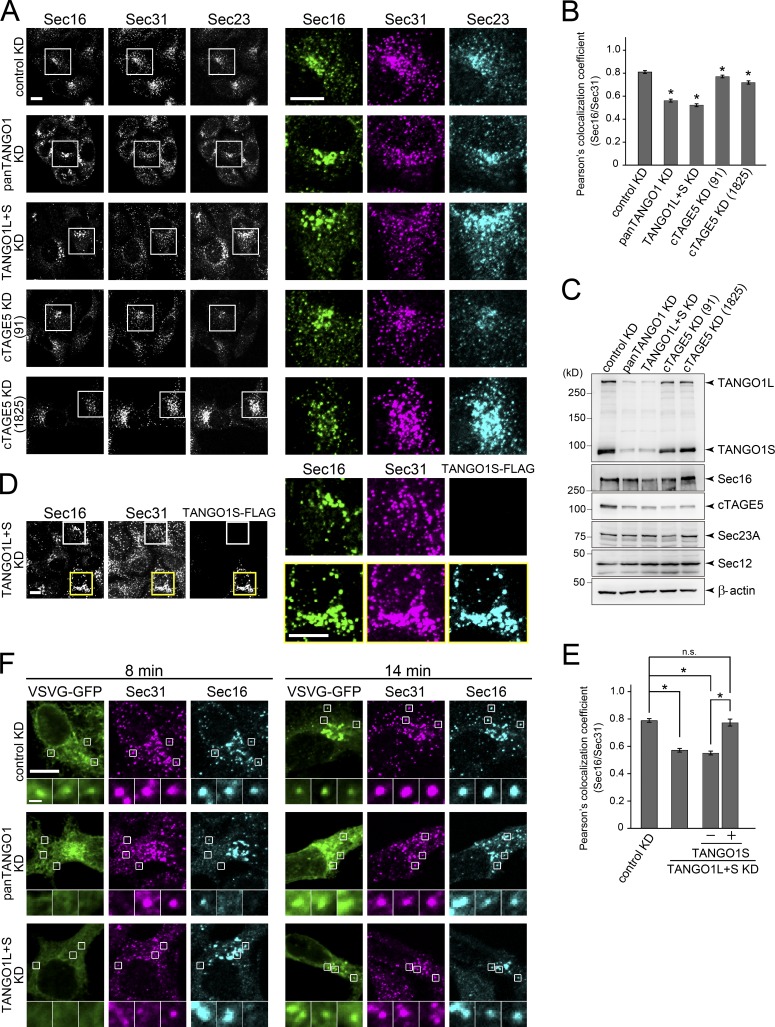
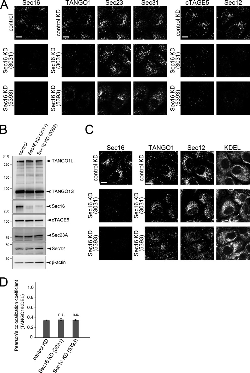
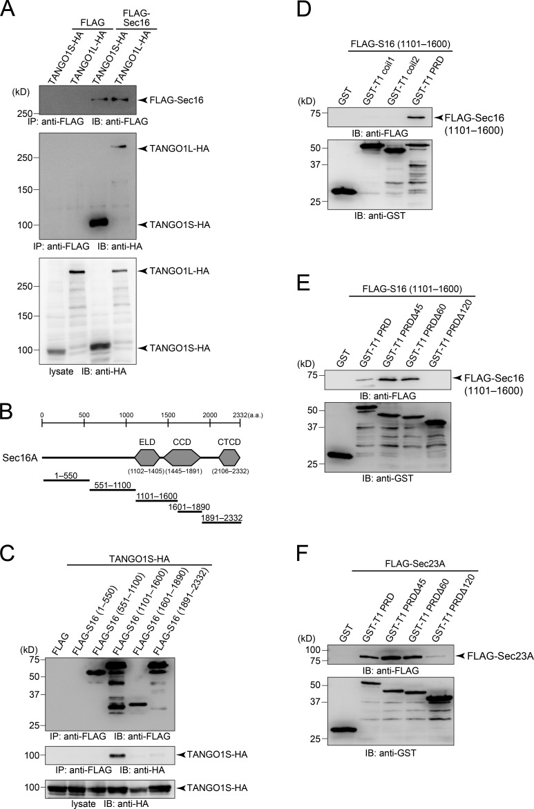
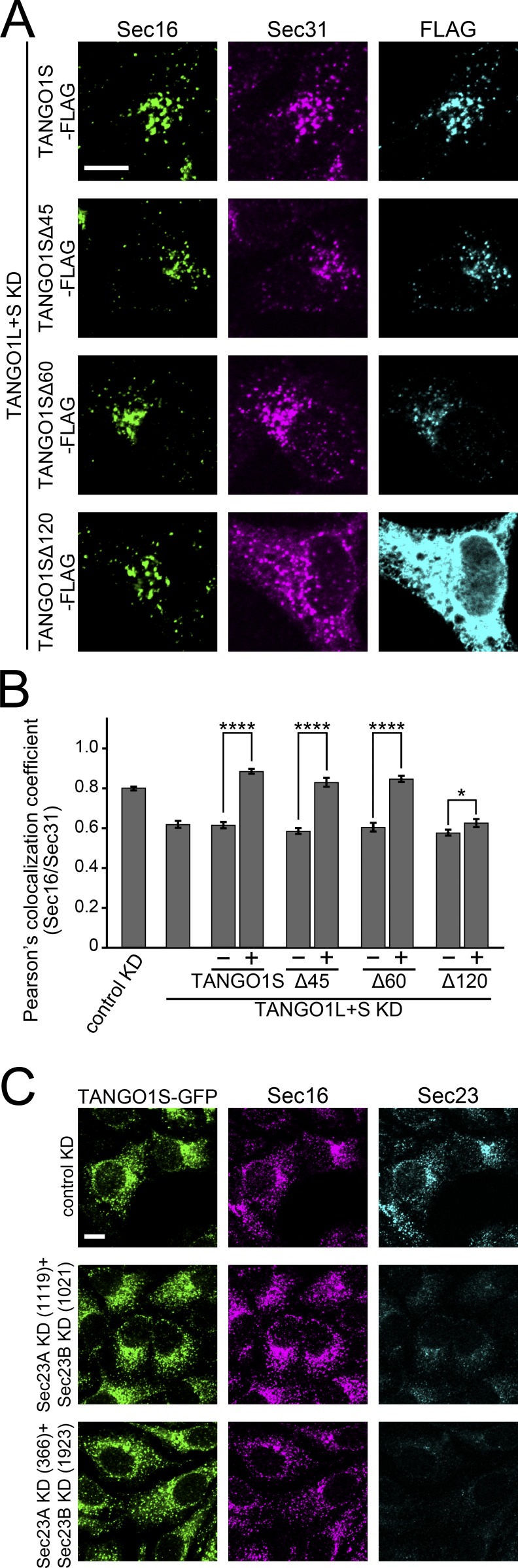
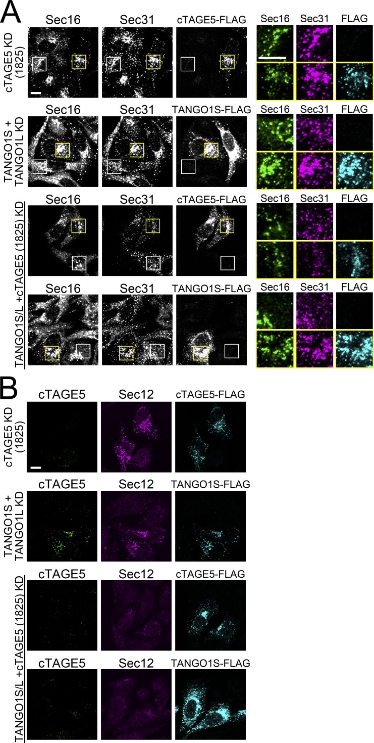
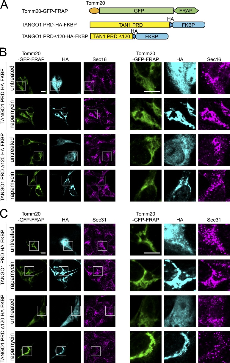
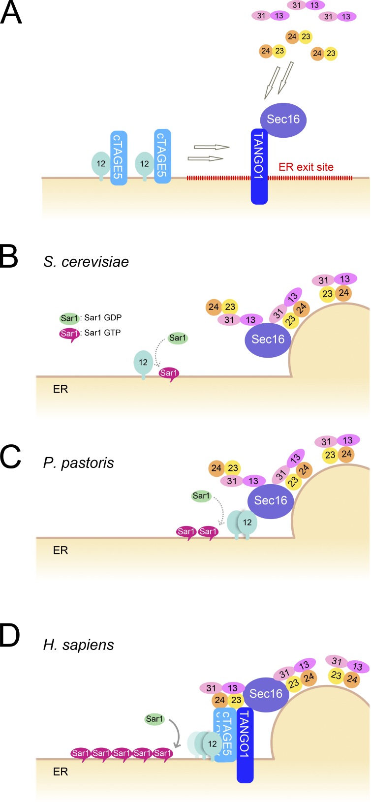
Mammalian endoplasmic reticulum (ER) exit sites export a variety of cargo molecules including oversized cargoes such as collagens. However, the mechanisms of their assembly and organization are not fully understood. TANGO1L is characterized as a collagen receptor, but the function of TANGO1S remains to be investigated. Here, we show that direct interaction between both isoforms of TANGO1 and Sec16 is not only important for their correct localization but also critical for the organization of ER exit sites. The depletion of TANGO1 disassembles COPII components as well as membrane-bound ER-resident complexes, resulting in fewer functional ER exit sites and delayed secretion. The ectopically expressed TANGO1 C-terminal domain responsible for Sec16 binding in mitochondria is capable of recruiting Sec16 and other COPII components. Moreover, TANGO1 recruits membrane-bound macromolecular complexes consisting of cTAGE5 and Sec12 to the ER exit sites. These data suggest that mammalian ER exit sites are organized by TANGO1 acting as a scaffold, in cooperation with Sec16 for efficient secretion.


Figures

Figure 1.
Figure 1.
TANGO1 depletion dissociates COPII components and delays secretion. (A) HeLa cells were transfected with the indicated siRNAs. After 48 h, cells were fixed and costained with anti-Sec16-C, anti-Sec31, and anti-Sec23 (5H2) antibodies. Right, magnifications of the indicated regions on the left. Bars, 10 µm. (B) Quantification of Pearson’s colocalization coefficient of A. Cells treated with control siRNA, n = 13; with panTANGO1 siRNA, n = 18; with TANGO1L and TANGO1S siRNAs, n = 17; with cTAGE5 (91) siRNA, n = 16; with cTAGE5 (1825) siRNA, n = 18. Error bars represent means ± SEM. *, P < 0.05 compared with control KD. (C) HeLa cells were transfected with the indicated siRNAs. After 48 h, proteins were extracted and subjected to SDS-PAGE, followed by Western blotting with anti-TANGO1 CC1, anti-Sec16-N, anti-cTAGE5 CC1, anti-Sec23A (11D8), anti-Sec12 (6B3), and anti β-actin antibodies. (D) HeLa cells were treated with TANGO1L and TANGO1S siRNAs. After 24 h, they were transfected with TANGO1S-FLAG construct and further cultured for 24 h. The cells were fixed and stained with anti-Sec16-C, anti-Sec31, and anti-FLAG antibodies. Right, magnifications of the indicated regions on the left (white square, TANGO1S-FLAG is not expressed; yellow square, TANGO1S-FLAG is expressed). Bars, 10 µm. (E) Quantification of Pearson’s colocalization coefficient of D. For cells treated with control siRNA, n = 17; with TANGO1L and TANGO1S siRNAs, n = 17; with TANGO1L and TANGO1S siRNAs, in which TANGO1S-FLAG is not expressed, n = 20; with TANGO1L and TANGO1S siRNAs, in which TANGO1S-FLAG is expressed, n = 12 (analysis of variance). *, P < 0.05; n.s., not significant. Error bars represent means ± SEM. (F) HeLa cells twice transfected with the indicated siRNAs were transfected with VSVG-ts045-GFP. The cells were cultured at 39.5°C to accumulate the protein in the ER, then incubated for the indicated times at 37°C before fixation. Fixed cells were stained with anti-Sec16-C and anti-Sec31 antibodies. Bars: (main) 10 µm; (insets) 1 µm.
Figure 2.
Figure 2.
TANGO1 is dispersed through the ER after Sec16 knockdown. (A) HeLa cells were transfected with the indicated siRNA. After 72 h, cells were fixed with cold methanol and costained with anti-Sec16-C, anti-TANGO1 CT, anti-Sec23 (5H2), anti-Sec31, anti-cTAGE5 CT, and anti-Sec12 (6B3) antibodies. Bars, 10 µm. (B) HeLa cells were transfected with the indicated siRNAs. After 72 h, proteins were extracted and subjected to SDS-PAGE, followed by Western blotting with anti-TANGO1 CC1, anti-Sec16-N, anti-cTAGE5 CC1, anti-Sec23A (11D8), anti-Sec12 (6B3), and anti β-actin antibodies. (C) HeLa cells were transfected with the indicated siRNAs. After 72 h, cells were fixed with PFA and costained with anti-Sec16-C, anti-TANGO1 CT, anti-Sec12 (6B3), and anti-KDEL antibodies. Bars, 10 µm. (D) Quantification of Pearson’s colocalization coefficient of C. For cells treated with control siRNA, n = 13; with Sec16 (3031) siRNA, n = 12; with Sec16 (5393) siRNAs, n = 10. Error bars represent means ± SEM. n.s., not significant compared with control KD.
Figure 3.
Figure 3.
TANGO1 interacts with Sec16. (A) 293T cells were transfected with TANGO1S-HA or TANGO1L-HA with FLAG-Sec16 constructs as indicated. Cell lysates were immunoprecipitated with anti-FLAG antibody and eluted with a FLAG peptide. Eluates were then subjected to SDS-PAGE followed by Western blotting with anti-FLAG and anti-HA antibodies. (B) Schematic representation of human Sec16A domain organization. CCD, central conserved domain; CTCD, C-terminal conserved domain. (C) 293T cells were transfected with FLAG-Sec16 (1–550 aa), FLAG-Sec16 (551–1,100 aa), FLAG-Sec16 (1,101–1,600 aa), FLAG-Sec16 (1,601–1,890 aa), or FLAG-Sec16 (1,891–2,332 aa) with TANGO1S-HA constructs as indicated. Cell lysates were immunoprecipitated with anti-FLAG antibody and eluted with a FLAG peptide. Eluates were then subjected to SDS-PAGE followed by Western blotting with anti-FLAG and anti-HA antibodies. (D) Recombinant GST, GST-tagged TANGO1-coil1 (1,211–1,440 aa), GST-tagged TANGO1-coil2 (1,441–1,650 aa), or GST-tagged TANGO1-PRD (1,651–1,907 aa) were immobilized to glutathione Sepharose resin and incubated with FLAG-Sec16 (1,101–1,600 aa). Resins were washed and eluted with glutathione. Eluted proteins were subjected to SDS-PAGE followed by Western blotting with anti-FLAG and anti-GST antibodies. (E and F) GST, GST-tagged TANGO1-PRD (1,651–1,907 aa), GST-tagged TANGO1-PRDΔ45 (1,651–1,862 aa), GST-tagged TANGO1-PRDΔ60 (1,651–1,847 aa), or GST-tagged TANGO1-PRDΔ120 (1,651–1,787 aa) were immobilized to glutathione Sepharose resin and incubated with FLAG-Sec16 (1,101–1,600 aa; E) or FLAG-Sec23A (F). Resins were washed and eluted with glutathione. Eluted proteins were subject to SDS-PAGE followed by Western blotting with anti-FLAG and anti-GST antibodies.
Figure 4.
Figure 4.
Interaction between TANGO1 and Sec16 is required for correct localization of both proteins and other COPII components. (A) HeLa cells were treated with TANGO1L and TANGO1S siRNAs. After 24 h, TANGO1S-FLAG, TANGO1SΔ45-FLAG, TANGO1SΔ60-FLAG, or TANGO1SΔ120-FLAG constructs were transfected and further cultured for 24 h. The cells were fixed and stained with anti-Sec16-C, anti-Sec31, and anti-FLAG antibodies. Bars, 10 µm. (B) Quantification of Pearson’s colocalization coefficient of A. For cells treated with control siRNA, n = 18; with TANGO1L and TANGO1S siRNAs, n = 16; with TANGO1L and TANGO1S siRNAs, in which TANGO1S-FLAG is not expressed, n = 18; with TANGO1L and TANGO1S siRNAs, in which TANGO1S-FLAG is expressed, n = 10; with TANGO1L and TANGO1S siRNAs, in which TANGO1SΔ45-FLAG is not expressed, n = 25; with TANGO1L and TANGO1S siRNAs, in which TANGO1SΔ45-FLAG is expressed, n = 10; with TANGO1L and TANGO1S siRNAs, in which TANGO1SΔ60-FLAG is not expressed, n = 19; with TANGO1L and TANGO1S siRNAs in which TANGO1SΔ60-FLAG is expressed, n = 10; with TANGO1L and TANGO1S siRNAs, in which TANGO1SΔ120-FLAG is not expressed, n = 27; with TANGO1L and TANGO1S siRNAs, in which TANGO1SΔ120-FLAG is expressed, n = 10 (analysis of variance). *, P < 0.05; ****, P < 0.0001. Error bars represent means ± SEM. (C) Doxycycline-inducible stable HeLa cell line for TANGO1S-GFP was treated with Sec23A and Sec23B siRNAs. After 24 h, TANGO1S-GFP was induced and cells were further cultured for 24 h. The cells were fixed and stained with anti–Sec16-C and anti-Sec23 (5H2) antibodies. Bars, 10 µm.
Figure 5.
Figure 5.
TANGO1 organizes membrane-bound ER exit site components. (A) HeLa cells were transfected with the indicated siRNAs. After 24 h, TANGO1S-FLAG or cTAGE5-FLAG constructs were transfected and further cultured for 24 h. The cells were fixed and stained with anti-Sec16-C, anti-Sec31, and anti-FLAG antibodies. Right, magnifications of the indicated regions on the left (white square, TANGO1S-FLAG or cTAGE5-FLAG are not expressed; yellow square, TANGO1S-FLAG or cTAGE5-FLAG are expressed). Bars, 10 µm. (B) HeLa cells were transfected with the indicated siRNAs. After 24 h, TANGO1S-FLAG or cTAGE5-FLAG constructs were transfected and further cultured for 24 h. The cells were fixed and stained with anti-cTAGE5 CT, anti-Sec12 (6B3), and anti-FLAG antibodies. Bars, 10 µm.
Figure 6.
Figure 6.
TANGO1 proline-rich domain recruits Sec16 and other COPII components. (A) Schematic representation of the constructs used in this study. Tomm20 (1–33 aa)-GFP-FRAP, TANGO1 PRD domain (1,651–1,907 aa)-HA-FKBP, TANGO1 PRD Δ120 (1,651–1,787 aa)-HA-FKBP. (B and C) HeLa cells were treated with TANGO1L and TANGO1S siRNAs. After 24 h, the Tomm20-GFP-FRAP construct and TANGO1 PRD-HA-FKBP or TANGO1 PRD Δ120-HA-FKBP constructs were transfected, and cells were further cultured for 24 h. The cells were incubated for another 2 h with or without 200 nM rapamycin. After fixation, the cells were stained with anti-HA and anti–Sec16-C (B) or anti-Sec31 (C) antibodies. Right, magnifications of the indicated regions on the left. Bars, 10 µm.
Figure 7.
Figure 7.
A model for mammalian ER exit site organization by TANGO1 and Sec16. (A) TANGO1 and Sec16 are mutually required for their localization to the ER exit sites. TANGO1 recruits membrane-bound ER-resident proteins to form macromolecular complexes. TANGO1 and Sec16 then interact with COPII components and localize them to the ER exit sites. (B) A model depicting ER exit sites of S. cerevisiae. Sec16 is recruited to the ER exit sites via the UCR. Sec12 is dispersed throughout the ER. Sar1 shows some accumulation at the rim of COPII-coated vesicles. (C) A model depicting ER exit sites of P. pastoris. Sec16 is recruited to the ER exit sites via interaction with COPII proteins. Sec12 is concentrated to the ER exit sites via interaction with Sec16. Sar1 is accumulated to the ER exit sites. (D) A model depicting ER exit sites of Homo sapiens. TANGO1 and Sec16 scaffold COPII components and ER-resident complexes at the ER exit sites. Sec16 is recruited to the ER exit sites via ELD by direct interaction with SIR of TANGO1. Sar1 is efficiently activated by multiple Sec12 molecules recruited to the ER exit sites by cTAGE5 and TANGO1.

References

    1. Aguilera-Gomez A., van Oorschot M.M., Veenendaal T., and Rabouille C.. 2016. In vivo vizualisation of mono-ADP-ribosylation by dPARP16 upon amino-acid starvation. eLife. 5:e21475 10.7554/eLife.21475 - DOI - PMC - PubMed
    1. Antonny B., Madden D., Hamamoto S., Orci L., and Schekman R.. 2001. Dynamics of the COPII coat with GTP and stable analogues. Nat. Cell Biol. 3:531–537. 10.1038/35078500 - DOI - PubMed
    1. Aridor M., Weissman J., Bannykh S., Nuoffer C., and Balch W.E.. 1998. Cargo selection by the COPII budding machinery during export from the ER. J. Cell Biol. 141:61–70. 10.1083/jcb.141.1.61 - DOI - PMC - PubMed
    1. Bannykh S.I., Rowe T., and Balch W.E.. 1996. The organization of endoplasmic reticulum export complexes. J. Cell Biol. 135:19–35. 10.1083/jcb.135.1.19 - DOI - PMC - PubMed
    1. Bard F., Casano L., Mallabiabarrena A., Wallace E., Saito K., Kitayama H., Guizzunti G., Hu Y., Wendler F., Dasgupta R., et al. . 2006. Functional genomics reveals genes involved in protein secretion and Golgi organization. Nature. 439:604–607. 10.1038/nature04377 - DOI - PubMed

MeSH terms