. 2017 Jun 16;292(24):9967-9974.
doi: 10.1074/jbc.M117.787119. Epub 2017 Apr 25.

β2-Adrenergic receptor activation mobilizes intracellular calcium via a non-canonical cAMP-independent signaling pathway

Affiliations

β2-Adrenergic receptor activation mobilizes intracellular calcium via a non-canonical cAMP-independent signaling pathway

Monica Galaz-Montoya et al. J Biol Chem. .

Abstract

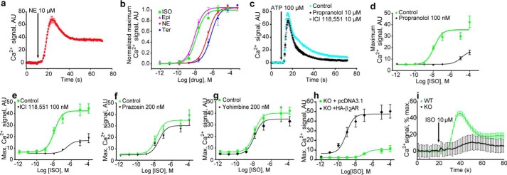
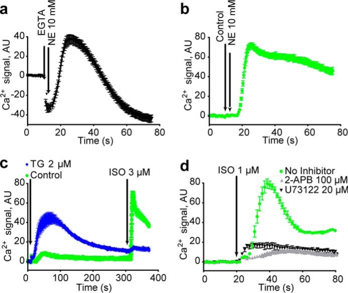
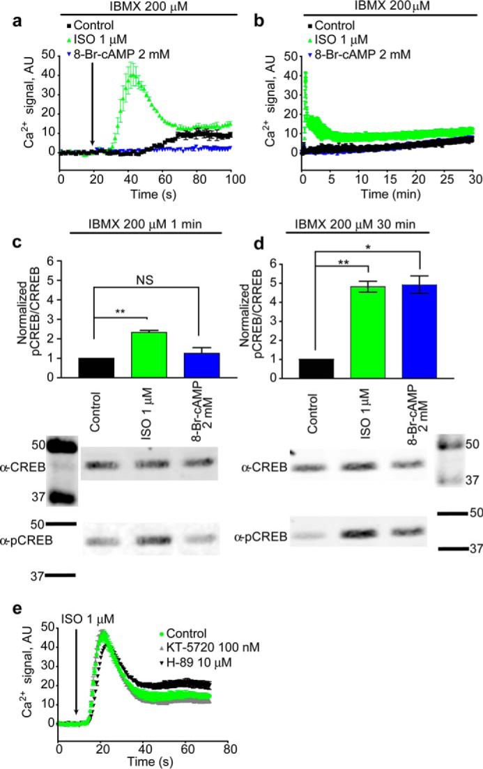
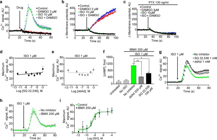
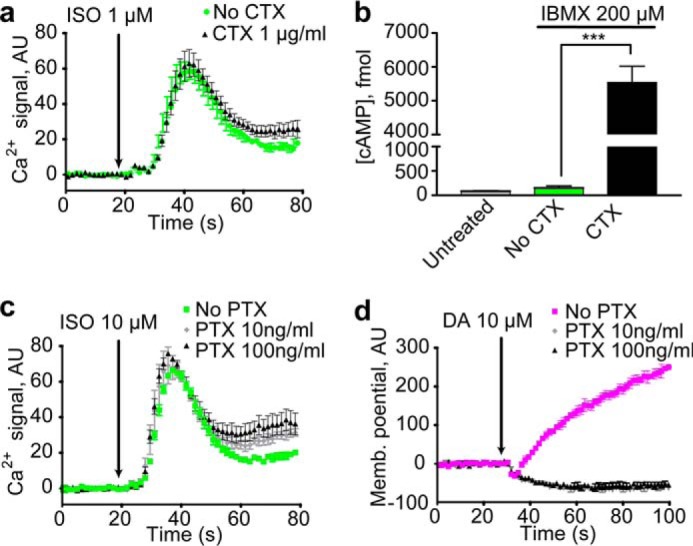
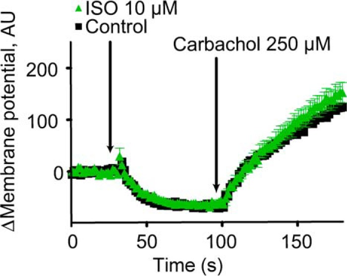
Beta adrenergic receptors (βARs) are G-protein-coupled receptors essential for physiological responses to the hormones/neurotransmitters epinephrine and norepinephrine which are found in the nervous system and throughout the body. They are the targets of numerous widely used drugs, especially in the case of the most extensively studied βAR, β2AR, whose ligands are used for asthma and cardiovascular disease. βARs signal through Gαs G-proteins and via activation of adenylyl cyclase and cAMP-dependent protein kinase, but some alternative downstream pathways have also been proposed that could be important for understanding normal physiological functioning of βAR signaling and its disruption in disease. Using fluorescence-based Ca2+ flux assays combined with pharmacology and gene knock-out methods, we discovered a previously unrecognized endogenous pathway in HEK-293 cells whereby β2AR activation leads to robust Ca2+ mobilization from intracellular stores via activation of phospholipase C and opening of inositol trisphosphate (InsP3) receptors. This pathway did not involve cAMP, Gαs, or Gαi or the participation of the other members of the canonical β2AR signaling cascade and, therefore, constitutes a novel signaling mechanism for this receptor. This newly uncovered mechanism for Ca2+ mobilization by β2AR has broad implications for adrenergic signaling, cross-talk with other signaling pathways, and the effects of βAR-directed drugs.

Keywords: G-protein-coupled receptor (GPCR); adrenergic receptor; calcium intracellular release; cell signaling; cyclic AMP (cAMP); β2-adrenergic receptor.


Conflict of interest statement

The authors declare that they have no conflicts of interest with the contents of this article. The content is solely the responsibility of the authors and does not necessarily represent the official views of the National Institutes of Health.

Figures

Figure 1.
Figure 1.
Endogenous β2AR activation increased cytoplasmic [Ca2+] in HEK cells. Continuous changes in Fluo-4 fluorescence intensity with time (a and c) or peak increases in intensity as a function of drug concentration (b and d–h) are plotted. a, NE treatment increased cytoplasmic [Ca2+]. b, NE response is mimicked by AR agonists epinephrine (Epi) and isoproterenol (ISO) and the β2AR-selective terbutaline (Ter). c, the β-AR inhibitor propranolol and the β2AR-selective ICI 118,551 do not suppress P2Y receptor signaling. d and e, β-adrenergic inhibitors suppress responses to ISO. f and g, α-adrenergic inhibitors do not suppress ISO responses. h and i, loss of Ca2+ response in cells lacking β2AR and restoration by β2AR expression. h, β2AR deletion mutant cells (KO) were transfected with pcDNA3.1 or HA-tagged β2AR and tested for Ca2+ responses over a range of ISO concentrations. i, β2AR deletion mutant cells (KO) or wildtype (WT) cells were treated with 10 μm ISO at the indicated times, and Ca2+ responses were monitored over time. For all panels Ca2+ traces represent three or more independent experiments; error bars indicate internal replicate S.E. Dose responses are the averages of three or more independent experiments, and error bars indicate S.E. AU, absorbance units.
Figure 2.
Figure 2.
The β2AR induced calcium response is due to release from the endoplasmic reticulum. a and b, chelation of extracellular Ca2+ with EGTA does not eliminate β2AR-mediated Ca2+ mobilization. c, depletion of endoplasmic reticulum Ca2+ by treatment with thapsigargin (TG) for 5 min nearly eliminates signaling with ISO. d, treatment with InsP3R inhibitor 2-APB and PLC inhibitor U73122 for 1 h suppresses signaling with ISO. Ca2+ traces represent three or more independent experiments, and error bars indicate internal replicate S.E. AU, absorbance units.
Figure 3.
Figure 3.
PKA did not mediate the β2AR calcium response. a, 1 min of 8-Br-cAMP treatment did not mimic ISO response. b, 30 min of 8-Br-cAMP treatment did not mimic ISO response. c, 1 min of ISO significantly raised pCREB levels (**, p = 0.0065), whereas 1 min of 8-Br-cAMP treatment did not significantly raise pCREB levels (NS). d, 30 min of ISO (**, p = 0.0056) and 8-Br-cAMP treatment (*, p = 0.0133) significantly elevated intracellular pCREB. e, treatment with PKA inhibitors KT-5720 and H-89 for 1 h did not suppress signaling with ISO. Ca2+ traces represent three or more independent experiments, and error bars indicate internal replicate S.E. pCREB/CREB graphs are the averages of three independent experiments; error bars indicate S.E. Full blot images are shown in supplemental Fig. S1. AU, absorbance units.
Figure 4.
Figure 4.
The β2AR calcium response was not mediated by AC or cAMP. a, Gαi activation did not inhibit ISO signals. b, Gαi activation induced changes in membrane potential. c, treatment with PTX abolished μOR-induced membrane potential changes. d–g, treatment with AC inhibitors SQ 22,536 and ddAd did not suppress signaling with ISO, even at 1 mm, although AC inhibitor treatment significantly suppressed cAMP formation (* = p = 0.0298 and ** = p = 0.0059) (f). h and i, IBMX treatment did not potentiate signaling with ISO. Ca2+ traces represent three or more independent experiments, and error bars indicate internal replicate S.E. Dose responses and bar graphs are the averages of three or more independent experiments, and error bars indicate S.E. AU, absorbance units.
Figure 5.
Figure 5.
β2AR did not couple to Gαs or Gαi to initiate the calcium response. a, treatment with the Gαs CTX did not potentiate signaling with ISO. b, treatment with CTX significantly increased cytoplasmic cAMP (***, p = 0.0004). c, treatment with the Gαi PTX did not suppress signaling with ISO. d, treatment with PTX completely eliminated changes in membrane potential induced by the dopamine-2 receptor upon the addition of dopamine (DA). Ca2+ and membrane potential traces represent three or more independent experiments, and error bars indicate internal replicate S.E. The bar graphs are the averages of three or more independent experiments, and the error bars indicate S.E. AU, absorbance units.
Figure 6.
Figure 6.
Supporting evidence against Gαq coupling to β2AR in TRPC4 β-expressing HEK-293 cells. Treatment with ISO did not mimic the membrane potential response of the Gαq-coupled M3 muscarinic receptors activated with carbachol. Ca2+ traces represent three or more independent experiments, and error bars indicate internal replicate S.E. Fig. 4 demonstrates robust membrane potential changes in response to μOR stimulation with DAMGO under these conditions without PTX treatment and robust Ca2+ release in response to isoproterenol stimulation under these conditions in these cells. AU, absorbance units.

Similar articles

Cited by

References

    1. Milligan G., Svoboda P., and Brown C. M. (1994) Why are there so many adrenoceptor subtypes? Biochem. Pharmacol. 48, 1059–1071 - PubMed
    1. Hoffmann C., Leitz M. R., Oberdorf-Maass S., Lohse M. J., and Klotz K. N. (2004) Comparative pharmacology of human β-adrenergic receptor subtypes: characterization of stably transfected receptors in CHO cells. Naunyn Schmiedebergs Arch. Pharmacol. 369, 151–159 - PubMed
    1. Weitl N., and Seifert R. (2008) Distinct interactions of human β1- and β2-adrenoceptors with isoproterenol, epinephrine, norepinephrine, and dopamine. J. Pharmacol. Exp. Ther. 327, 760–769 - PubMed
    1. Bylund D. B., Eikenberg D. C., Hieble J. P., Langer S. Z., Lefkowitz R. J., Minneman K. P., Molinoff P. B., Ruffolo R. R. Jr., and Trendelenburg U. (1994) International Union of Pharmacology nomenclature of adrenoceptors. Pharmacol. Rev. 46, 121–136 - PubMed
    1. Summers R. J., Kompa A., and Roberts S. J. (1997) β-adrenoceptor subtypes and their desensitization mechanisms. J. Auton. Pharmacol. 17, 331–343 - PubMed

Publication types

MeSH terms

Substances