. 2017 Jun 23;292(25):10672-10684.
doi: 10.1074/jbc.M117.783068. Epub 2017 Apr 25.

The phospholipase iPLA2γ is a major mediator releasing oxidized aliphatic chains from cardiolipin, integrating mitochondrial bioenergetics and signaling

Affiliations

The phospholipase iPLA2γ is a major mediator releasing oxidized aliphatic chains from cardiolipin, integrating mitochondrial bioenergetics and signaling

Gao-Yuan Liu et al. J Biol Chem. .

Abstract

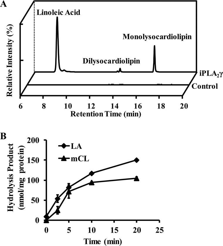
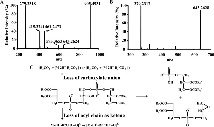
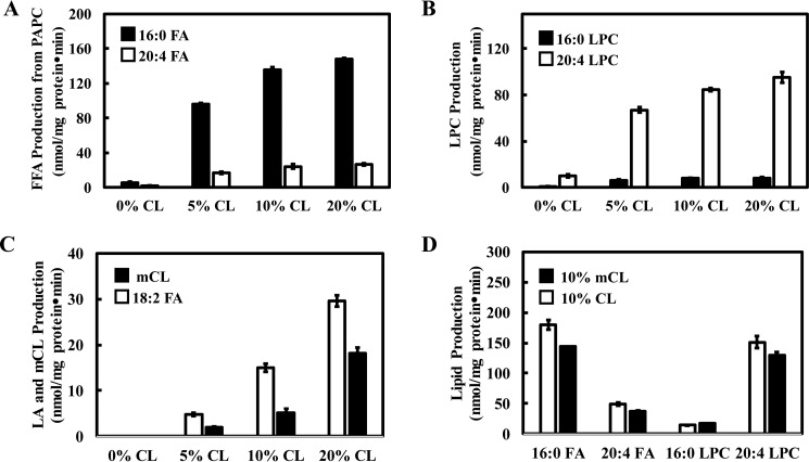
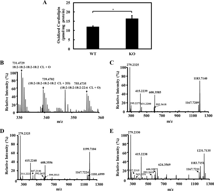
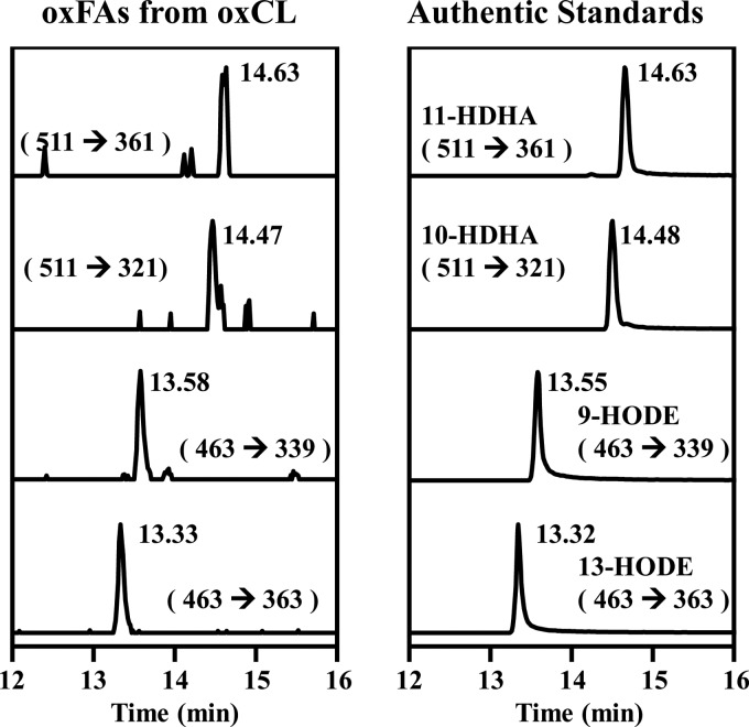
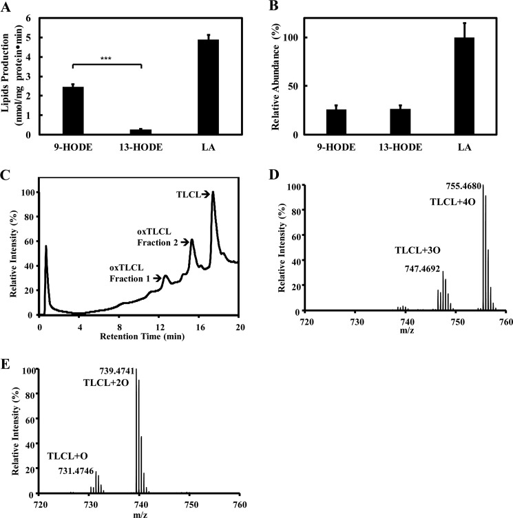
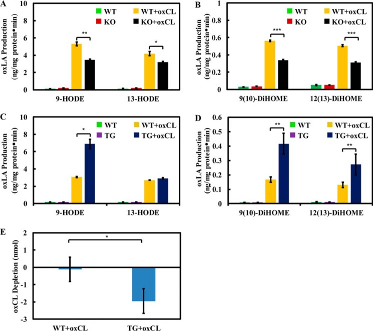
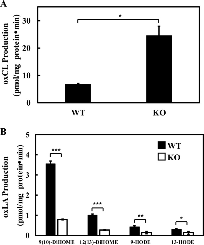
Cardiolipin (CL) is a dimeric phospholipid with critical roles in mitochondrial bioenergetics and signaling. Recently, inhibition of the release of oxidized fatty acyl chains from CL by the calcium-independent phospholipase A2γ (iPLA2γ)-selective inhibitor (R)-BEL suggested that iPLA2γ is responsible for the hydrolysis of oxidized CL and subsequent signaling mediated by the released oxidized fatty acids. However, chemical inhibition by BEL is subject to off-target pharmacologic effects. Accordingly, to unambiguously determine the role of iPLA2γ in the hydrolysis of oxidized CL, we compared alterations in oxidized CLs and the release of oxidized aliphatic chains from CL in experiments with purified recombinant iPLA2γ, germ-line iPLA2γ-/- mice, cardiac myocyte-specific iPLA2γ transgenic mice, and wild-type mice. Using charge-switch high mass accuracy LC-MS/MS with selected reaction monitoring and product ion accurate masses, we demonstrated that iPLA2γ is the major enzyme responsible for the release of oxidized aliphatic chains from CL. Our results also indicated that iPLA2γ selectively hydrolyzes 9-hydroxy-octadecenoic acid in comparison to 13-hydroxy-octadecenoic acid from oxidized CLs. Moreover, oxidative stress (ADP, NADPH, and Fe3+) resulted in the robust production of oxidized CLs in intact mitochondria from iPLA2γ-/- mice. In sharp contrast, oxidized CLs were readily hydrolyzed in mitochondria from wild-type mice during oxidative stress. Finally, we demonstrated that CL activates the iPLA2γ-mediated hydrolysis of arachidonic acid from phosphatidylcholine, thereby integrating the production of lipid messengers from different lipid classes in mitochondria. Collectively, these results demonstrate the integrated roles of CL and iPLA2γ in lipid second-messenger production and mitochondrial bioenergetics during oxidative stress.

Keywords: cardiolipin; lipid metabolism; lipid oxidation; lipid signaling; mass spectrometry (MS).


Conflict of interest statement

The content is solely the responsibility of the authors and does not necessarily represent the official views of the National Institutes of Health

Figures

Figure 1.
Figure 1.
iPLA2γ mediated hydrolysis of cardiolipin to produce free fatty acids, monolysocardiolipin, and dilysocardiolipin. A, extracted ion chromatogram of iPLA2γ-hydrolyzed products from TLCL. Purified recombinant iPLA2γ (6 μg) was incubated with 6 μm TLCL (10 mol%) and 54 μm PAPC at 37 °C for 15 min in 20 mm HEPES, pH 7.2, containing 2 mm EGTA and 1 mm DTT. The reaction was terminated by adding chloroform/methanol (1:1, v/v), and the resultant lipids were extracted in the presence of internal standards (14:0–14:0–14:0 monolysocardiolipin (mCL), d4-16:0-FFA). The chloroform phase was separated and dried under a nitrogen stream. The dried residue was reconstituted in methanol, separated on a C18 HPLC column, and analyzed using a LTQ-Orbitrap mass spectrometer with a mass resolution of 30,000 at m/z = 400 and in the negative ion mode. The extracted ion chromatograms of linoleic acid (279.2325), monolysocardiolipin (592.3632), and dilysocardiolipin (461.2479) are as shown. B, production of linoleic acid (LA) and monolysocardiolipin (mCL) from TLCL hydrolysis by iPLA2γ at different incubation times. Values are the average of three independent preparations ± S.E.
Figure 2.
Figure 2.
Identification of monolysocardiolipin and dilysocardiolipin released from TLCL by purified recombinant human iPLA2γ. The lysocardiolipins generated by iPLA2γ-mediated hydrolysis of TLCL were separated on a C18 HPLC column and analyzed by mass spectrometry. Fragmentations were performed in an LTQ ion trap with a collision energy of 30 eV, and the resultant fragment ions were detected in Orbitrap with a mass resolution of 30,000 at m/z = 400 and a mass accuracy within 5 ppm. A, MS2 spectra of parent ion [M-2H+]2− at m/z 592 (corresponding monolysocardiolipin, the chromatographic peak at 17 min in Fig. 1A). The major fragment ions at m/z 279 and m/z 905 resulting from 18:2 carboxyl anion loss of [M-2H+]2− are characteristic for monolysocardiolipin. B, MS2 spectra of the parent ion [M-2H+]2− at m/z 461 (corresponding dilysocardiolipin, the chromatographic peak at 14 min in Fig. 1A). The major fragment ions at m/z 279 and m/z 643 resulting from 18:2 carboxyl anion loss of [M-2H+]2− are characteristic for dilysocardiolipin. C, scheme of the fragmentation pathways of doubly charged cardiolipin.
Figure 3.
Figure 3.
Cardiolipin activated iPLA2γ phospholipase activity resulting in increased release of free fatty acids and lysolipids. A–C, effect of increasing CL content on PAPC and CL hydrolysis. Purified recombinant iPLA2γ (6 μg) was incubated with PAPC SUVs (60 μm) containing either 0, 3, 6, or 12 μm TLCL (0, 5, 10, 20 mol % of PAPC) at 37 °C for 15 min in 20 mm HEPES, pH 7.2, containing 2 mm EGTA and 1 mm DTT. The reaction was terminated by adding chloroform/methanol (1:1, v/v), and the resultant lipids were extracted in the presence of internal standards (17:0-LPC and d4-16:0-FFA). The chloroform phase was separated and dried under nitrogen stream. The dried residue was reconstituted in methanol, separated on a C18 HPLC column, and analyzed by an LTQ-Orbitrap mass spectrometer. The palmitic acid and arachidonic acid released from PAPC (A), lysophosphatidylcholine released from PAPC (B), and linoleic acid and monolysocardiolipin (mCL) released from TLCL (C) were quantified. D, purified recombinant iPLA2γ (6 μg) was incubated with 6 μm TLCL or 6 μm 18:2–18:2–18:2 monolysocardiolipin and 60 μm PAPC at 37 °C for 15 min in 20 mm HEPES, pH 7.2, containing 2 mm EGTA and 1 mm DTT. The lipids released from PAPC were quantified and comparatively shown. Values are the average of three independent preparations ± S.E.
Figure 4.
Figure 4.
Genetic ablation of iPLA2γ caused the accumulation of oxidized cardiolipin. A, oxidized cardiolipin levels (i.e. the sum of the three predominant oxidized CL species) in wild-type and iPLA2γ−/− myocardium tissue. Freshly isolated heart tissues from wild-type and iPLA2γ−/− mice were flash-frozen in liquid nitrogen, homogenized using a Teflon pestle grinder, and extracted in the presence of TMCL internal standard. The extracts were purified by aminopropyl solid phase extraction column and analyzed by LC-MS/MS in negative ion mode as described under “Experimental procedures.” Values are the average of four independent preparations ± S.E. *, p < 0.05. B, mass spectrum of oxidized cardiolipin from wild-type mouse myocardium tissue. C–E, aminopropyl solid phase extraction purified lipid extract (from two mouse hearts) was separated on a C18 HPLC column, and the fraction containing oxidized cardiolipin was collected and dried. The dried residue was reconstituted in 50 μl of methanol and analyzed by LC-MS/MS. Fragmentations were performed in the LTQ ion trap with collision energy of 30 eV, and the resultant fragment ions were detected in Orbitrap with a mass resolution of 30,000 at m/z = 400 and a mass accuracy within 5 ppm. MS2 spectra of parent ion [M-2H+]2− at m/z 731 (corresponding 18:2–18:2–18:2–18:2-CL-OH) (C), parent ion [M-2H+]2− at m/z 739 (corresponding 18:2–18:2–18:2–18:2-CL-OOH) (D), and parent ion [M-2H+]2− at m/z 755 (corresponding 18:2–18:2–18:2–22:6-CL-OH) (E) are shown here.
Figure 5.
Figure 5.
Identification of the oxidized fatty acyl chains in oxidized cardiolipin from mouse myocardial tissue. Lipids extracts from mouse myocardial tissue were first purified using an aminopropyl solid phase extraction column, and the oxidized cardiolipin was then separated by a C18 HPLC column. The purified oxidized cardiolipin was then completely hydrolyzed by porcine pancreas PLA2 and T. lanuginosus PLA1, derivatized with AMPP, and analyzed by LC-MS/MS. The left panel displays the transitions of the detected oxidized fatty acids in the hydrolyzed oxidized cardiolipin sample, which have identical retention times as authentic standards (right panel).
Figure 6.
Figure 6.
Recombinant human iPLA2γ selectively hydrolyzed 9-HODE, but not 13-HODE, from oxidized TLCL. A, specific activity of iPLA2γ hydrolysis of oxidized cardiolipin. Purified recombinant iPLA2γ (2.5 μg) was incubated with 6 μm TLCL, 6 μm oxTLCL (fraction 1 from HPLC purification of oxCL), and 48 μm PAPC at 37 °C for 10 min in 20 mm HEPES, pH 7.2, containing 2 mm EGTA and 1 mm DTT. The products including 9-HODE, 13-HODE, and linoleic acid (LA) were extracted in the presence of internal standards (d4-16:0 FFA, 13-HODE-d4), derivatized with AMPP, and analyzed by LC-MS/MS. B, relative amounts of 9-HODE, 13-HODE, and LA in TLCL/oxTLCL/PAPC vesicles after complete hydrolysis by porcine pancreas PLA2 and T. lanuginosus PLA1, derivatization with AMPP, and analysis by LC-MS/MS. C–E, oxidized cardiolipin used as substrate for iPLA2γ hydrolysis was prepared by cytochrome c-mediated oxidation in the presence of hydrogen peroxide as described previously (52) and purified by reversed-phase HPLC (C). Fraction 1 (D) and fraction 2 (E) containing oxTLCL were collected and analyzed by mass spectrometry as described under “Experimental procedures.” Values are the average of three independent preparations ± S.E. ***, p < 0.001.
Figure 7.
Figure 7.
Oxidized linoleic acid production and oxidized cardiolipin consumption in wild-type, iPLA2γ−/−, and cardiac myocyte-specific iPLA2γ transgenic mitochondrial homogenates in the presence of exogenous oxTLCL. A and B, mitochondria were isolated from wild-type and iPLA2γ−/− mouse liver and homogenized by sonication. Mitochondria homogenates (1 mg protein/ml) were incubated with 20 μm oxTLCL or ethanol vehicle alone at 37 °C for 15 min. The reactions were terminated by the addition of methanol (25% total volume) containing internal standards (13-HODE-d4, 12(13)-DiHOME-d4). The released oxidized fatty acids were purified by reversed phase solid phase extraction, derivatized with AMPP, and finally analyzed by LC-MS/MS. C and D, the same experiments were performed with heart mitochondria isolated from wild-type and cardiac myocyte specific iPLA2γ transgenic mice. E, heart mitochondria were isolated from wild-type and cardiac myocyte-specific iPLA2γ transgenic mice. Mitochondria homogenates (1 mg protein/ml) were incubated with 20 μm oxTLCL or ethanol vehicle alone at 37 °C for 15 min. The reactions were terminated by adding chloroform/methanol 1:1 (v/v) in the presence of TMCL internal standard. The chloroform phase was separated, dried, and redissolved in chloroform. The oxidized cardiolipin was purified by aminopropyl solid phase extraction column and analyzed by LC-MS/MS in the negative ion mode. The results here show the consumption of oxidized cardiolipin per mg of protein in 30-min incubations. Values are the average of three independent preparations ± S.E. *, p < 0.05; **, p < 0.01; ***, p < 0.001.
Figure 8.
Figure 8.
Accumulation of oxidized cardiolipin (A) and production of oxidized linoleic acids (B) in intact wild-type and iPLA2γ−/− mitochondria stimulated by oxidative stress. Myocardial mitochondria were isolated from wild-type and iPLA2γ−/− mice and reconstituted in isotonic buffer. Intact mitochondria (0.8 mg/ml) were incubated with 2 mm ADP, 0.3 mm NADPH, 0.012 mm Fe3+, and 2.5 mm phosphate at 37 °C for 15 min. The reactions were terminated by adding chloroform/methanol (1:1, v/v). The chloroform phase was separated and dried under a nitrogen stream. A, for analysis of oxidized cardiolipin, the dried residues were redissolved in chloroform, purified by aminopropyl solid phase extraction, and analyzed by LC-MS/MS in the negative ion mode. B, for analysis of oxidized linoleic acids (oxLAs), the dried residues were redissolved in water/methanol 4:1, purified by reversed phase solid phase extraction, derivatized with AMPP, and finally analyzed by LC-MS/MS in the positive ion mode. Values are the average of four independent preparations ± S.E. *, p < 0.05; **, p < 0.01; ***, p < 0.001.

References

    1. Patil V. A., and Greenberg M. L. (2013) Cardiolipin-mediated cellular signaling. Adv. Exp. Med. Biol. 991, 195–213 - PubMed
    1. Shen Z., Li Y., Gasparski A. N., Abeliovich H., and Greenberg M. L. (2017) Cardiolipin regulates mitophagy through the PKC pathway. J. Biol. Chem. 292, 2916–2923 - PMC - PubMed
    1. Raja V., Joshi A. S., Li G., Maddipati K. R., and Greenberg M. L. (2017) Loss of cardiolipin leads to perturbation of acetyl-CoA synthesis. J. Biol. Chem. 292, 1092–1102 - PMC - PubMed
    1. Ren M., Phoon C. K., and Schlame M. (2014) Metabolism and function of mitochondrial cardiolipin. Prog. Lipid. Res. 55, 1–16 - PubMed
    1. Acehan D., Malhotra A., Xu Y., Ren M., Stokes D. L., and Schlame M. (2011) Cardiolipin affects the supramolecular organization of ATP synthase in mitochondria. Biophys. J. 100, 2184–2192 - PMC - PubMed

Publication types