. 2017 Apr 25;8(2):e00333-17.
doi: 10.1128/mBio.00333-17.

The EXIT Strategy: an Approach for Identifying Bacterial Proteins Exported during Host Infection

Affiliations

The EXIT Strategy: an Approach for Identifying Bacterial Proteins Exported during Host Infection

E F Perkowski et al. mBio. .

Erratum in

Abstract

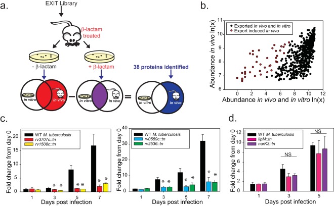
Exported proteins of bacterial pathogens function both in essential physiological processes and in virulence. Past efforts to identify exported proteins were limited by the use of bacteria growing under laboratory (in vitro) conditions. Thus, exported proteins that are exported only or preferentially in the context of infection may be overlooked. To solve this problem, we developed a genome-wide method, named EXIT (exported in vivotechnology), to identify proteins that are exported by bacteria during infection and applied it to Mycobacterium tuberculosis during murine infection. Our studies validate the power of EXIT to identify proteins exported during infection on an unprecedented scale (593 proteins) and to reveal in vivo induced exported proteins (i.e., proteins exported significantly more during in vivo infection than in vitro). Our EXIT data also provide an unmatched resource for mapping the topology of M. tuberculosis membrane proteins. As a new approach for identifying exported proteins, EXIT has potential applicability to other pathogens and experimental conditions.IMPORTANCE There is long-standing interest in identifying exported proteins of bacteria as they play critical roles in physiology and virulence and are commonly immunogenic antigens and targets of antibiotics. While significant effort has been made to identify the bacterial proteins that are exported beyond the cytoplasm to the membrane, cell wall, or host environment, current methods to identify exported proteins are limited by their use of bacteria growing under laboratory (in vitro) conditions. Because in vitro conditions do not mimic the complexity of the host environment, critical exported proteins that are preferentially exported in the context of infection may be overlooked. We developed a novel method to identify proteins that are exported by bacteria during host infection and applied it to identify Mycobacterium tuberculosis proteins exported in a mouse model of tuberculosis.

Keywords: EXIT; Mycobacterium tuberculosis; beta-lactamase reporter; in vivo; membrane proteins; protein export; protein secretion; virulence.


Figures

FIG 1
FIG 1
(a) The ‘BlaTEM reporter. The ‘BlaTEM reporter is compatible with proteins localized to the bacterial cytoplasmic membrane or cell wall or secreted from the bacterial cell. The right panel indicates in-frame fusions to categories of exported proteins that confer β-lactam resistance (red). In-frame fusions to cytoplasmic proteins or the cytoplasmic domain of integral membrane proteins (purple) do not confer β-lactam resistance. (b) EXIT strategy. In step 1, a comprehensive library of 5 × 10⁶ plasmids containing fragments of M. tuberculosis (Mtb) genomic DNA fused to the ‘blaTEM reporter was constructed. The plasmid library was transformed into the ΔblaC β-lactamase-sensitive mutant of M. tuberculosis, and 5 × 10⁶ transformants were pooled to generate the EXIT library. In step 2, mice were infected by intravenous injection with the EXIT library and treated with β-lactam antibiotics (oral gavage twice daily) to select for EXIT clones exporting ‘BlaTEM fusion proteins. β-lactam treatment began 1 day after infection and continued to 2 weeks after infection. Mice were sacrificed, and spleens and lungs were harvested and homogenized. In step 3, organ homogenates were plated on 7H10 agar and grown to recover M. tuberculosis clones that survived β-lactam treatment during infection. Plates were scraped, and colonies were pooled separately for lungs and spleens. In step 4, plasmids from the recovered bacteria and the input samples were isolated and the fusion junction was sequenced using next-generation sequencing. Sequencing primers were designed to read out of the ‘blaTEM reporter and sequence the immediately adjacent M. tuberculosis DNA. Sequences were aligned to the M. tuberculosis genome. Unique sequences were counted to identify the abundance of each fusion junction site within the population. The genes that were most highly abundant after in vivo β-lactam treatment were identified, and the results corresponded to plasmids producing in-frame exported ‘BlaTEM fusion proteins.
FIG 2
FIG 2
EXIT identified 593 proteins as exported during murine infection. (a) The most abundant fusion site within each annotated gene in the M. tuberculosis genome was identified individually within the output for each of two replicate experiments. The lower of these two numbers was plotted on a histogram. A two-component Gaussian mixture model (black line overlay) was used to generate a statistical model distinguishing between high-abundance genes (right) and low-abundance genes (left), with a statistical cutoff of log10 = 2.90 (red line). A total of 593 genes were identified in the high-abundance population corresponding to EXIT exported proteins. (b) Genes identified as encoding exported proteins were analyzed for the number of statistically enriched unique fusion sites after in vivo β-lactam treatment. On average, 4 unique fusion sites were enriched for each exported protein. Percentiles are shown with dotted lines representing the 25th and 75th percentiles and a solid line representing the 50th percentile. (c) The input EXIT library was composed of fusions in 99% of M. tuberculosis genes, with 74% encoding proteins with no predicted export signal (yellow), 15% encoding predicted integral membrane proteins (blue), and 11% encoding proteins containing predicted signal peptides (black). In contrast, 95% of the proteins in the EXIT output contained an export signal. The 593 proteins identified as exported in EXIT were composed of 57% predicted integral membrane proteins (blue), 38% proteins containing a predicted signal peptide (black), and 5% proteins with no predicted export signal (yellow). By analysis of all ORFs of M. tuberculosis H37Rv for in silico predicted export signals (see Materials and Methods), 26% (1,040 proteins) of the M. tuberculosis proteome were predicted to be exported. This compares well to predictions of exported proteins in other bacteria, which usually predict 20% to 30% of the proteome to be exported (77).
FIG 3
FIG 3
Validation of EXIT-identified exported proteins with no in silico predicted export signal. Three proteins with no in silico predicted export signal (Rv1728c, Rv3707c, and Rv3811) were engineered with C-terminal HA tags and expressed from the constitutive hsp60 promoter in M. tuberculosis. Cells were irradiated, lysed by the use of a French pressure cell into whole-cell lysate (WCL), equalized by bicinchoninic acid (BCA) protein quantification, and fractionated by differential ultracentrifugation into cell wall (CW), membrane (MEM), and soluble/cytoplasmic (SOL) fractions. Fractions derived from equivalent amounts of starting cellular material were separated by SDS-PAGE, and HA-tagged proteins were detected by immunoblotting performed with anti-HA antibodies. The cell wall protein (HbhA), membrane protein (19-kDa lipoprotein), and cytoplasmic protein (SigA) were included as fractionation controls.
FIG 4
FIG 4
MmpL3 topology mapping using EXIT fusion site data. A total of 37 unique fusion sites in MmpL3 were represented in the input library (black hexagons). Of these, 13 fusion sites were enriched during β-lactam treatment of mice, indicating an extracytoplasmic location (red hexagons) corresponding to two large exported domains of the MmpL3 protein. Exported fusion sites were mapped onto the in silico topology prediction generated by TopPred (21).
FIG 5
FIG 5
Strategy for identification of in vivo induced exported proteins. (a) Identification of in vivo induced exported proteins. Spleens from β-lactam-treated mice infected with the EXIT library were harvested after 2 weeks of infection. Spleen homogenates were plated in parallel on 7H10 agar without β-lactam to recover all clones (red Venn diagram) and on 7H10 agar containing β-lactam to recover clones exporting ‘BlaTEM fusion proteins during in vivo growth and in vitro growth (purple Venn diagram). The population of clones identified only or in significantly greater abundance on media lacking β-lactams represents proteins whose export was induced during infection (blue). (b) Sequenced read count values recovered from agar with or without β-lactam for the 593 EXIT proteins were plotted to compare abundances after β-lactam treatment in vivo, with the abundance after dual β-lactam treatment in vivo and in vitro indicated. The majority of proteins identified as exported in vivo remained highly abundant after additional β-lactam treatment in vitro (black). A total of 38 genes (highlighted in red) were identified as statistically less abundant after in vitro β-lactam selection, representing proteins exported significantly more in vivo than in vitro (see Materials and Methods for details on statistical analysis). (c) In vivo induced exported proteins with roles promoting growth in macrophages (rv1508::tn, rv3707c:tn, rv0559c::tn, and rv2536::tn). Murine bone marrow-derived macrophages were infected with M. tuberculosis CDC1551 transposon mutants lacking individual in vivo induced exported proteins. At specific times postinfection, macrophage lysates were plated to measure intracellular CFU. The fold change in CFU over the course of the infection is plotted relative to the bacterial burden at day 0 postinfection. Statistical significance was determined by one-way analysis of variance (ANOVA) with multiple comparisons performed by the use of the Holm-Sidak (normal by Shapiro-Wilk) or Student-Newman-Keuls (nonnormal) test (*, P < 0.05 [compared to wild-type {WT} CDC1551]). These data are representative of results of four independent experiments, each performed with triplicate wells of infected macrophages. (d) NarK3 and LipM [lipM::tn (rv2284::tn) and narK3::tn (rv0261c::tn)] mutants did not exhibit intracellular growth defects in macrophages. NS, not significant.

Similar articles

Cited by

References

    1. McCann JR, Kurtz S, Braunstein M. 2009. Secreted and exported proteins important to Mycobacterium tuberculosis pathogenesis, p 265–298. In Wooldridge K (ed), Bacterial secreted proteins: secretory mechanisms and role in pathogenesis. Caister Academic Press, Norfolk, United Kingdom.
    1. Jackson M, McNeil MR, Brennan PJ. 2013. Progress in targeting cell envelope biogenesis in Mycobacterium tuberculosis. Future Microbiol 8:855–875. doi:10.2217/fmb.13.52. - DOI - PMC - PubMed
    1. Chandramouli K, Qian PY. 2009. Proteomics: challenges, techniques and possibilities to overcome biological sample complexity. Hum Genomics Proteomics 2009:239204. doi:10.4061/2009/239204. - DOI - PMC - PubMed
    1. de Souza GA, Wiker HG. 2011. A proteomic view of mycobacteria. Proteomics 11:3118–3127. doi:10.1002/pmic.201100043. - DOI - PubMed
    1. Seo KS, Kim JW, Park JY, Viall AK, Minnich SS, Rohde HN, Schnider DR, Lim SY, Hong JB, Hinnebusch BJ, O’Loughlin JL, Deobald CF, Bohach GA, Hovde CJ, Minnich SA. 2012. Role of a new intimin/invasin-like protein in Yersinia pestis virulence. Infect Immun 80:3559–3569. doi:10.1128/IAI.00294-12. - DOI - PMC - PubMed