. 2017 Apr 25;7(1):1126.
doi: 10.1038/s41598-017-01262-w.

Chlamydia trachomatis neither exerts deleterious effects on spermatozoa nor impairs male fertility

Affiliations

Chlamydia trachomatis neither exerts deleterious effects on spermatozoa nor impairs male fertility

Jenniffer Puerta Suarez et al. Sci Rep. .

Erratum in

Abstract

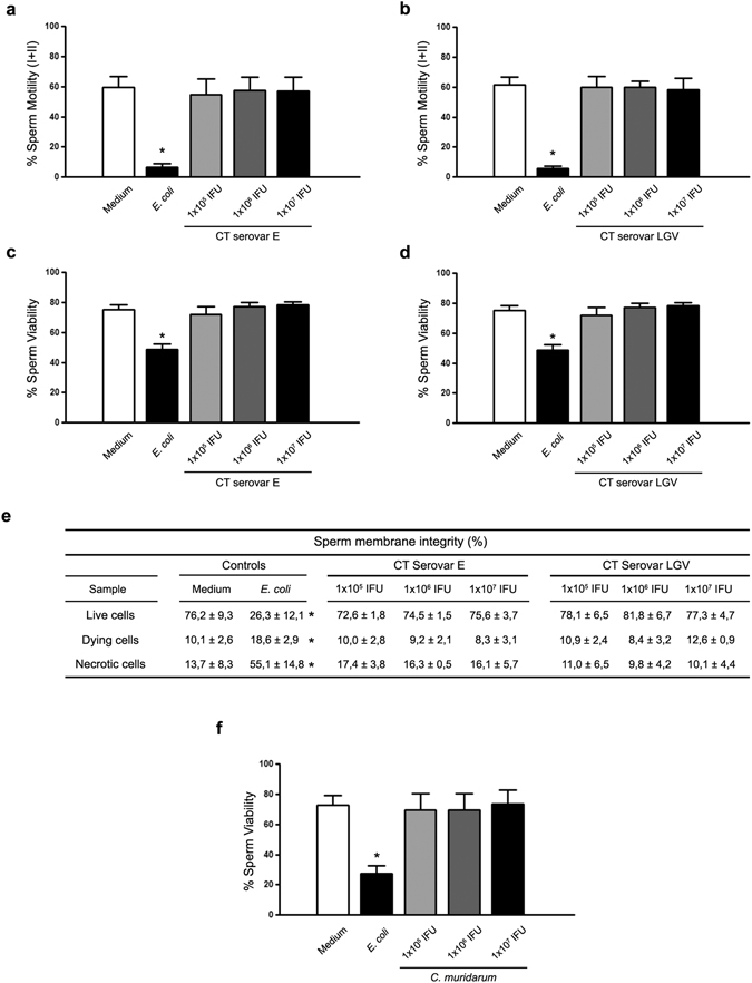
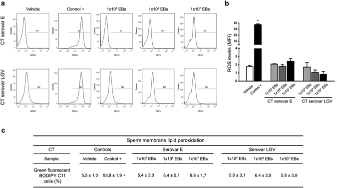
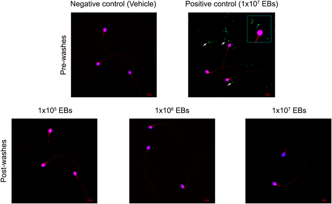
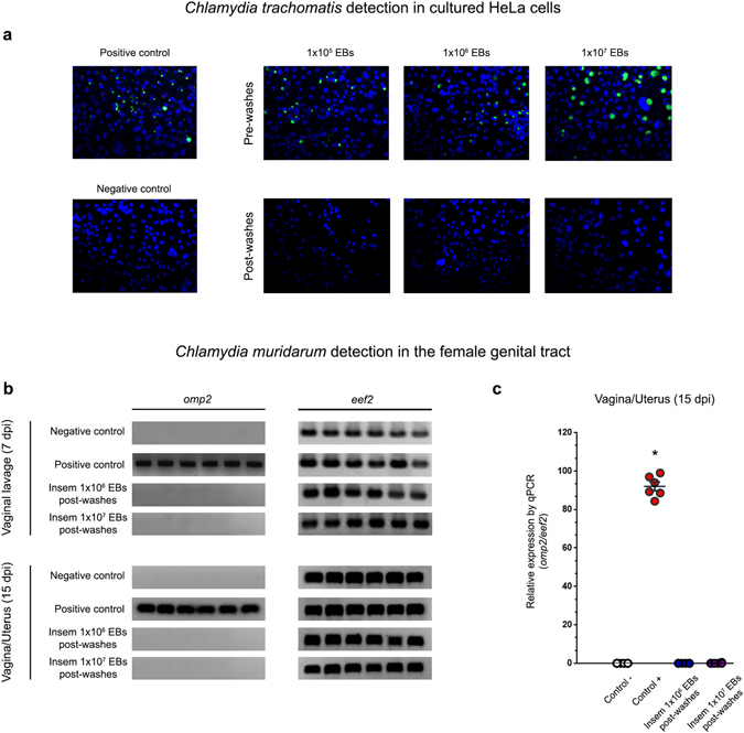
Chlamydia trachomatis is the most prevalent sexually transmitted bacterial infection. However, whether Chlamydia trachomatis has a negative impact on sperm quality and male fertility is still controversial. Herein, we report the effects on sperm quality of the in vitro exposure of spermatozoa to Chlamydia trachomatis, and also the effects of male genital infection on male fertility using an animal model. Human and mouse sperm were obtained from healthy donors and cauda epididimys from C57BL/6 mice, respectively. Highly motile human or mouse spermatozoa were in vitro exposed to C. trachomatis (serovar E or LGV) or C. muridarum, respectively. Then, sperm quality parameters were analyzed. Moreover, male fertility of Chlamydia muridarum infected male C57BL/6 mice was assessed. Human or murine sperm in vitro exposed to increasing bacterial concentrations or soluble factors from C. trachomatis or C. muridarum, respectively, did not show differences in sperm motility and viability, apoptosis, mitochondrial membrane potential, DNA fragmentation, ROS production and lipid peroxidation levels, when compared with control sperm (p > 0.05). Moreover, no differences in fertility parameters (potency, fecundity, fertility index, pre- and post-implantation loss) were observed between control and infected males. In conclusion, our results indicate that Chlamydia spp. neither directly exerts deleterious effects on spermatozoa nor impairs male fertility.


Conflict of interest statement

The authors declare that they have no competing interests.

Figures

Figure 1
Figure 1
Effects of Chlamydia spp. on sperm motility and viability. Human sperm motility (%) after 6 h of in vitro incubation without bacteria (Control), or with increasing concentrations of EBs of CT serovar E (a) or serovar LGV (b) per million spermatozoa. Human sperm viability (%) after 6 h of in vitro incubation without bacteria (Control), or with increasing concentrations of EBs of CT serovar E (c) or serovar LGV (d) per million spermatozoa. (e) Sperm membrane integrity (%) in human spermatozoa after in vitro incubation without bacteria (Control), or with increasing concentrations of EBs of CT serovar E or serovar LGV per million spermatozoa. (f) Murine sperm viability (%) after 30 min of in vitro incubation without bacteria (Control) or with increasing concentrations of EBs of C. muridarum per million spermatozoa. As positive controls, sperm fractions were incubated with uropathogenic E. coli (1 × 106 CFU/mL). Data are shown as mean ± SD. Fractions of human (n = 54) and mouse (n = 24) sperm samples were tested separately and maintained at 37 °C throughout all procedures. Statistical analysis was performed using one-way ANOVA with Bonferroni post hoc test analysis and no significant differences were found in any condition (p < 0.05).
Figure 2
Figure 2
ROS production and lipid peroxidation in human spermatozoa exposed to CT. Analysis of ROS production by human spermatozoa after 6 h of in vitro incubation without bacteria (Vehicle), 100 nM PMA (positive control) or with increasing concentrations of EBs of CT serovar E or serovar LGV per million spermatozoa. ROS production was assessed by flow cytometry using the probe DCFH-DA that fluoresces when oxidized to DCFH. (a) Histograms show sperm with ROS production (M1). (b) ROS production levels (MFI) are shown in bars. (c) Human sperm membrane lipid peroxidation (%) after 6 h of in vitro incubation without bacteria (Vehicle), 100 mM TBHP (positive control) or with increasing concentrations of EBs of CT serovar E or serovar LGV per million spermatozoa. Lipid peroxidation was analyzed using BODIPY C11. Data are shown as mean ± SD. Fractions of sperm samples (n = 36) were tested separately. Statistical analysis was performed using one-way ANOVA with Bonferroni post hoc test analysis. *p < 0.05.
Figure 3
Figure 3
CT does not attach to spermatozoa in vitro. Fractions of highly motile human sperm samples containing 1 × 106 sperm/ml were in vitro incubated with 3% BSA supplemented BWW medium alone or with 1 × 105, 1 × 106 and 1 × 107 EBs/mL of GFP-CT (green fluorescent) during 6 h at 37 °C. Sperm suspensions were then subjected to five consecutive washes and centrifuged at 300 × g for 5 min to remove free bacteria (Post-washes). Sperm suspensions were smeared onto glass slides, stained with DAPI (stain for DNA) and counterstained with 0.01% Evans blue and analyzed by confocal microscopy. Negative control corresponds to fractions of sperm samples incubated with supplemented BWW medium alone [Negative control (Vehicle), Pre-washes]. Positive control corresponds to fractions of sperm samples incubated with 1 × 107 EBs of GFP-CT without subsequent washings [Positive control (1 × 107 EBs), Pre-washes]. Green fluorescent EBs of GFP-CT are observed as green/blue co-localizing dots (marked with arrows). Results shown are from one representative experiment out of three performed (using fractions of n = 10 different sperm samples) with essentially the same results. Images were captured using an Olympus FV1200 laser scanning confocal microscope with an objective PLAPON 60X (1.42 NA). Fluorophore signals were acquired in sequential mode.
Figure 4
Figure 4
Spermatozoa do not carry attached EBs of Chlamydia spp. (a) In vitro cultured confluent HeLa cells were inoculated with human spermatozoa in vitro pre-incubated with different concentration rates (1 × 105, 1 × 106 or 1 × 107 EBs/million sperm) of CT serovar E, immediately after incubation without removing bacteria (Pre-washes), or after five consecutive washes post-incubation (Post-washes) in order to remove free bacteria. Some cultured cells were directly inoculated with 1 × 106 EBs of CT serovar E/mL (positive control) or with supplemented BWW medium alone (negative control). After 48 h of culture, infection of cultures was assessed by the detection of chlamydial inclusion bodies by direct immunofluorescence using a FITC-labeled anti-cLPS monoclonal antibody. Representative microphotographs of 1 out of 4 independent experiments performed with essentially the same results. (b and c) C. muridarum detection (omp2 gene) by conventional (b) or real time PCR (qPCR) (c) in vaginal lavages (at 7 days post insemination) and female genital tract tissue samples (at 15 days post insemination) from female C57BL/6 mice that were intravaginally inseminated with murine sperm in vitro pre-incubated with C. muridarum in capacitating conditions and then washed 5 times to remove free bacteria. Four experimental groups were included: the sham infected group (n = 6) inseminated with 30 µl of a solution containing 1 × 106 sperm that were pre-incubated with vehicle alone (BWW buffer); the positive control group (n = 6) inseminated with 30 µl of a solution containing 1 × 106 sperm that were pre-incubated with 1 × 107 EBs of C. muridarum without subsequent washings; one group (n = 6) inseminated with 30 µl of a solution containing 1 × 106 sperm that were pre-incubated with 1 × 106 EBs of C. muridarum and then washed 5 times; and one group (n = 6) inseminated with 30 µl of a solution containing 1 × 106 sperm that were pre-incubated with 1 × 107 EBs of C. muridarum and then washed 5 times. The expression of the housekeeping gene eef2 was assayed. Representative data from 1 out of 3 independent experiments performed with essentially the same results. Data are shown as mean ± SD. Statistical analysis was performed using two-way ANOVA with Bonferroni post hoc test analysis. *p < 0.01.

References

    1. Brunham RC, Rappuoli R. Chlamydia trachomatis control requires a vaccine. Vaccine. 2013;31:1892–1897. doi: 10.1016/j.vaccine.2013.01.024. - DOI - PMC - PubMed
    1. Senior K. Chlamydia: a much underestimated STI. Lancet Infect. Dis. 2012;12:517–518. doi: 10.1016/S1473-3099(12)70161-5. - DOI - PubMed
    1. Dimech W, et al. ACCESS collaboration. Analysis of laboratory testing results collected in an enhanced chlamydia surveillance system in Australia, 2008–2010. BMC Infect. Dis. 2014;14:325. doi: 10.1186/1471-2334-14-325. - DOI - PMC - PubMed
    1. Paavonen J. Chlamydia trachomatis infections of the female genital tract: state of the art. Ann. Med. 2012;44:18–28. doi: 10.3109/07853890.2010.546365. - DOI - PubMed
    1. Wagenlehner FM, Weidner W, Pilatz A, Naber KG. Urinary tract infections and bacterial prostatitis in men. Curr. Opin. Infect. Dis. 2014;27:97–101. doi: 10.1097/QCO.0000000000000024. - DOI - PubMed

Publication types

Substances