. 2017 Apr 25;7(1):1131.
doi: 10.1038/s41598-017-01258-6.

Phosphorylation of the mitochondrial autophagy receptor Nix enhances its interaction with LC3 proteins

Affiliations

Phosphorylation of the mitochondrial autophagy receptor Nix enhances its interaction with LC3 proteins

Vladimir V Rogov et al. Sci Rep. .

Abstract

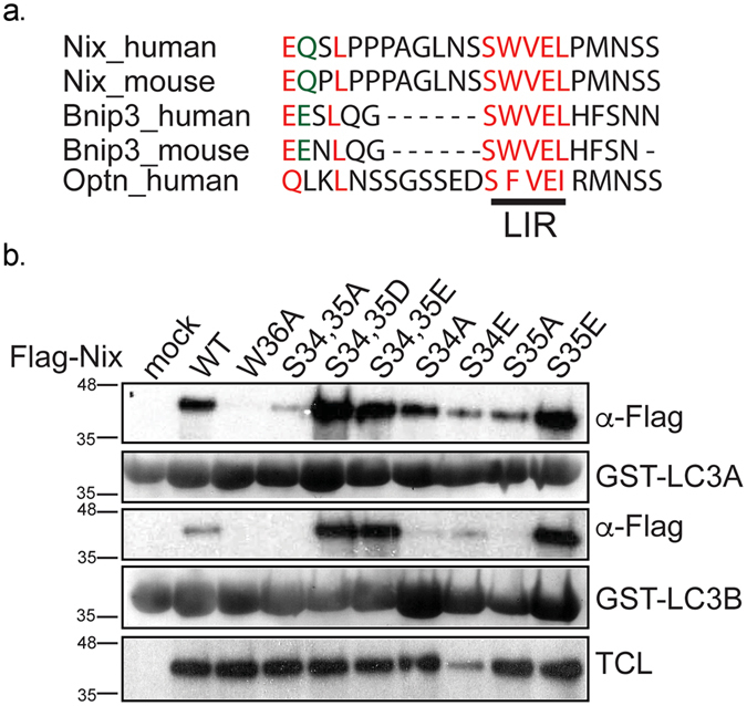
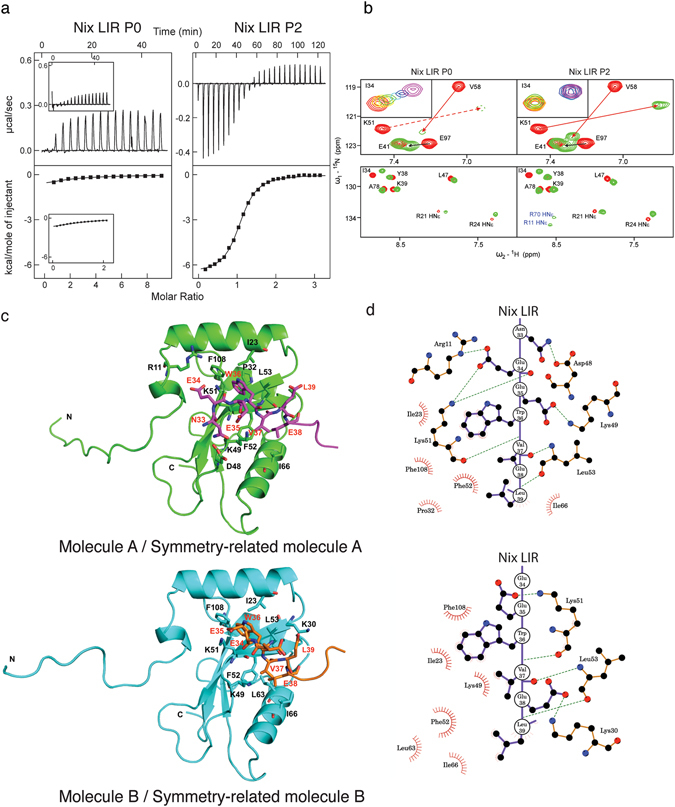
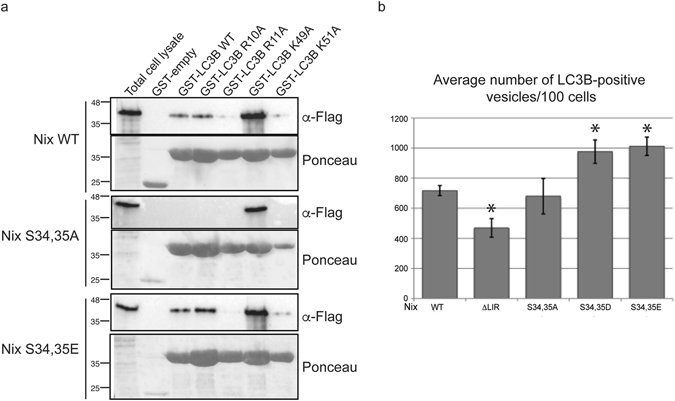
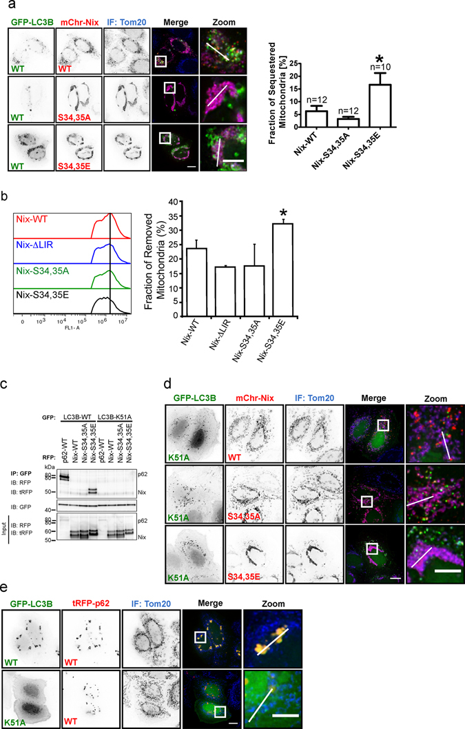
The mitophagy receptor Nix interacts with LC3/GABARAP proteins, targeting mitochondria into autophagosomes for degradation. Here we present evidence for phosphorylation-driven regulation of the Nix:LC3B interaction. Isothermal titration calorimetry and NMR indicate a ~100 fold enhanced affinity of the serine 34/35-phosphorylated Nix LC3-interacting region (LIR) to LC3B and formation of a very rigid complex compared to the non-phosphorylated sequence. Moreover, the crystal structure of LC3B in complex with the Nix LIR peptide containing glutamic acids as phosphomimetic residues and NMR experiments revealed that LIR phosphorylation stabilizes the Nix:LC3B complex via formation of two additional hydrogen bonds between phosphorylated serines of Nix LIR and Arg11, Lys49 and Lys51 in LC3B. Substitution of Lys51 to Ala in LC3B abrogates binding of a phosphomimetic Nix mutant. Functionally, serine 34/35 phosphorylation enhances autophagosome recruitment to mitochondria in HeLa cells. Together, this study provides cellular, biochemical and biophysical evidence that phosphorylation of the LIR domain of Nix enhances mitophagy receptor engagement.


Conflict of interest statement

The authors declare that they have no competing interests.

Figures

Figure 1
Figure 1
Phosphorylation of serine residues juxtaposed to LIR of Nix enhances interaction of Nix to LC3 protein family. (a) Alignment of LIR motif of Nix, Bnip3 and Optineurin. (b) GST pull-down of GST-LC3A and GST-LC3B and Nix wt and Nix W36A, double Nix S34,35A, Nix S34,35D, Nix S34,35E mutants and single Nix S34A, Nix S34E, Nix S35A, Nix S35E mutants. TCL corresponds to total cell lysate.
Figure 2
Figure 2
Influence of Nix phosphorylation on its interaction with LC3B studied by ITC, NMR and crystallography. (a) ITC profiles for the titration of Nix LIR P0 (left) and Nix LIR P2 (right) peptides into LC3B are shown at the main plots in the same scales of energy. For comparison, ITC data from Novak et al., representing LC3B titration with P0, are shown in the small windows within corresponding plot. In each plot the top diagram displays the raw measurements and the bottom diagram shows the integrated heat per titration step. (b) Upper plots: Representative sections (“fingerprint area”) of 15N-labeled LC3B [15N,1H]-TROSY HSQC spectra in the free LC3B state (red contours) and in complex with Nix LIR P0 and P2 peptides (green contours). Arrows show direction of CSP for selected residues. Dashed lines represent CSP of the K51 HN resonance, which cannot be visualized at this experimental series. Small windows within the plots represent exchange modes for the characteristic LC3B residues Ile34 upon P0 and P2 peptides titrations (rainbow color-code). Low plots: Representative spectral section showing arginine HNε side-chain resonances. They are aliased in the ω1 dimension and appear at a 15N chemical shift 50 ppm downfield from their true position. Tentative assignment for the HNε side-chain resonances appeared upon P2 titration is given in blue. (c) Structure of Nix/LC3B complex. Structures of Nix-LIRS34,35E-LC3B2–119 molecules A and B are represented by ribbon diagrams and colored in green (molecule A) and cyan (molecule B). Nix LIR portions in symmetry-related molecules (molecule A, magenta; molecule B, orange) and its interacting residues are shown in stick representation. Oxygen, nitrogen and sulfur atoms are shown in red, blue and yellow, respectively. Schematic representations showing the interaction between LC3B and Nix LIRS34,35E on level of residues were generated by LIGPLOT.
Figure 3
Figure 3
LC3B Arg10, Arg11 and Lys49 residues are important for interaction with phosphorylated Nix. (a) GST pull-downs with GST-LC3B wild type GST-LC3B, R10A, R11A, K49A and K51A were performed against Flag-Nix wild type, S34,35A and S34,35E. (b) Cells cotransfected with Nix wild type, ΔLIR, S34,35A, S34,35E or S34,35D and GFP-LC3B were screened using immunofluorescent microscopy. Three independent experiments were performed and average numbers of LC3B positive vesicles per 100 cells in each experiment were calculated. Error bars are standard deviation. *Significantly different from the control Nix wild type, p < 0.05.
Figure 4
Figure 4
Phosphorylation of the Nix LIR influences the initation of mitophagy and Nix:LC3B interaction depends on LC3B Lys51. (a) Hela cells coexpressing GFP-LC3B wild type and Nix wild type or mutants, immunostained (IF) for mitochondrial marker Tom20. GFP-LC3B shows colocalization with RFP-Nix and enhanced colocalization with S34,35E mutant. RFP-Nix S34,35A does not colocalize with GFP-LC3B. Analysis of fraction of sequestered mitochondria is depicted on the graph. (b) Analysis of mitochondrial removal is determined by flow cytometry using GFP-Nix, ΔLIR, S34,35A and S34,35E mutants (c) Immunoprecipitation of GFP-LC3B wild type or GFP-LC3B K51A in cells co-expressing tRFP-p62 wild type, RFP-Nix wild type, S34,35A and S34,35E mutant. GFP-LC3B K51A fails to coimmunoprecipitate with p62, Nix wild type and mutants. (d) GFP-LC3B K51A does not colocalize with Nix wild type or mutants. (e) Hela cells coexpressing GFP-LC3B wild type or K51A mutant and RFP-p62, immunostained (IF) for Tom20. Scale bar 10 µm.

References

    1. Yang Z, Klionsky DJ. Eaten alive: a history of macroautophagy. Nat Cell Biol. 2010;12:814–822. doi: 10.1038/ncb0910-814. - DOI - PMC - PubMed
    1. Choi AM, Ryter SW, Levine B. Autophagy in human health and disease. N Engl J Med. 2013;368:651–662. doi: 10.1056/NEJMra1205406. - DOI - PubMed
    1. Pankiv S, et al. p62/SQSTM1 binds directly to Atg8/LC3 to facilitate degradation of ubiquitinated protein aggregates by autophagy. J Biol Chem. 2007;282:24131–24145. doi: 10.1074/jbc.M702824200. - DOI - PubMed
    1. Kirkin V, et al. A role for NBR1 in autophagosomal degradation of ubiquitinated substrates. Mol Cell. 2009;33:505–516. doi: 10.1016/j.molcel.2009.01.020. - DOI - PubMed
    1. Wild P, et al. Phosphorylation of the autophagy receptor optineurin restricts Salmonella growth. Science. 2011;333:228–233. doi: 10.1126/science.1205405. - DOI - PMC - PubMed

Publication types

MeSH terms