. 2017 Apr 25;7(1):1112.
doi: 10.1038/s41598-017-01148-x.

Double-stranded DNA induces a prothrombotic phenotype in the vascular endothelium

Affiliations

Double-stranded DNA induces a prothrombotic phenotype in the vascular endothelium

Erik Gaitzsch et al. Sci Rep. .

Abstract

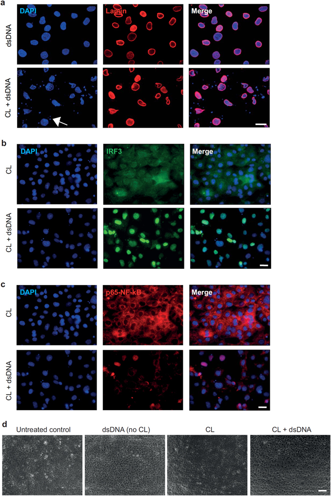
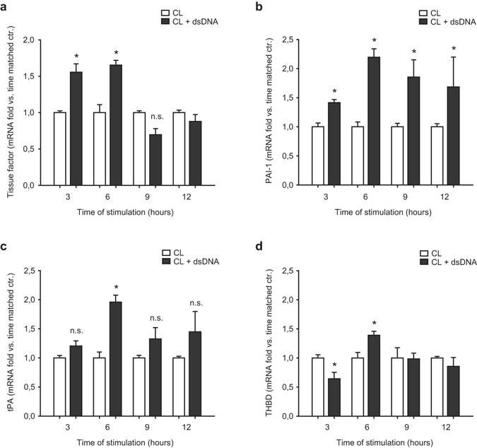
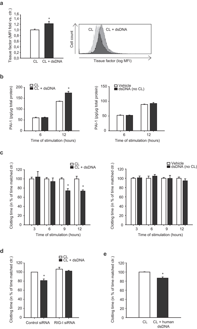
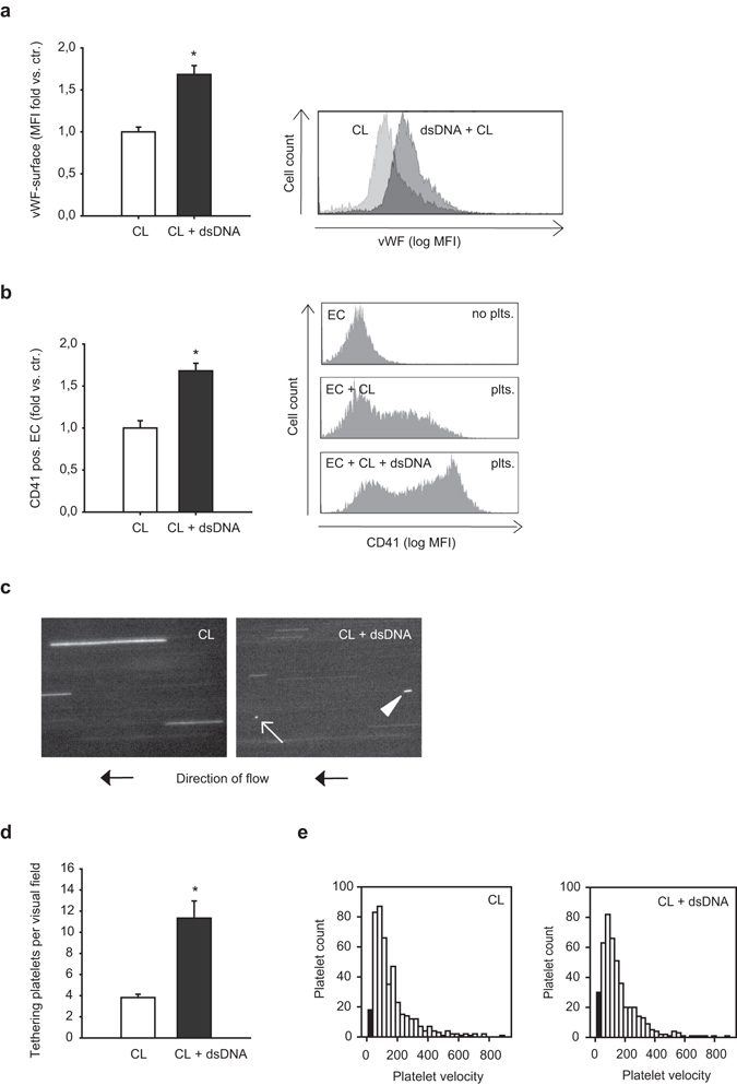
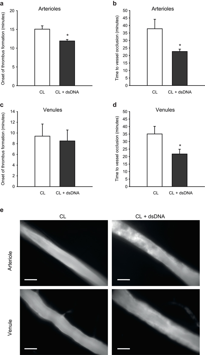
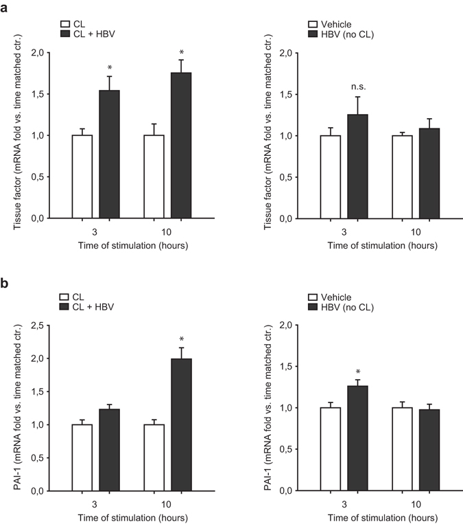
Double-stranded DNA (dsDNA) constitutes a potent activator of innate immunity, given its ability to bind intracellular pattern recognition receptors during viral infections or sterile tissue damage. While effects of dsDNA in immune cells have been extensively studied, dsDNA signalling and its pathophysiological implications in non-immune cells, such as the vascular endothelium, remain poorly understood. The aim of this study was to characterize prothrombotic effects of dsDNA in vascular endothelial cells. Transfection of cultured human endothelial cells with the synthetic dsDNA poly(dA:dT) induced upregulation of the prothrombotic molecules tissue factor and PAI-1, resulting in accelerated blood clotting in vitro, which was partly dependent on RIG-I signalling. Prothrombotic effects were also observed upon transfection of endothelial cells with hepatitis B virus DNA-containing immunoprecipitates as well human genomic DNA. In addition, dsDNA led to surface expression of von Willebrand factor resulting in increased platelet-endothelium-interactions under flow. Eventually, intrascrotal injection of dsDNA resulted in accelerated thrombus formation upon light/dye-induced endothelial injury in mouse cremaster arterioles and venules in vivo. In conclusion, we show that viral or endogenous dsDNA induces a prothrombotic phenotype in the vascular endothelium. These findings represent a novel link between pathogen- and danger-associated patterns within innate immunity and thrombosis.


Conflict of interest statement

The authors declare that they have no competing interests.

Figures

Figure 1
Figure 1
Double-stranded DNA led to nuclear translocation of transcription factors IRF3 and NF-κB. (a) Immunofluorescent staining of cultured endothelial cells with DAPI (blue) and anti-Lamin-antibody (red) showed transfection of poly(dA:dT) into the intracellular compartment after complex formation with cationic lipids. Scale bar is 10 µm. (b,c) Transfection of endothelial cells with poly(dA:dT) led to nuclear translocation of transcription factors IRF3 (b) and to a lesser extent of NF-κB (c). Cells were stained with antibodies against IRF3 (green) and p65-subunit of NF-κB (red). Scale bar is 10 µm. (d) Representative bright field images upon treatment of endothelial cells with poly(dA:dT) with or without cationic lipids. Scale bar is 100 µm.
Figure 2
Figure 2
Double-stranded DNA induces expression of prothrombotic genes in vascular endothelial cells. (a,b) Expression of the prothrombotic molecules tissue factor (a) and Plasminogen activator inhibitor-1 (PAI-1, b) as assessed by RT-PCR upon transfection of vascular endothelial cells with poly(dA:dT). (c,d) Expression of the antithrombotic molecules tissue plasminogen activator (tPA, (c)) and thrombomodulin (THBD, (d)) upon transfection of vascular endothelial cells with poly(dA:dT); (*P < 0.05 vs. respective time-matched control, n = 4).
Figure 3
Figure 3
Double stranded DNA induces prothrombotic proteins and accelerates endothelial dependent blood clotting in vitro. (a) Tissue factor surface expression was assessed by flow cytometry 12 hours after transfection of endothelial cells with poly(dA:dT) (*P < 0.05 vs. control, n = 8, MFI mean fluorescence intensity). Representative histogram is shown on the right. (b) PAI-1 release by endothelial cells was measured by ELISA 6 and 12 hours after transfection with poly(dA:dT), PAI-1 release after stimulation of endothelial cells with poly(dA:dT) alone (i.e. without cationic lipids) is shown on the right (*P < 0.05 vs. time matched control, n = 5). (c) Stimulation of whole blood with endothelial cell lysates transfected with poly(dA:dT) accelerated blood clotting time compared to stimulation with untreated cell lysates as measured by thromboelastometry. Stimulation of whole blood with lysates of endothelial cells treated with poly(dA:dT) alone (i.e. without cationic lipids) had no effect on blood clotting time (right) (*P < 0.05 vs. time matched control, n = 4). (d) The prothrombotic effect of poly(dA:dT) after 12 hours was partly reversed by siRNA-silencing of RIG-I (*P < 0.05 vs. time matched control, n = 4). (e) A similar prothrombotic effect was observed 12 hours after transfection of endothelial cells with human genomic DNA (*P < 0.05 vs. time-matched control, n = 4).
Figure 4
Figure 4
Double-stranded DNA induces vWF upregulation and platelet tethering in vitro. (a) Transfection of endothelial cells with poly(dA:dT) for 12 hours caused upregulation of von Willebrand factor on the endothelial cell surface (*P < 0.05 vs. control, n = 6; MFI mean fluorescence intensity). Representative histogram is shown on the right. (b) Co-cultivation of endothelial cells transfected with poly(dA:dT) for 12 hours and non-stimulated isolated platelets resulted in increased adhesion of platelets to endothelial cells, as assessed by FACS analysis (*P < 0.05, vs. control, n = 6). Representative histograms are shown on the right. (c) Representative snap shots of flow chamber assays with transfusion of fluorescently labeled platelets simulating vascular shear stress of 1 dyn/cm2. Platelet tethering (white arrow) and very slow rolling platelets (white arrowhead) were analyzed after stimulation of endothelial cells with poly(dA:dT) for 12 hours. (d) Quantitative analysis showed increased number of tethering platelets (*P < 0.05 vs. control, n = 9) as well as (e) increased number of very slow rolling platelets (as displayed by the black columns in the histograms showing platelet velocity profiles) upon transfection with poly(dA:dT).
Figure 5
Figure 5
Double-stranded DNA accelerates microvascular thrombosis in vivo. Thrombus formation in vivo was investigated by phototoxic (light/dye-induced) vessel injury of microvessels in the mouse cremaster muscle. Intrascrotal injection of poly(dA:dT) (5 µg DNA 12 hours prior to the experiment) resulted in a more rapid onset of thrombus formation in arterioles (a) but not in venules (c). Time to complete vessel occlusion was significantly accelerated after poly(dA:dT) injection in both arterioles (b) and venules (d). (e) Representative images show thrombus formation in mouse cremaster arterioles (20 min after start of injury) and mouse cremaster venules (10 min after start of injury) with markedly increased thrombus formation in poly(dA:dT) treated animals (right pictures) as compared to control animals (left pictures). Scale bar is 50 µm. (*P < 0.05 vs. control, n = 6 animals each).
Figure 6
Figure 6
Hepatitis B virus DNA-containing immunoprecipitates induce a prothrombotic phenotype. Endothelial cells were transfected with HBV-DNA containing immunoprecipitates isolated from a patient with ongoing hepatitis B infection and associated polyarteritis nodosa with a high viral load. HBV-DNA containing immunoprecipitates resulted in upregulation of tissue factor starting 3 hours after transfection (a) as well as PAI-I at 10 hours after transfection (b) as analyzed by RT-PCR. Expression of tissue factor and PAI-1 after stimulation of endothelial cells with HBV-DNA alone (i.e. without cationic lipids) is shown on the right ((a and b), respectively). (*P < 0.05 vs. control).

References

    1. Stetson DB, Medzhitov R. Recognition of cytosolic DNA activates an IRF3-dependent innate immune response. Immunity. 2006;24:93–103. doi: 10.1016/j.immuni.2005.12.003. - DOI - PubMed
    1. Takaoka A, Taniguchi T. Cytosolic DNA recognition for triggering innate immune responses. Advanced drug delivery reviews. 2008;60:847–857. doi: 10.1016/j.addr.2007.12.002. - DOI - PubMed
    1. Takaoka A, et al. DAI (DLM-1/ZBP1) is a cytosolic DNA sensor and an activator of innate immune response. Nature. 2007;448:501–505. doi: 10.1038/nature06013. - DOI - PubMed
    1. Hornung V, Latz E. Intracellular DNA recognition. Nature reviews. Immunology. 2010;10:123–130. doi: 10.1038/nri2690. - DOI - PubMed
    1. Ablasser A, et al. RIG-I-dependent sensing of poly(dA:dT) through the induction of an RNA polymerase III-transcribed RNA intermediate. Nat Immunol. 2009;10:1065–1072. doi: 10.1038/ni.1779. - DOI - PMC - PubMed

Publication types

LinkOut - more resources