. 2017 Apr 25;7(1):1117.
doi: 10.1038/s41598-017-01320-3.

MicroRNA-30a Regulation of Epithelial-Mesenchymal Transition in Diabetic Cataracts Through Targeting SNAI1

Affiliations

MicroRNA-30a Regulation of Epithelial-Mesenchymal Transition in Diabetic Cataracts Through Targeting SNAI1

Lu Zhang et al. Sci Rep. .

Abstract

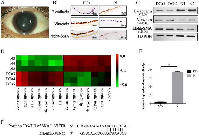
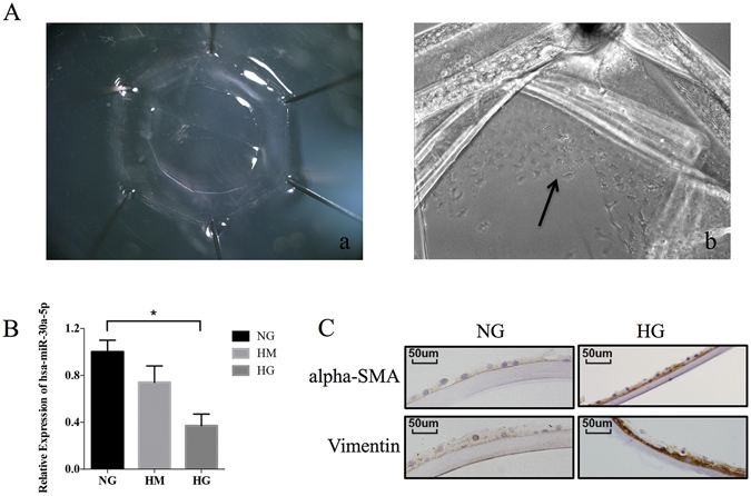
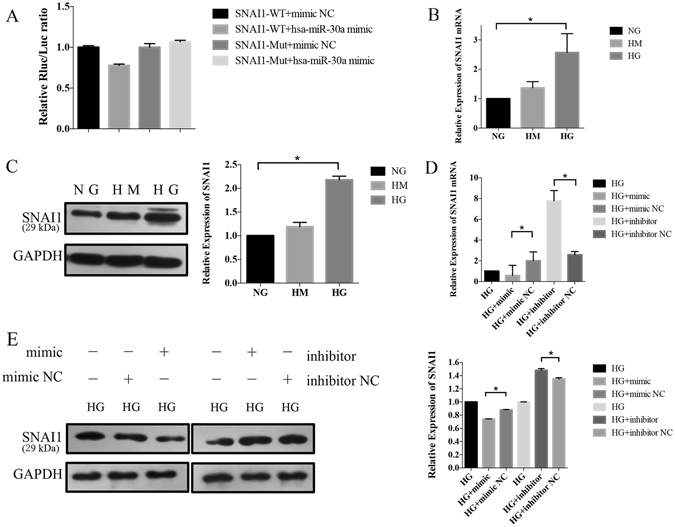
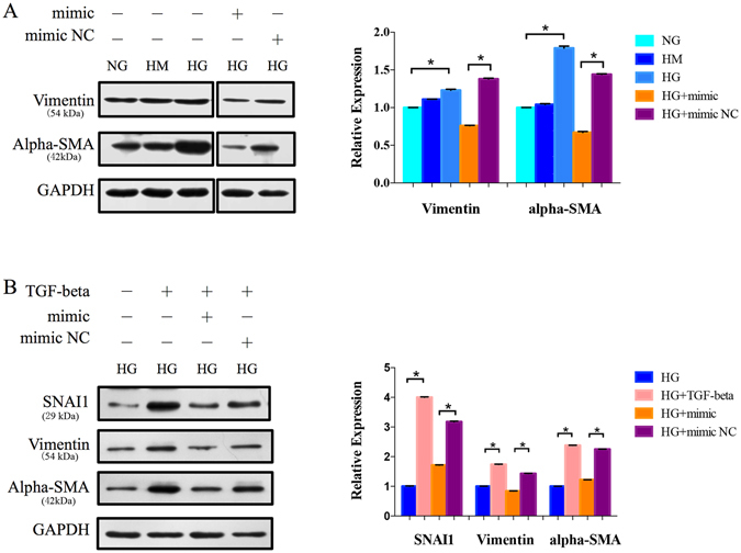
Epithelial-mesenchymal transition (EMT) is a highly conserved and fundamental process in development, fibrosis, and metastasis. During the process, epithelial cells lose their morphology and transcriptional program, and transdifferentiate to mesenchymal cells. It has been reported that lens epithelial cells undergo EMT during cataract formation, and regulation of microRNAs on genes is associated with lens development. However, the molecular mechanisms of this regulation in diabetic cataract still need to be investigated. In the present study, the expression of E-cadherin was downregulated, while the expression of alpha-SMA and vimentin was upregulated in diabetic cataract tissues and the in vitro model, suggesting the involvement of EMT in diabetic cataract formation. Results of miRNA profiling demonstrated that miR-30a was markedly downregulated in diabetic cataract tissues. Overexpression of miR-30a-5p decreased SNAI1, a known modulator of EMT, and the expression of vimentin and alpha-SMA in our diabetic cataract model in vitro. It is concluded that EMT is involved in human diabetic cataract, and upregulation of miR-30a can repress EMT through its targeting of SNAI1 in lens epithelial cells, which make miR-30a a novel target of therapeutic intervention for human diabetic cataract.


Conflict of interest statement

The authors declare that they have no competing interests.

Figures

Figure 1
Figure 1
Expression of the epithelial marker (E-cadherin) and EMT markers (vimentin and alpha-SMA) and miRNA profiles in the normal and diabetic cataract lens epithelial cells (LECs). (A) Opacity of lens subcapsular tissues in diabetic cataract patients. (B) Immunohistochemistry demonstrated a high expression of E-cadherin and a low expression of vimentin and alpha-SMA in normal LECS, and a significantly downregulated expression of E-cadherin and an upregulated expression of vimentin and alpha-SMA in diabetic cataract LECs. (C) Western blotting for quantification of the protein expression (top, data from the gels; bottom, normalization to GAPDH). Two normal samples (N1 and N2) and two diabetic cataract samples (DCa1 and DCa2) were included. (D) Hierarchical clustering was performed [Rows, miRNA; Column, three normal samples (N3, N4, and N5) and three diabetic cataract samples (Dca3, Dca4, and Dca5)]. A total of 272 miRNAs were identified as having an altered expression more than 2-fold between the diabetic cataract and normal LECs, and 14 regulated miRNAs were depicted in this figure. The red color indicates the high expression, and the green color denotes the low expression. (E) qRT-PCR was performed by using the same extracted total RNA for the microarray analysis to validate the microarray data. The relative amount of hsa-miR-30a-5p was normalized to the U6 expression. The relative expression levels of hsa-miR-30a-5p were downregulated significantly in the diabetic cataract LECs (N as control, *P < 0.05). Significant differences are indicated by t-test (*P < 0.05). (F) Position of the miR-30a-5p target sequence in the 3′-UTR of SNAI1 mRNA.
Figure 2
Figure 2
Human donor capsular bags as a diabetic cataract model in vitro. (A) Capsular bags were immersed in the culture medium: (a) immediately after pinning; (b) after 24 hours, when the LECs were observed to be migrating (arrow). (B) The expression of miR-30a-5p was detected by qRT-PCR. MiR-30a-5p increased in the presence of HG (*P < 0.05, NG as control, N = 1), instead of HM (osmotic control). (C) Immunohistochemistry analysis was performed to detect the protein expression of EMT markers. The staining intensity of vimentin and alpha-SMA expression increased in HG compared to NG.
Figure 3
Figure 3
MiR-30a-5p directly targeted and regulated SNAI1 in the diabetic cataract model in vitro. (A) A dual luciferase reporter assay. When pmiR-RB-REPORT-SNAI1-3′UTR and miR-30a-5p mimic were cotransfected, a significant decrease in relative luciferase activity was observed. The mutation of the perfectly complementary sites in the SNAI1-3′UTR disrupted the interaction between miR-30a-5p and Snai11 and abolished the suppressive effect. (B) The mRNA expression of SNAIl. qRT-PCR showed the SNAI1 mRNA expression increased significantly in HG (NG as control, *P < 0.05, N = 1). (C) The protein expression of Snail in Western blot. Left: The representative gel results (top, data from the gels; bottom, normalization to GAPDH); Right: The normalized graphs results. The result showed an increased expression of SNAI1 in HG conditions (NG as control, *P < 0.05, N = 2). (D) Results of qRT-PCR in the transfected LECs. There were five groups in this part: 1, capsular bags cultured in HG for 3 days were used as the control group; 2, the cells were transfected with a mimic negative control (NC) of miR-30a-5p; 3, the cells were transfected with a mimic of miR-30a-5p; 4, the cells were transfected with an inhibitor of miR-30a-5p; and 5, the cells were transfected with an inhibitor NC of miR-30a-5p. SNAI1 mRNA levels were significantly increased by the miR-30a-5p mimic (*P < 0.05, mimic NC as control, N = 1) and decreased by the miR-30a-5p inhibitor (*P < 0.05, inhibitor NC as control, N = 1). (E) Results of Western blotting in the transfected LECs (top, data from the gels; bottom, normalization to GAPDH). SNAI1 protein expression was decreased by transfection with the miR-30a-5p mimic (*P < 0.05, mimic NC as control, N = 2) but restored by transfection with the miR-30a-5p inhibitor (*P < 0.05, inhibitor NC as control, N = 2) in the LECs significantly.
Figure 4
Figure 4
Upregulation of miR-30a-5p repressed EMT. (A) Protein expression levels of the EMT markers. The protein expression of vimentin and alpha-SMA was decreased when the LECs were transfected with the miR-30a-5p mimic in HG conditions (*P < 0.05, mimic NC as control, N = 2). (B) The capsular bags were treated with TGF-beta and a mimic of miR-30a-5p. In the presence of TGF-beta, EMT was induced with the increased protein levels of vimentin and alpha-SMA, as well as SNAI1 (*P < 0.05, HG without TFG-beta as control, N = 2), while the miR-30a- 5p mimic repressed vimentin and alpha-SMA (P < 0.05, mimic NC as control, N = 2).

Similar articles

Cited by

References

    1. Pascolini D, Mariotti SP. Global estimates of visual impairment: 2010. The British journal of ophthalmology. 2012;96:614–618. doi: 10.1136/bjophthalmol-2011-300539. - DOI - PubMed
    1. Harding JJ, Egerton M, van Heyningen R, Harding RS. Diabetes, glaucoma, sex, and cataract: analysis of combined data from two case control studies. The British journal of ophthalmology. 1993;77:2–6. doi: 10.1136/bjo.77.1.2. - DOI - PMC - PubMed
    1. Kahn HA, et al. The Framingham Eye Study. II. Association of ophthalmic pathology with single variables previously measured in the Framingham Heart Study. American journal of epidemiology. 1977;106:33–41. doi: 10.1093/oxfordjournals.aje.a112429. - DOI - PubMed
    1. Pollreisz A, Schmidt-Erfurth U. Diabetic cataract-pathogenesis, epidemiology and treatment. Journal of ophthalmology. 2010;2010:608751–8. doi: 10.1155/2010/608751. - DOI - PMC - PubMed
    1. Worgul BV, Merriam GR, Jr., Medvedovsky C. Cortical cataract development–an expression of primary damage to the lens epithelium. Lens and eye toxicity research. 1989;6:559–571. - PubMed

Publication types

MeSH terms