

Dermatofibrosarcoma Protuberance of the Breast: a Diagnostic Challenge
- PMID: 28442848
- PMCID: PMC5386944
- DOI: 10.1007/s12262-016-1502-1
Dermatofibrosarcoma Protuberance of the Breast: a Diagnostic Challenge
Abstract
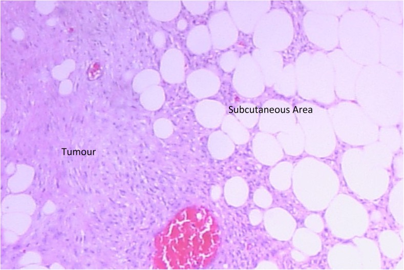
Dermatofibrosarcoma protuberans (DFSP) is an uncommon slow growing neoplasm of the dermis with tendency to invade the subcutaneous tissues. It presents during the third to fourth decade of life and is commonly seen over the trunk, extremities and head and neck. DFSP presenting as a breast lump is rare but few cases have been reported in the literature. Pre-operative diagnosis with mammography, ultrasonography and FNAC is challenging. We report a case of a DFSP of the right breast in a middle aged lady with history of recurrent breast lumps excised and diagnosed in the past as benign. She presented with progressively increasing right breast lump of 2 months duration. She underwent wide local excision and histology revealed dermatofibrosarcoma protuberans. In view of its local aggressiveness with incomplete surgical margin, mastectomy was performed.
Keywords: Breast; Dermatofibrosarcoma protuberans; Immunohistochemical stains; Mastectomy.
Conflict of interest statement
There is no conflict of interest or any financial support involved in this case report. It has not been published or presented in printed or electronic form.
Figures


References
-
- Nggada H, Bali B, Na’aya H. A clinicopathological study of dermatofibrosarcoma protuberans in Maiduguri, northeastern Nigeria. Niger J Surg Res. 2006;8(1–2):78–80.
LinkOut - more resources
Full Text Sources
Other Literature Sources
Miscellaneous