

Observer Variability in Evaluating Pisotriquetral Osteoarthritis using Pisotriquetral View
- PMID: 28442858
- PMCID: PMC5403736
- DOI: 10.1055/s-0037-1602127
Observer Variability in Evaluating Pisotriquetral Osteoarthritis using Pisotriquetral View
Abstract
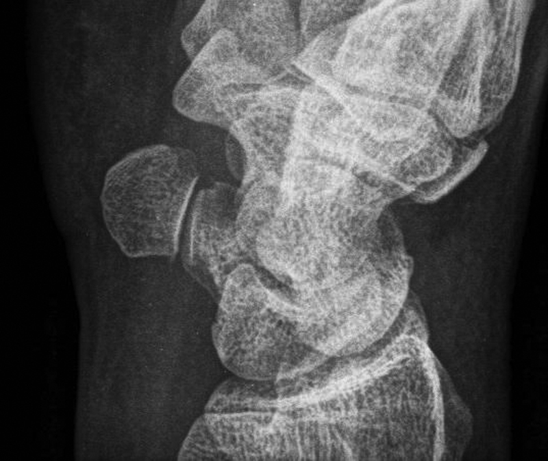
A pisotriquetral (semilateral) view of the wrist may improve the assessment of pisotriquetral osteoarthritis (OA), but its reliability and reproducibility are unclear. The purpose of this cross-sectional observer study was to investigate (1) the inter- and intraobserver agreement of evaluating pisotriquetral OA using pisotriquetral views with a special focus on sclerosis, joint space width (JSW) narrowing and osteophyte formation, and (2) the incidence of these latter radiographic features in patients suspected for pisotriquetral OA. Five independent observers rated independently at two different occasions 27 pisotriquetral views from patients treated for ulnar-sided wrist pain suspected for pisotriquetral OA requiring a pisiform resection. The agreement was calculated using kappa statistic. Agreement between observers ranged from 0.38 (fair) to 0.56 (moderate). Average intraobserver agreement ranged from 0.43 (moderate) to 0.52 (moderate). In 36% of the ratings, JSW narrowing was observed, followed by osteophyte formation (30%) and sclerosis (28%). Observers found it especially difficult to detect JSW narrowing. Despite the availability of a pisotriquetral view to enhance visualization of the pisotriquetral joint, assessment of the specific features indicating pisotriquetral OA leads to only fair-to-moderate agreement. This limits the applicability of a radiographic assessment. A rationale for a more reliable radiologic approach in assessing the level of pisotriquetral OA is needed, which may require the use of more advanced imaging techniques.
Keywords: observer; pisiform; pisotriquetral joint; pisotriquetral osteoarthritis; triquetral.
Conflict of interest statement
Figures
Similar articles
-
Joint Space Narrowing in Patients With Pisotriquetral Osteoarthritis.Hand (N Y). 2017 Sep;12(5):490-492. doi: 10.1177/1558944716677542. Epub 2016 Nov 10. Hand (N Y). 2017. PMID: 28832198 Free PMC article.
-
Reproducibility and sensitivity to change of a new method of computer measurement of joint space width in hip osteoarthritis. Performance of three radiographic views obtained at a 3-year interval.Osteoarthritis Cartilage. 2009 Jul;17(7):864-70. doi: 10.1016/j.joca.2008.12.003. Epub 2008 Dec 24. Osteoarthritis Cartilage. 2009. PMID: 19138537 Clinical Trial.
-
Inter- and Intraobserver Reliabilities of Four Different Radiographic Grading Scales of Osteoarthritis of the Knee Joint.J Knee Surg. 2018 Mar;31(3):247-253. doi: 10.1055/s-0037-1602249. Epub 2017 May 1. J Knee Surg. 2018. PMID: 28460407
-
Atlas of radiographic features of osteoarthritis of the ankle and hindfoot.Osteoarthritis Cartilage. 2015 Dec;23(12):2059-2085. doi: 10.1016/j.joca.2015.08.008. Epub 2015 Aug 28. Osteoarthritis Cartilage. 2015. PMID: 26318654 Free PMC article. Review.
-
Management of Pisotriquetral Instability.J Hand Surg Am. 2018 Jan;43(1):54-60. doi: 10.1016/j.jhsa.2017.10.020. Epub 2017 Nov 21. J Hand Surg Am. 2018. PMID: 29169722 Review.
References
-
- Moraux A, Lefebvre G, Pansini V. et al.Pisotriquetral joint disorders: an under-recognized cause of ulnar side wrist pain. Skeletal Radiol. 2014;43(6):761–773. - PubMed
-
- Paley D, McMurtry R Y, Cruickshank B. Pathologic conditions of the pisiform and pisotriquetral joint. J Hand Surg Am. 1987;12(1):110–119. - PubMed
-
- van Eijzeren J, Karthaus R P. The effect of pisiform excision on wrist function. J Hand Surg Am. 2014;39(7):1258–1263. - PubMed
-
- Lautenbach M, Eisenschenk A, Langner I, Arntz U, Millrose M. Comparison of clinical results after pisiformectomy in patients with rheumatic versus posttraumatic osteoarthritis. Orthopedics. 2013;36(10):e1239–e1243. - PubMed
LinkOut - more resources
Full Text Sources
Other Literature Sources
