. 2017 Apr 10:12:2733-2748.
doi: 10.2147/IJN.S127528. eCollection 2017.

Magnetic and pH dual-responsive mesoporous silica nanocomposites for effective and low-toxic photodynamic therapy

Affiliations

Magnetic and pH dual-responsive mesoporous silica nanocomposites for effective and low-toxic photodynamic therapy

Jieqiong Zhan et al. Int J Nanomedicine. .

Abstract

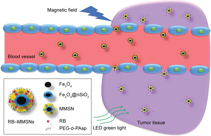
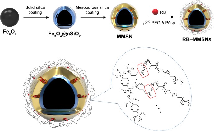
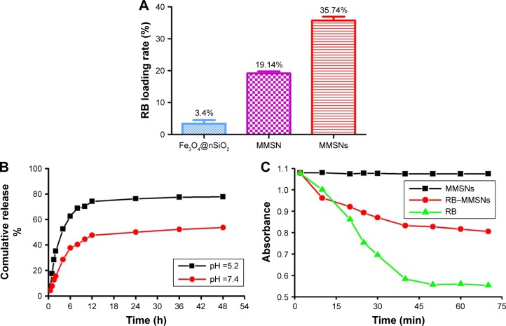
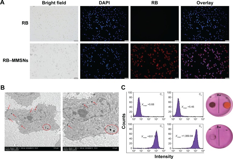
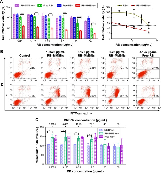
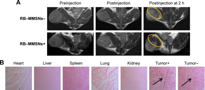
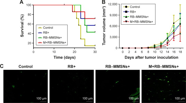
Nonspecific targeting, large doses and phototoxicity severely hamper the clinical effect of photodynamic therapy (PDT). In this work, superparamagnetic Fe3O4 mesoporous silica nanoparticles grafted by pH-responsive block polymer polyethylene glycol-b-poly(aspartic acid) (PEG-b-PAsp) were fabricated to load the model photosensitizer rose bengal (RB) in the aim of enhancing the efficiency of PDT. Compared to free RB, the nanocomposites (polyethylene glycol-b-polyaspartate-modified rose bengal-loaded magnetic mesoporous silica [RB-MMSNs]) could greatly enhance the cellular uptake due to their effective endocytosis by mouse melanoma B16 cell and exhibited higher induced apoptosis although with little dark toxicity. RB-MMSNs had little dark toxicity and even much could be facilitated by magnetic field in vitro. RB-MMSNs demonstrated 10 times induced apoptosis efficiency than that of free RB at the same RB concentration, both by cell counting kit-8 (CCK-8) result and apoptosis detection. Furthermore, RB-MMSNs-mediated PDT in vivo on tumor-bearing mice showed steady physical targeting of RB-MMSNs to the tumor site; tumor volumes were significantly reduced in the magnetic field with green light irradiation. More importantly, the survival time of tumor-bearing mice treated with RB-MMSNs was much prolonged. Henceforth, polyethylene glycol-b-polyaspartate-modified magnetic mesoporous silica (MMSNs) probably have great potential in clinical cancer photodynamic treatment because of their effective and low-toxic performance as photosensitizers' vesicles.

Keywords: magnetic mesoporous silica; magnetic targeting; pH responsive; photodynamic therapy; polymer polyethylene glycol-b-poly(aspartic acid); rose bengal.


Conflict of interest statement

Disclosure The authors report no conflicts of interest in this work.

Figures

Figure 1
Figure 1
Illustration of MMSN-mediated treatment in a magnetic field. Note: RB was loaded into the mesoporous silica pores and surrounded by PEG-b-PAsp in blood vessels, when they were released in tumor sites. Abbreviations: MMSN, magnetic mesoporous silica nanoparticle; RB, rose bengal; PEG-b-PAsp, polyethylene glycol-b-poly(aspartic acid); RB−MMSNs, polyethylene glycol-b-polyaspartate-modified rose bengal-loaded magnetic mesoporous silica.
Figure 2
Figure 2
Schematic illustration for the synthesis of core–shell RB−MMSNs. Abbreviations: RB−MMSNs, polyethylene glycol-b-polyaspartate-modified rose bengal-loaded magnetic mesoporous silica; MMSN, magnetic mesoporous silica nanoparticle; RB, rose bengal; PEG-b-PAsp, polyethylene glycol-b-poly(aspartic acid).
Figure 3
Figure 3
The structural characterization of samples. Notes: (A) The TEM images of Fe3O4, Fe3O4@nSiO2 and MMSN. (B) BET nitrogen absorption/desorption isotherms for MMSN. Inset shows BJH pore size distributions of MMSN. (C) Wide-angel XRD patterns of Fe3O4, Fe3O4@nSiO2 and MMSN. Abbreviations: TEM, transmission electron microscope; MMSN, magnetic mesoporous silica nanoparticle; BET, Brunauer–Emmett–Teller; BJH, Barrett–Joyner–Halenda; XRD, X-ray diffraction; au, atomic unit.
Figure 4
Figure 4
Characterization on the magnetic properties of nanoparticles. Notes: (A) FTIR spectra of MMSNs and PEG-b-PAsp. (B) Hysteresis loop of Fe3O4, MMSNs and RB−MMSNs normalized to per gram of the dry powder by the means of VSM. Inset: digital photos of RB−MMSNs aqueous solution without (left) and with (right) an external magnetic field. (C and D) T2-weighted MRI (left) and T2 relaxation rates (right) of Fe3O4 or RB−MMSNs gel solutions at different Fe concentrations. Abbreviations: FTIR, Fourier transform infrared spectroscopy; MMSNs, polyethylene glycol-b-polyaspartate-modified magnetic mesoporous silica; PEG-b-PAsp, polyethylene glycol-b-poly(aspartic acid); RB−MMSNs, polyethylene glycol-b-polyaspartate-modified rose bengal-loaded magnetic mesoporous silica; VSM, vibrating sample magnetometer; MRI, magnetic resonance image.
Figure 5
Figure 5
RB loading and release performance of nanoparticles. Notes: (A) The columnar contrast diagram of Fe3O4@nSiO2, MMSN and MMSNs in terms of RB loading rate. (B) pH responsiveness release curve of drugs in vitro. (C) Decay curves of the UA absorption band at 292 nm as a function of the irradiation time in the presence of MMSNs, RB−MMSNs and free RB. Abbreviations: MMSN, magnetic mesoporous silica nanoparticle; MMSNs, polyethylene glycol-b-polyaspartate-modified magnetic mesoporous silica; RB, rose bengal; UA, uric acid; RB−MMSNs, polyethylene glycol-b-polyaspartate-modified rose bengal-loaded magnetic mesoporous silica.
Figure 6
Figure 6
Cellular uptake and magnetic targeting of RB-MMSNs in vitro. Notes: (A) Fluorescence images of B16 cells after co-incubation with 10 μg/mL RB and RB−MMSNs for 2 h. Scale bar =50 μm. (B) Low magnification (left) and high magnification (right) biological TEM images of B16 cells treated with MMSNs using the concentration of 50 μg/mL for 2 h. Arrows and circles point out the intercellular locations for nanoparticles. (C) Flow cytometry showing the fluorescence intensity of B16 cells under different culture conditions for 2 h (left) and photos of B16 cells culture dish (right). Abbreviations: RB, rose bengal; RB−MMSNs, polyethylene glycol-b-polyaspartate-modified rose bengal-loaded magnetic mesoporous silica; TEM, transmission electron microscope; MMSNs, polyethylene glycol-b-polyaspartate-modified magnetic mesoporous silica; DAPI, 4′,6-diamidinio-2-phenylindole.
Figure 7
Figure 7
The therapeutical effect of the RB-MMSNs in vitro. Notes: (A) Relative viabilities (left) and half inhibition curve (right) of B16 cells treated with various concentrations of MMSNs, RB−MMSNs and RB with or without green light irradiation. The wavelength was 535 nm, and the optical dose was 25 mW/cm2 for 3 min (mean ± SD, n=3; **P<0.01, ***P<0.001.) (B) Flow cytometry showing the apoptosis of B16 cells treated with different concentrations of free RB and RB−MMSNs without (up) or with (down) 535 nm green light irradiation for 3 min (25 mW/cm2). (C) ROS generation of B16 cells incubated with MMSNs, RB−MMSNs and free RB with green light irradiation (mean ± SD, n=3; *P<0.05, **P<0.01). The wavelength was 535 nm, and the optical dose was 25 mW/cm2 for 3 min. Abbreviations: FITC, fluorescein isothiocyanate; MMSNs, polyethylene glycol-b-polyaspartate-modified magnetic mesoporous silica; PI, propidium iodide; RB−MMSNs, polyethylene glycol-b-polyaspartate-modified rose bengal-loaded magnetic mesoporous silica; RB, rose bengal; ROS, reactive oxygen species.
Figure 8
Figure 8
Magnetic targeting of RB-MMSNs in vivo. Notes: (A) T2-weighted MRI of mice: preinjection, injection and 2 h postinjection with RB−MMSNs. Circles point out the locations of nanocomposites in tumor site. (B) Prussian blue-stained images of major organs and tumor tissues (black arrows indicate RB−MMSNs deposition). Scale bar =50 μm. Abbreviations: MRI, magnetic resonance image; RB−MMSNs, polyethylene glycol-b-polyaspartate-modified rose bengal-loaded magnetic mesoporous silica.
Figure 9
Figure 9
Antitumor experiments in vivo. Notes: (A and B) The survival, tumor growth and body weight change curves of different treatment groups (control, RB+, RB−MMSNs+ and M+RB−MMSNs+). (C) TUNEL analysis of tumor sections from the C57BL/6J xenografts mice treated with different therapeutics (control, RB+, RB−MMSNs+ and M+RB−MMSNs+). *Indicates P<0.05, ***P<0.001, they are all the statistically significant differences between the control group and the treatment group of M+RB-MMSNs+. &Indicates the treatment group of M+RB-MMSNs+ shows significant differences with the other three groups (control, RB+ and RB-MMSNs+) at day 14. Abbreviations: RB, rose bengal; RB−MMSNs, polyethylene glycol-b-polyaspartate-modified rose bengal-loaded magnetic mesoporous silica; TUNEL, terminal deoxynucleotidyl transferase dUTP nick-end labeling.

Similar articles

Cited by

References

    1. Monge-Fuentes V, Muehlmann LA, de Azevedo RB. Perspectives on the application of nanotechnology in photodynamic therapy for the treatment of melanoma. Nano Rev. 2014;5 - PMC - PubMed
    1. Zhao B, Yin JJ, Bilski PJ, Chignell CF, Roberts JE, He YY. Enhanced photodynamic efficacy towards melanoma cells by encapsulation of Pc4 in silica nanoparticles. Toxicol Appl Pharmacol. 2009;241(2):163–172. - PMC - PubMed
    1. Fan W, Huang P, Chen X. Overcoming the Achilles’ heel of photodynamic therapy. Chem Soc Rev. 2016;45(23):6488–6519. - PubMed
    1. Inada NM, Kurachi C, Ferreira J, et al. Proc SPIE. 2009. Treatment of vulvar/vaginal condyloma by HPV: developed instrumentation and clinical report; p. 738054. (Photodynamic Therapy: Back to the Future).
    1. Atchison J, Kamila S, McEwan C, et al. Modulation of ROS production in photodynamic therapy using a pH controlled photoinduced electron transfer (PET) based sensitiser. Chem Commun (Camb) 2015;51(94):16832–16835. - PubMed

MeSH terms

LinkOut - more resources