. 2017 Apr;26(2):82-89.
doi: 10.5607/en.2017.26.2.82. Epub 2017 Mar 7.

Immunohistochemical Localization of Translationally Controlled Tumor Protein in Axon Terminals of Mouse Hippocampal Neurons

Affiliations

Immunohistochemical Localization of Translationally Controlled Tumor Protein in Axon Terminals of Mouse Hippocampal Neurons

Seong-Yeon Bae et al. Exp Neurobiol. 2017 Apr.

Abstract

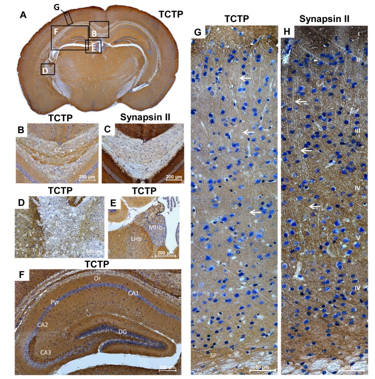
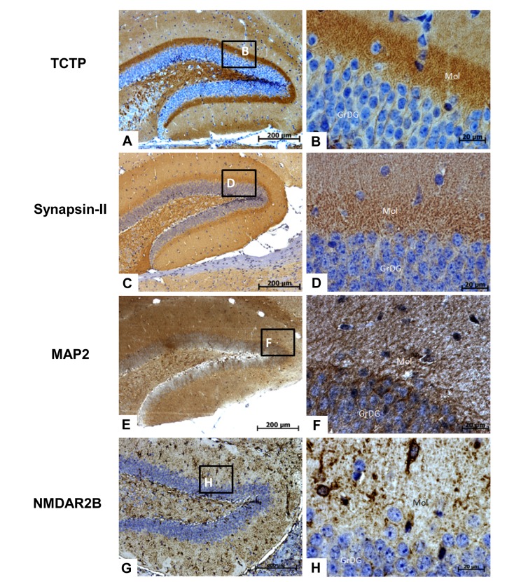
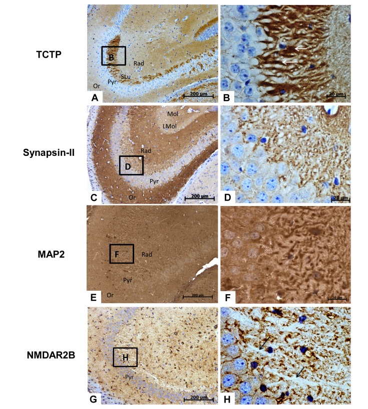
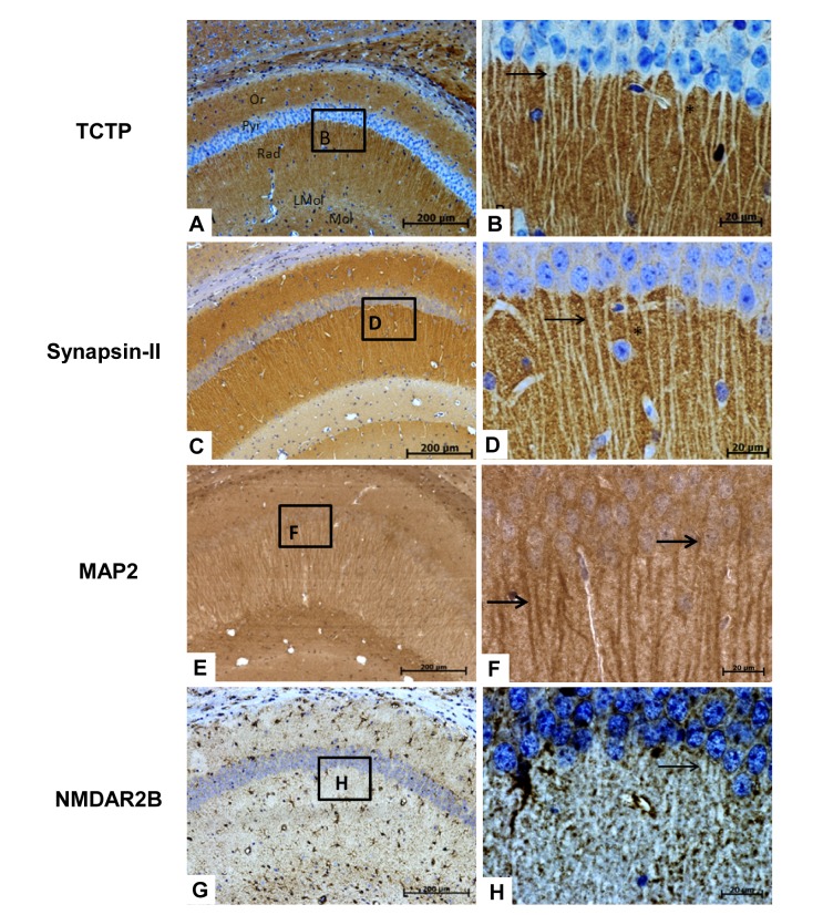
Translationally controlled tumor protein (TCTP) is a cytosolic protein with microtubule stabilization and calcium-binding activities. TCTP is expressed in most organs including the nervous system. However, detailed distribution and functional significance of TCTP in the brain remain unexplored. In this study, we investigated the global and subcellular distributions of TCTP in the mouse brain. Immunohistochemical analyses with anti-TCTP revealed that TCTP was widely distributed in almost all regions of the brain including the cerebral cortex, thalamus, hypothalamus, hippocampus, and amygdala, wherein it was localized in axon tracts and axon terminals. In the hippocampus, TCTP was prominently localized to axon terminals of the perforant path in the dentate gyrus, the mossy fibers in the cornu ammonis (CA)3 region, and the Schaffer collaterals in the CA1 field, but not in cell bodies of granule cells and pyramidal neurons, and in their dendritic processes. Widespread distribution of TCTP in axon tracts and axon terminals throughout the brain suggests that TCTP is likely involved in neurotransmitter release and/or maintaining synaptic structures in the brain, and that it might have a role in maintaining synaptic functions and synaptic configurations important for normal cognitive, stress and emotional functions.

Keywords: Cognition; Immunohistochemistry; Mossy fiber; Mouse hippocampus; Translationally Controlled Tumor Protein (TCTP).


Figures

Fig. 1
Fig. 1. Immunohistochemical localization of TCTP in coronal sections of the mouse brain. Sections were stained by anti-TCTP antibody (a, b, d, e, f, and g) or anti-synapsin II antibody (c, h). Immunohistochemical signals were visualized using DAB and cell nuclei were stained with hematoxylin. Anti-synapsin II staining was used for staining of axonal tracts (c, h). Anti-TCTP staining was widely distributed in the brain, along with distinct localizations in axon tracts and axon terminals. A subset of axon fibers in the corpus callosum (CC) were moderately stained by anti-TCTP, while the internal capsule (IC) was absent from TCTP (d). Axonal fibers running in the subplate (SP) underneath the parietal cortex were stained by anti-TCTP (g, h). Red arrows in the parietal cortex indicate dendritic shafts arising from pyramidal neurons in the layers 3-5 heading towards the layer 1 (g, h). CC, corpus callosum; MHb, medial habenula; LHb, lateral habenula; Or, stratum oriens; Pyr, stratum pyramidale; DG, the dentate gyrus; SP, subplate; I-VI, cortical layers 1-VI. Scale bars: 200 µm (B, C, D, E, F), and 50 µm (G, H).
Fig. 2
Fig. 2. Photomicrographs showing the localization of TCTP in the dentate gyrus. Sections were stained by anti-TCTP antibody (a, b), anti-synapsin II antibody (c, d), anti-MAP2 antibody (e, f), or anti-NMDAR2B (g, h). Immunohistochemical signals were visualized using DAB and cell nuclei were stained with hematoxylin. b, d, f, and h: high magnification of the boxed areas in a, c, e and f. Mol, molecular layer; GrDG, granule cell layer of the dentate gyrus. Scale bars: 200 µm (A, C, E, G), and 20 µm (B, D, F, H).
Fig. 3
Fig. 3. Photomicrographs showing the localization of TCTP in the CA3 region of the hippocampus. Sections were stained by anti-TCTP antibody (a, b), anti-synapsin II antibody (c, d), anti-MAP2 antibody (e, f), or anti-NMDAR2B (g, h). Immunohistochemical signals were visualized using DAB and cell nuclei were stained with hematoxylin. Arrows in b indicate “mossy fiber terminals”. b, d, f, and h: high magnification of the boxed areas in a, c, e and g. Or, stratum oriens; Pyr, stratum pyramidale; Rad, stratum radiatum; SLu, stratum lucidum; Lmol, stratum lacunosum-moleculare; Mol, stratum moleculare. Scale bars: 200 µm (A, C, E, G), and 20 µm (B, D, F, H).
Fig. 4
Fig. 4. Photomicrographs showing the localization of TCTP in the CA1 region of the hippocampus. Sections were stained by anti-TCTP antibody (a, b), anti-synapsin II antibody (c, d), anti-MAP2 antibody (e, f), or anti-NMDAR2B (g, h). Immunohistochemical signals were visualized using DAB and cell nuclei were stained with hematoxylin. Dendritic shafts of pyramidal cells in the CA1 were marked by arrows: In staining with anti-TCTP antibody (b) or anti-synapsin II (d), dendritic shafts from pyramidal cells were not stained at all, while in staining with anti-MAP2, dendritic shafts from pyramidal cells appeared with the internal side stainings (f). Anti-NMDAR2B immunoreactivity was detected in cell membrane of dendritic processes of CA1 pyramidal cells (h). b, d, f, and h: high magnification of the boxed areas in a, c, e and f. Or, stratum oriens; Pyr, stratum pyramidale; Rad, stratum radiatum; Lmol, stratum lacunosum moleculare; Mol, stratum moleculare. Scale bars: 200 µm (A, C, E, G), and 20 µm (B, D, F, H).

Similar articles

Cited by

References

    1. Bommer UA. Cellular function and regulation of the translationally controlled tumour protein TCTP. Open Allergy J. 2012;5:19–32.
    1. Wang D, Gao L. Proteomic analysis of neural differentiation of mouse embryonic stem cells. Proteomics. 2005;5:4414–4426. - PubMed
    1. Corti V, Sanchez-Ruiz Y, Piccoli G, Bergamaschi A, Cannistraci CV, Pattini L, Cerutti S, Bachi A, Alessio M, Malgaroli A. Protein fingerprints of cultured CA3-CA1 hippocampal neurons: comparative analysis of the distribution of synaptosomal and cytosolic proteins. BMC Neurosci. 2008;9:36. - PMC - PubMed
    1. Li F, Zhang D, Fujise K. Characterization of fortilin, a novel antiapoptotic protein. J Biol Chem. 2001;276:47542–47549. - PubMed
    1. Kim SH, Cairns N, Fountoulakisc M, Lubec G. Decreased brain histamine-releasing factor protein in patients with Down syndrome and Alzheimer's disease. Neurosci Lett. 2001;300:41–44. - PubMed