Review
. 2017 Apr 10:11:30.
doi: 10.3389/fnana.2017.00030. eCollection 2017.

Roles of Multiple Globus Pallidus Territories of Monkeys and Humans in Motivation, Cognition and Action: An Anatomical, Physiological and Pathophysiological Review

Affiliations
Review

Roles of Multiple Globus Pallidus Territories of Monkeys and Humans in Motivation, Cognition and Action: An Anatomical, Physiological and Pathophysiological Review

Yosuke Saga et al. Front Neuroanat. .

Abstract

The globus pallidus (GP) communicates with widespread cortical areas that support various functions, including motivation, cognition and action. Anatomical tract-tracing studies revealed that the anteroventral GP communicates with the medial prefrontal and orbitofrontal cortices, which are involved in motivational control; the anterodorsal GP communicates with the lateral prefrontal cortex, which is involved in cognitive control; and the posterior GP communicates with the frontal motor cortex, which is involved in action control. This organization suggests that distinct subdivisions within the GP play specific roles. Neurophysiological studies examining GP neurons in monkeys during behavior revealed that the types of information coding performed within these subdivisions differ greatly. The anteroventral GP is characterized by activities related to motivation, such as reward seeking and aversive avoidance; the anterodorsal GP is characterized by activity that reflects cognition, such as goal decision and action selection; and the posterior GP is characterized by activity associated with action preparation and execution. Pathophysiological studies have shown that GABA-related substances or GP lesions result in abnormal activity in the GP, which causes site-specific behavioral and motor symptoms. The present review article discusses the anatomical organization, physiology and pathophysiology of the three major GP territories in nonhuman primates and humans.

Keywords: GABA; cortico-basal ganglia circuit; functional territory; globus pallidus; human; nonhuman primate; rabies virus.


Figures
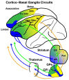
Figure 1
Figure 1
Schematic diagrams of cortico- basal ganglia (BG) anatomical loops with functional territories. The motor (yellow), associative (green) and limbic (blue) territories are represented in circuits between the frontal cortical areas and the BG.
Figure 2
Figure 2
Multiple output channels in the internal segment of the globus pallidus (GPi) projecting to cortical areas represented on a two-dimensional unfolded map. The horizontal and vertical axes indicate the anteroposterior and dorsoventral axes, respectively. Each colored circle in the map represents the output territory of the GPi confirmed by rabies virus (RV) studies. Tick mark intervals = 1 mm.
Figure 3
Figure 3
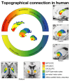
Diffusion tensor imaging (DTI) study showing topographical connections between the cortex and BG in humans. Each frontal cortical area projects to different territories in the BG. Limbic (red: medial and orbital frontal cortices), associative (yellow: dorsolateral prefrontal cortex), higher motor (green: premotor cortex) and motor (blue: motor cortex) cortical territories project to corresponding territories in the BG according to a topographical distribution. Data from Draganski et al. (2008) used with permission from the Society for Neuroscience.
Figure 4
Figure 4
Movement-related activity exhibiting directional preferences in the globus pallidus (GP). Movement-related population activity for the preferred direction (blue) and the anti-preferred direction (red: mean ± SEM) showing excitation and inhibition, respectively. The yellow zone represents the average time of the movement period and the histogram is aligned with touch the screen (“Touch”). Data from Saga et al. (2013) used with permission from the Society for Neuroscience.
Figure 5
Figure 5
Electrophysiological recordings from the GP with estimated functional territories. Neuronal activity recorded from the GP revealed task-related neurons during a variety of task conditions. (A) Movement-related activity mainly found in the ventral aspect of the GP (DeLong, ; data modified and used with the American Physiological Society). (B) Anti-saccade related neurons recorded in the dorsal aspect of the GP (Yoshida and Tanaka, ; data used with permission from Oxford University Press). (C) Recording sites of neuronal activity modulated by memory-guided reaching movements (Turner and Anderson, ; data modified and used with permission from the Society for Neuroscience). (D) Goal-monitoring neurons identified in the dorsal to middle aspect of the GP during movement in which monkeys monitor the behavioral goal (spatial or object, Saga et al., ; data modified and used with permission from the Society for Neuroscience). (E) Reward-related neurons in the ventral pallidum (VP), which also show reward expectation activity for larger rewards (Tachibana and Hikosaka, ; data modified and used with permission from Elsevier). (F) VP neurons showing positive or negative context preference (Saga et al., ; data modified and used with permission from Oxford University Press).
Figure 6
Figure 6
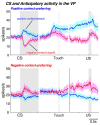
Neuronal population activities for positive motivational context-preferring (upper panel) and negative motivational context-preferring neurons (lower panel) in the VP for conditioned stimulus (CS)-related activity and anticipatory activity. Monkeys performed different motivational tasks in which they chose approach or avoidance behaviors to obtain a reward or prevent the delivery of an air puff depending on the motivational context (Saga et al., 2016). The blue histogram (mean ± SEM) indicates neuronal activity in trials in the positive motivational context and red represents activity in trials in the negative motivational context. Each panel is aligned with CS onset, touch screen and unconditioned stimulus (US) delivery and the gray zone represents the time period of the behavioral event. Data modified from Saga et al. (2016) used with permission from Oxford University Press.

Similar articles

Cited by

References

    1. Akkal D., Dum R. P., Strick P. L. (2007). Supplementary motor area and presupplementary motor area: targets of basal ganglia and cerebellar output. J. Neurosci. 27, 10659–10673. 10.1523/JNEUROSCI.3134-07.2007 - DOI - PMC - PubMed
    1. Alexander G. E., DeLong M. R., Strick P. L. (1986). Parallel organization of functionally segregated circuits linking basal ganglia and cortex. Annu. Rev. Neurosci. 9, 357–381. 10.1146/annurev.neuro.9.1.357 - DOI - PubMed
    1. Alheid G. F., Heimer L. (1988). New perspectives in basal forebrain organization of special relevance for neuropsychiatric disorders: the striatopallidal, amygdaloid, and corticopetal components of substantia innominata. Neuroscience 27, 1–39. 10.1016/0306-4522(88)90217-5 - DOI - PubMed
    1. Arimura N., Nakayama Y., Yamagata T., Tanji J., Hoshi E. (2013). Involvement of the globus pallidus in behavioral goal determination and action specification. J. Neurosci. 33, 13639–13653. 10.1523/JNEUROSCI.1620-13.2013 - DOI - PMC - PubMed
    1. Arkadir D., Morris G., Vaadia E., Bergman H. (2004). Independent coding of movement direction and reward prediction by single pallidal neurons. J. Neurosci. 24, 10047–10056. 10.1523/JNEUROSCI.2583-04.2004 - DOI - PMC - PubMed

LinkOut - more resources